16S rRNA Amplicon Sequencing: A Complete Guide for Microbiome Research in 2024
This comprehensive guide provides researchers and drug development professionals with a detailed, up-to-date overview of 16S rRNA amplicon sequencing.
16S rRNA Amplicon Sequencing: A Complete Guide for Microbiome Research in 2024
Abstract
This comprehensive guide provides researchers and drug development professionals with a detailed, up-to-date overview of 16S rRNA amplicon sequencing. The article covers foundational concepts of the microbial phylogenetic marker and its role in microbial ecology. It details modern methodological workflows from primer selection and library prep through to bioinformatics pipelines like QIIME 2 and DADA2, highlighting applications in drug discovery and clinical diagnostics. Practical troubleshooting sections address common pitfalls in contamination, PCR bias, and low biomass samples. Finally, the guide explores validation strategies, compares 16S sequencing to metagenomic shotgun and culturomics approaches, and discusses its critical role in validating therapeutic microbial consortia. This synthesis offers a complete resource for designing robust, reproducible microbiome studies.
The 16S rRNA Gene: Your Foundational Guide to Microbial Community Profiling
What is the 16S rRNA Gene and Why is it the Gold Standard for Microbial Taxonomy?
The 16S ribosomal RNA (rRNA) gene is a ~1,550 base pair component of the prokaryotic (bacterial and archaeal) 30S ribosomal subunit. It is encoded by the rrs gene and performs critical functions in protein synthesis. Its unique characteristics have cemented its role as the universal molecular chronometer for microbial identification and phylogenetic classification.
Core Properties Establishing it as the Gold Standard:
- Ubiquity and Essential Function: It is present in all prokaryotes, fulfilling an indispensable role in translation.
- Evolutionary Conservation: Specific regions of the gene are highly conserved across all domains of life, allowing for the design of universal PCR primers.
- Hypervariable Regions: Interspersed conserved regions are nine (V1-V9) hypervariable regions that provide genus- and species-specific signatures.
- Low Horizontal Gene Transfer: Its function is so fundamental that it is rarely transferred horizontally, providing a true vertical phylogenetic signal.
- Extensive Reference Databases: Large, curated databases (e.g., SILVA, RDP, Greengenes) contain hundreds of thousands of reference sequences.
Quantitative Comparison of Key 16S rRNA Gene Properties and Databases
Table 1: Characteristics of the Nine Hypervariable (V) Regions
| Region | Approx. Length (bp) | Taxonomic Resolution | Common Sequencing Platforms | Notes |
|---|---|---|---|---|
| V1-V2 | 350 | High for many bacteria | 454, Ion Torrent, MiSeq | Good for skin microbiota. |
| V3-V4 | 460 | High (most common) | MiSeq, NextSeq | Optimal for Illumina 2x250/300 bp runs. |
| V4 | 250-290 | Moderate to High | MiSeq, MiniSeq | Robust, minimal amplification bias. |
| V4-V5 | 390 | Moderate | MiSeq, NextSeq | Balanced resolution and length. |
| V6-V8 | 400+ | Moderate | 454, PacBio | Useful for certain archaea. |
| V9 | ~150 | Lower | All platforms | Short, useful for degraded samples. |
Table 2: Major Public 16S rRNA Gene Reference Databases (2024)
| Database | Latest Version (Year) | Number of High-Quality Sequences | Curated Taxonomy? | Update Frequency | Primary Use Case |
|---|---|---|---|---|---|
| SILVA | SIVA 138.1 (2023) | ~2.7 million aligned | Yes | Regular | Comprehensive phylogeny & taxonomy |
| RDP | RDP 11.5 (2022) | ~3.5 million | Yes (RDP classifier) | Slower | Rapid taxonomic classification |
| Greengenes | 13_8 (2013) | ~1.3 million | Yes | Frozen | Legacy comparisons, QIIME1 |
| NCBI RefSeq | 220 (2024) | ~2.4 million | Semi-automatic | Continuous | Broad, linked to GenBank records |
Detailed Experimental Protocol: 16S rRNA Gene Amplicon Sequencing from Sample to Data
This protocol outlines the standard workflow for Illumina MiSeq sequencing of the V3-V4 region.
A. Sample Preparation and DNA Extraction
- Key Reagent: Bead-beating lysis tubes, enzymatic lysis buffers (Lysozyme, Proteinase K), spin-column or magnetic bead-based purification kits.
- Protocol: For stool, soil, or biofilm samples, use a rigorous mechanical lysis step (bead beating for 2-5 min) combined with chemical/enzymatic lysis. Purify DNA using a kit validated for inhibitor removal (e.g., humic acids). Quantify DNA using fluorometry (e.g., Qubit). Store at -20°C.
B. PCR Amplification of Target Region
- Primers: Use barcoded versions of universal primers (e.g., 341F:
CCTACGGGNGGCWGCAG, 806R:GGACTACHVGGGTWTCTAAT). - Reaction Mix (25 µL):
- 12.5 µL 2x High-Fidelity Master Mix
- 1.0 µL each forward/reverse primer (10 µM)
- 1-10 ng template DNA
- Nuclease-free water to 25 µL
- Thermocycler Conditions:
- 98°C for 30 sec (initial denaturation)
- 25-35 cycles of: 98°C for 10 sec, 55°C for 30 sec, 72°C for 30 sec
- 72°C for 5 min (final extension)
- Purification: Clean amplified products using double-sided magnetic bead cleanup (e.g., 0.8x and 1.2x SPRI ratio).
C. Library Preparation and Sequencing
- Index PCR: Add Illumina flow cell adapters and dual indices via a second, limited-cycle (8 cycles) PCR.
- Pooling & Quantification: Quantify libraries (fluorometry), pool in equimolar ratios, and quantify the final pool (qPCR). Denature with NaOH and dilute to 4-6 pM for loading on a MiSeq with a 15% PhiX spike-in for low-diversity libraries.
- Run Parameters: Use a 2x250 bp or 2x300 bp paired-end run on a MiSeq v2 or v3 kit.
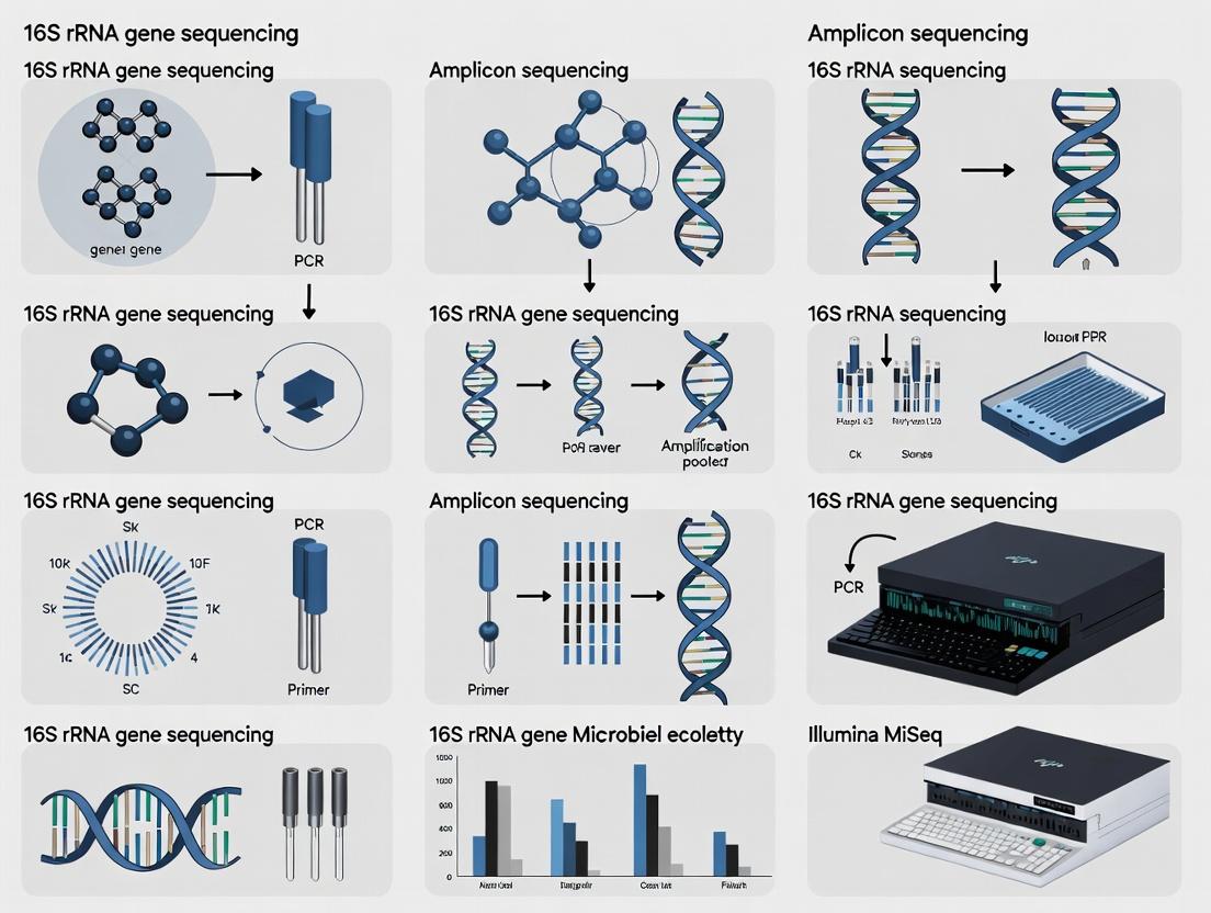
Visualization of Workflows and Concepts
16S Amplicon Sequencing Core Workflow
Primer Binding and Hypervariable Region Analysis
The Scientist's Toolkit: Key Research Reagent Solutions
Table 3: Essential Reagents and Kits for 16S rRNA Gene Sequencing
| Item | Function | Example Product(s) |
|---|---|---|
| Inhibitor-Removing DNA Extraction Kit | Isolate high-purity microbial DNA from complex samples (stool, soil) while removing PCR inhibitors. | DNeasy PowerSoil Pro Kit, MagMAX Microbiome Kit |
| High-Fidelity DNA Polymerase | Perform PCR amplification with low error rates to minimize sequencing artifacts. | Q5 Hot-Start (NEB), KAPA HiFi HotStart |
| Validated 16S Primer Panels | Pre-designed, barcoded primer sets targeting specific hypervariable regions. | Illumina 16S Metagenomic Library Prep, QIAGEN QIAseq 16S Panels |
| Magnetic Bead Cleanup Reagents | For size selection and purification of PCR products (removes primers, dimers). | AMPure XP Beads, Sera-Mag Select Beads |
| Library Quantification Kit | Accurate qPCR-based quantification of final library pool for precise sequencing loading. | KAPA Library Quant Kit |
| Positive Control (Mock Community) | Defined mix of genomic DNA from known species to assess run accuracy and bias. | ZymoBIOMICS Microbial Community Standard |
| Negative Control (No-Template) | PCR water control to identify reagent/lab-borne contamination. | Nuclease-Free Water |
| Bioinformatics Pipeline Software | Process raw sequences into taxonomic units and diversity metrics. | QIIME 2, mothur, DADA2 (R package) |
Application Notes on 16S rRNA Gene Regions
The bacterial 16S ribosomal RNA (rRNA) gene (~1,500 bp) consists of nine hypervariable regions (V1-V9) interspersed with conserved regions. The selection of which region(s) to sequence is the primary determinant of taxonomic resolution and experimental outcome in amplicon sequencing studies.
Table 1: Characteristics and Phylogenetic Resolution of 16S rRNA Hypervariable Regions
| Region | Approx. Length (bp) | Taxonomic Resolution (General) | Key Considerations & Common Use Cases |
|---|---|---|---|
| V1-V2 | 330-360 | High (Genus/Species) | High sequence diversity; good for distinguishing closely related species. Can be prone to chimeras. Common in human microbiome studies (e.g., Illumina MiSeq with 2x300bp). |
| V3-V4 | 460-480 | Moderate to High (Genus) | The current most widely adopted region (e.g., Illumina MiSeq 16S Metagenomic Sequencing Library Prep). Balanced resolution, robust primer sets, and well-curated databases (e.g., SILVA, Greengenes). |
| V4 | 250-260 | Moderate (Genus/Family) | Shorter, highly accurate. Used by the Earth Microbiome Project. Excellent for high-throughput sequencing but may lack resolution for some closely related species. |
| V4-V5 | ~400 | Moderate (Genus) | A compromise offering slightly more information than V4 alone. Useful for environmental samples with high diversity. |
| V6-V8 / V7-V9 | 380-500 | Lower (Family/Phylum) | Often used with long-read platforms (e.g., PacBio, Oxford Nanopore) for full-length or near-full-length 16S sequencing. V9 alone is very short and rarely used. |
| Full-length (V1-V9) | ~1,500 | Highest (Species/Strain) | Provides maximum phylogenetic resolution. Enabled by third-generation sequencing. Essential for novel species discovery and high-resolution phylogenetics. |
Core Principle: The conserved regions flanking hypervariable segments enable the design of universal PCR primers that amplify target sequences from a vast range of bacteria. The hypervariable regions contain the phylogenetic signal. The number of informative variable sites sequenced directly correlates with potential phylogenetic resolution. Therefore, sequencing a single hypervariable region (e.g., V4) is cost-effective for community profiling but may collapse distinct species into the same operational taxonomic unit (OTU) or amplicon sequence variant (ASV). In contrast, sequencing multiple or all variable regions increases discrimination power.
Protocol: Comparative Analysis of V4 vs. V1-V9 Amplicons for High-Resolution Phylogenetics
Objective: To evaluate the trade-off between read depth/breadth (short-amplicon) and phylogenetic resolution (long-amplicon) in a complex microbial community sample (e.g., gut microbiome, soil).
I. Experimental Design & Sample Preparation
- Sample: Use a well-characterized mock microbial community (e.g., ZymoBIOMICS Microbial Community Standard) alongside environmental samples.
- DNA Extraction: Perform extraction using a standardized kit (e.g., DNeasy PowerSoil Pro Kit) to ensure uniform lysis across cell types.
- PCR Amplification:
- Short-Amplicon (V4): Amplify using primers 515F (5′-GTGYCAGCMGCCGCGGTAA-3′) and 806R (5′-GGACTACNVGGGTWTCTAAT-3′).
- Long-Amplicon (V1-V9): Amplify using primers 27F (5′-AGRGTTTGATYMTGGCTCAG-3′) and 1492R (5′-RGYTACCTTGTTACGACTT-3′).
- Sequencing Platform: Sequence V4 amplicons on an Illumina MiSeq (2x250bp). Sequence V1-V9 amplicons on a PacBio Sequel IIe system (Circular Consensus Sequencing mode) or an Oxford Nanopore MinION.
II. Bioinformatic Analysis Workflow
Diagram Title: Bioinformatic Workflow for Short vs. Long 16S Amplicons
III. Key Metrics for Comparison Table 2: Comparative Analysis Metrics for V4 vs. V1-V9 Protocols
| Metric | V4 Illumina Protocol | V1-V9 Long-Read Protocol | Interpretation for Thesis |
|---|---|---|---|
| Mean Read Depth per Sample | Very High (~50,000-100,000) | Moderate (~10,000-50,000) | V4 better for detecting rare taxa. |
| Observed ASVs/OTUs in Mock Community | Accurate at genus, may merge species. | Should resolve all expected species/strains. | Quantifies resolution loss in short-amplicon. |
| Distance to Reference Phylogeny (e.g., Robinson-Foulds distance) | Higher (Less accurate tree) | Lower (More accurate tree) | Direct measure of phylogenetic fidelity. |
| Beta Diversity Stability (PERMANOVA on Bray-Curtis) | May show inflated technical variation between regions. | Community differences more aligned with biology. | Informs choice for longitudinal studies. |
| Computational Load & Cost | Lower cost, faster processing. | Higher cost, specialized tools needed. | Practical consideration for study design. |
The Scientist's Toolkit: Key Research Reagent Solutions
Table 3: Essential Materials for 16S rRNA Amplicon Sequencing Studies
| Item | Function & Rationale |
|---|---|
| Standardized Mock Community (e.g., ZymoBIOMICS D6300) | Contains known abundances of bacterial/fungal strains. Serves as a positive control to benchmark primer bias, resolution, and bioinformatic pipeline accuracy. |
| Bias-Reduced Polymerase (e.g., KAPA HiFi HotStart) | High-fidelity polymerase with minimal GC-bias is critical for accurate representation of community composition during PCR amplification. |
| Dual-Indexed PCR Primer Kits (e.g., Nextera XT Index Kit) | Allows multiplexing of hundreds of samples in one sequencing run by attaching unique barcodes to each sample during PCR. |
| Magnetic Bead-Based Cleanup System (e.g., AMPure XP Beads) | For reproducible size selection and purification of PCR amplicons, removing primer dimers and contaminants. |
| Quantification Kit (e.g., Qubit dsDNA HS Assay) | Fluorometric quantification is essential for accurate normalization and pooling of amplicon libraries, unlike absorbance-based methods. |
| Platform-Specific Sequencing Kit | Illumina MiSeq Reagent Kit v3 (600-cycle) for V4. PacBio SMRTbell Express Template Prep Kit 2.0 for V1-V9. |
| Curated Reference Database (e.g., SILVA, GTDB, RDP) | Essential for taxonomic assignment. Choice impacts results; GTDB offers modern phylogeny, SILVA is widely used for V4. Full-length sequences improve long-read analysis. |
Application Notes: Comparative Analysis of Sequencing Eras
The evolution from Sanger to Next-Generation Sequencing (NGS) for 16S rRNA gene amplicon sequencing represents a paradigm shift in microbial ecology and drug discovery research. This transition underpins a broader thesis on how technological advancement has exponentially increased the scale, resolution, and application of microbiome research, directly impacting biomarker discovery and therapeutic development.
Key Evolutionary Milestones:
- Sanger Era (1977-2005): Characterized by single-amplicon, clone-based sequencing. Provided high accuracy but was low-throughput, expensive, and limited in its ability to describe complex communities.
- NGS Era (2005-Present): Marked by massively parallel sequencing of amplicon libraries. Enabled high-throughput, cost-effective profiling of entire microbial communities from complex samples, revealing unprecedented diversity.
Quantitative Comparison of Technologies:
Table 1: Technical and Performance Comparison of 16S Sequencing Technologies
| Parameter | Sanger Sequencing | Next-Generation Sequencing (Illumina MiSeq) |
|---|---|---|
| Reads/Run | 96 (per capillary array) | 25 million |
| Read Length | ~900-1000 bp (full-length 16S) | 2x300 bp (V3-V4 hypervariable regions) |
| Cost per Sample | High (~$10-$20 per read) | Low (<$10 per sample for multiplexed run) |
| Throughput Time | Days for cloning + sequencing | < 3 days (library prep to data) |
| Primary Application | Isolate identification, phylogenetic studies | Complex community profiling, alpha/beta diversity |
| Key Limitation | Low depth, cannot capture rare taxa | Shorter reads, PCR/sequencing errors requiring robust bioinformatics |
Table 2: Impact on Microbial Community Analysis
| Metric | Sanger (Clone Library) | NGS (Amplicon Seq) |
|---|---|---|
| Observed OTUs per sample | 10s - 100s | 1000s - 10,000s |
| Coverage of Rare Biosphere | Minimal | Significant |
| Statistical Power | Low for complex comparisons | High, enables multivariate analysis |
| Suitability for Longitudinal Studies | Poor (cost/depth) | Excellent |
Experimental Protocols
Protocol 2.1: Historical Sanger Sequencing of 16S rRNA Gene Clones
This protocol outlines the traditional method for obtaining full-length 16S sequences from environmental samples, critical for foundational phylogenetic trees.
Materials:
- Genomic DNA from microbial isolate or environmental sample.
- Universal 16S rRNA gene primers (e.g., 27F: 5'-AGAGTTTGATCMTGGCTCAG-3', 1492R: 5'-GGTTACCTTGTTACGACTT-3').
- PCR reagents, TA Cloning Kit, competent E. coli, LB-Amp plates.
- Plasmid purification kit, BigDye Terminator v3.1 Cycle Sequencing Kit.
- Capillary sequencer.
Procedure:
- PCR Amplification: Amplify the ~1500 bp 16S gene using universal primers. Verify amplicon on agarose gel.
- Cloning: Ligate purified PCR product into a TA cloning vector. Transform into competent E. coli. Plate on selective media.
- Colony Screening: Pick 96-384 colonies. Perform colony PCR with vector-specific primers to confirm insert size.
- Plasmid Preparation: Inoculate positive clones in liquid culture. Purify plasmid DNA.
- Sanger Sequencing: Set up sequencing reactions for each plasmid using BigDye chemistry and primers (M13F/R). Purify reactions.
- Capillary Electrophoresis: Run purified reactions on the sequencer.
- Analysis: Manually curate and assemble contiguous sequences. Perform BLAST against NCBI database for identification.
Protocol 2.2: Contemporary NGS Amplicon Sequencing (Illumina 2x300 bp)
This is the current standard workflow for high-throughput 16S community profiling, generating millions of reads for complex sample sets.
Materials:
- Extracted genomic DNA.
- 16S V3-V4 region primers with overhang adapters (e.g., 341F: 5'-CCTACGGGNGGCWGCAG-3', 805R: 5'-GACTACHVGGGTATCTAATCC-3').
- High-fidelity DNA polymerase, AMPure XP beads.
- Indexing primers (Nextera XT Index Kit), PCR reagents.
- Quantification kit (Qubit), Library Normalization Beads.
- MiSeq Reagent Kit v3 (600-cycle).
Procedure:
- First-Stage PCR (Amplicon with Overhangs): Amplify the target V3-V4 region using primers containing Illumina adapter overhangs. Clean up with AMPure XP beads.
- Indexing PCR (Dual Indexing): Attach unique dual indices and full Illumina adapters to the amplicon using a limited-cycle PCR. Clean up with AMPure XP beads.
- Library Quantification & Normalization: Quantify each library fluorometrically. Normalize to equal molarity.
- Pooling: Combine normalized libraries into a single pool.
- Denature & Dilute: Denature the pooled library with NaOH and dilute to optimal loading concentration in hybridization buffer.
- Sequencing: Load onto MiSeq flow cell. Run with 2x300 bp paired-end chemistry.
- Bioinformatics Processing: Demultiplex reads. Merge paired-ends. Perform quality filtering (DADA2 or Deblur), chimera removal, assign taxonomy against a reference database (e.g., SILVA, Greengenes).
Visualizations
Evolution of 16S Sequencing: Two Parallel Workflows
Technological Evolution Drives Thesis Research Scope
The Scientist's Toolkit: Essential Research Reagents & Materials
Table 3: Key Reagent Solutions for Modern 16S NGS Workflow
| Item | Function | Example Product/Kit |
|---|---|---|
| Magnetic Bead Cleanup | Size selection and purification of PCR products; removes primers, dNTPs, and salts. | AMPure XP Beads |
| High-Fidelity DNA Polymerase | Reduces PCR errors during initial amplicon generation, crucial for accurate variant calling. | Q5 Hot Start Polymerase, KAPA HiFi |
| Dual-Indexed Adapter Kit | Attaches unique barcode combinations to each sample for multiplexing, enabling sample identification post-sequencing. | Illumina Nextera XT Index Kit, 16S Metagenomic Kit |
| Library Quantification Kit | Accurate fluorometric measurement of library concentration for precise pooling. | Qubit dsDNA HS Assay |
| Normalization Beads | Simplifies library pooling by automating equalization of library concentrations. | Illumina Library Normalization Beads |
| PhiX Control v3 | Serves as a quality control for cluster generation, sequencing, and alignment; essential for low-diversity 16S libraries. | Illumina PhiX Control |
| Sequencing Reagent Cartridge | Contains enzymes, buffers, and nucleotides for the sequencing-by-synthesis chemistry. | MiSeq Reagent Kit v3 |
| Bioinformatics Pipeline | Software for processing raw reads into biological insights (QC, clustering, taxonomy). | QIIME 2, Mothur, DADA2 |
1. Introduction within 16S rRNA Amplicon Sequencing Research This Application Note details protocols for leveraging 16S rRNA gene sequencing to establish causative and diagnostic links between gut microbial dysbiosis, specific disease states, and variability in therapeutic drug response. Framed within a thesis on amplicon sequencing, it provides actionable methodologies for researchers and drug development professionals to translate taxonomic profiles into mechanistic insights and predictive biomarkers.
2. Quantitative Summary of Dysbiosis-Disease-Drug Associations Table 1: Key Disease-Associated Dysbiosis Signatures and Drug Metabolism Impacts
| Disease State | Dysbiosis Signature (Common 16S Findings) | Linked Microbial Function | Impact on Drug/Response | Reported Effect Size (e.g., Odds Ratio/Change) |
|---|---|---|---|---|
| Inflammatory Bowel Disease (IBD) | ↓ Faecalibacterium prausnitzii (Firmicutes), ↑ Escherichia/Shigella (Proteobacteria) | Reduced SCFA (butyrate) production; increased mucosal inflammation. | Altered anti-TNFα (infliximab) response. | Non-responders show 2.3x lower microbial diversity at baseline. |
| Colorectal Cancer (CRC) | ↑ Fusobacterium nucleatum, ↑ Bacteroides fragilis (enterotoxic), ↓ Roseburia spp. | Pro-inflammatory; activation of oncogenic signaling (β-catenin). | Affects efficacy of 5-fluorouracil and immunotherapy (checkpoint inhibitors). | High F. nucleatum associated with 3.5x increased cancer recurrence risk. |
| Type 2 Diabetes | ↓ Akkermansia muciniphila, ↑ Lactobacillus gasseri, altered Firmicutes/Bacteroidetes ratio. | Impaired gut barrier function; metabolic endotoxemia. | Modifies metformin efficacy; influences pharmacokinetics. | A. muciniphila abundance inversely correlates (r=-0.42) with HbA1c levels. |
| Checkpoint Inhibitor Immunotherapy | ↑ Akkermansia muciniphila, ↑ Faecalibacterium spp., ↑ Bifidobacterium spp. | Enhanced antigen presentation and T-cell priming. | Predicts response to PD-1 inhibitors (pembrolizumab, nivolumab). | Responders have 4-5x higher abundance of predictive taxa. |
| Cardiovascular Disease | ↑ Trimethylamine (TMA)-producing bacteria (e.g., Clostridium, Emergencia), ↓ SCFA producers. | Increased TMAO production from dietary choline/carnitine. | Reduces efficacy of statins; TMAO is a independent risk factor. | High TMAO levels correlate with 2.5x increased major adverse cardiac event risk. |
3. Detailed Experimental Protocols
Protocol 3.1: Longitudinal Cohort Study for Linking Dysbiosis to Drug Response Objective: To identify pre-treatment microbial biomarkers predictive of drug efficacy or adverse events. Workflow:
- Cohort & Sampling: Recruit patients (n ≥ 50 per arm) prior to initiating therapy. Collect stool, blood, and clinical metadata at baseline (T0).
- DNA Extraction & 16S Sequencing: Use bead-beating mechanical lysis kit (e.g., QIAamp PowerFecal Pro DNA Kit). Amplify the V3-V4 hypervariable region with primers 341F/806R. Sequence on Illumina MiSeq (2x300 bp).
- Bioinformatics: Process raw reads via QIIME 2 (2024.2). Denoise with DADA2. Assign taxonomy using SILVA v138 reference database. Generate ASV (Amplicon Sequence Variant) table.
- Statistical Integration: Correlate baseline ASV relative abundance (α/β-diversity) with primary clinical endpoint (e.g., drug response at 12 weeks) using multivariate models (PERMANOVA, LEfSe, Random Forest).
- Validation: Validate predictive taxa in an independent validation cohort using targeted qPCR.
Protocol 3.2: In Vitro Functional Validation of Microbial Drug Metabolism Objective: To characterize direct microbial biotransformation of a target drug. Workflow:
- Bacterial Culture: Anaerobically culture candidate bacterial strain(s) in pre-reduced medium.
- Drug Incubation: Add therapeutic drug at physiologically relevant concentration (e.g., 100 μM) to mid-log phase culture. Include sterile medium + drug control.
- Sampling & Quenching: Collect aliquots at T=0, 2, 6, 24h. Centrifuge immediately (13,000 x g, 5 min, 4°C). Store supernatant at -80°C.
- Metabolite Analysis: Analyze supernatants via LC-MS/MS. Quantify parent drug and suspected metabolites using authentic standards.
- Enzyme Identification: Perform comparative genomics on active vs. inactive strains. Express putative microbial enzymes heterologously in E. coli to confirm metabolic activity.
4. Visualization of Key Pathways and Workflows
Title: Dysbiosis Drives Inflammation and Modulates Drug Response in IBD
Title: 16S-Based Prediction of Immunotherapy Outcome
5. The Scientist's Toolkit: Research Reagent Solutions Table 2: Essential Materials for 16S-Based Dysbiosis-Drug Studies
| Item / Reagent Solution | Function / Purpose | Example Product |
|---|---|---|
| Stabilization Buffer | Preserves microbial community structure at room temperature for transport/storage. | OMNIgene•GUT, Zymo DNA/RNA Shield |
| Mechanical Lysis DNA Kit | Robust cell wall disruption for Gram-positive bacteria; yields high-quality, unbiased genomic DNA. | QIAamp PowerFecal Pro DNA Kit, MP Biomedicals FastDNA Spin Kit |
| PCR Inhibitor Removal Beads | Critical for stool samples; removes humic acids and other PCR inhibitors. | OneStep PCR Inhibitor Removal Kit, Zymo-Spin IC Columns |
| 16S PCR Primers (Barcoded) | Amplifies target hypervariable region with unique sample indexes for multiplexing. | Illumina 16S Metagenomic Library Prep, Earth Microbiome Project primers |
| Positive Control Mock Community | Validates entire wet-lab and bioinformatics pipeline; assesses bias and sensitivity. | ZymoBIOMICS Microbial Community Standard, ATCC MSA-1003 |
| Bioinformatics Pipeline | Standardized analysis from raw reads to taxonomic profiles and diversity metrics. | QIIME 2, mothur, DADA2 (R package) |
| Statistical Analysis Software | Performs multivariate analysis linking microbiome data to clinical covariates. | R (vegan, phyloseq, LEfSe packages), SIMCA (PLS-DA) |
In 16S rRNA gene amplicon sequencing research, characterizing microbial communities requires standardized metrics. Alpha diversity, beta diversity, and taxonomic composition form the foundational triad for interpreting ecological structure, stability, and responses to perturbation. This application note details their definitions, calculation protocols, and integration within a drug development research framework.
Key Metrics: Definitions and Quantitative Comparisons
Table 1: Core Diversity Metrics in 16S rRNA Amplicon Analysis
| Metric Category | Specific Metric | Formula/Description | Interpretation | Typical Value Range | ||||
|---|---|---|---|---|---|---|---|---|
| Alpha Diversity | Observed ASVs/OTUs | Count of distinct sequences in a sample. | Simple richness. | 10s - 1000s | ||||
| Chao1 | $$S{Chao1} = S{obs} + \frac{F1^2}{2F2}$$ | Estimates total richness, correcting for rare species. | ≥ Observed count | |||||
| Shannon Index (H') | $$H' = -\sum{i=1}^{S} pi \ln(p_i)$$ | Combines richness and evenness. Higher = more diverse. | Typically 1.5 - 7 | |||||
| Simpson Index (λ) | $$\lambda = \sum{i=1}^{S} pi^2$$ | Probability two random reads are same species. Lower = more diverse. | 0 - 1 | |||||
| Beta Diversity | Jaccard Distance | $$D_{J} = 1 - \frac{ | A \cap B | }{ | A \cup B | }$$ (presence/absence) | Dissimilarity based on shared features. | 0 (identical) to 1 (no overlap) |
| Bray-Curtis Dissimilarity | $$D{BC} = \frac{\sumi |xi - yi|}{\sumi (xi + y_i)}$$ (abundance-aware) | Most common for microbial ecology. | 0 (identical) to 1 (no overlap) | |||||
| Weighted UniFrac | Phylogenetic distance weighted by abundance. | Differences driven by abundant lineages. | 0 to 1 | |||||
| Unweighted UniFrac | Phylogenetic distance based on presence/absence. | Differences driven by rare lineages. | 0 to 1 | |||||
| Taxonomic Composition | Relative Abundance | Proportion of reads assigned to a taxon. | Community profile. | 0 - 1 (per taxon) |
Detailed Experimental Protocols
Protocol 1: Standardized Alpha & Beta Diversity Analysis Pipeline (QIIME 2)
Objective: To calculate alpha and beta diversity metrics from a filtered ASV/OTU table. Reagents & Software: QIIME 2 (2024.5+), rarefied feature table, rooted phylogenetic tree. Procedure:
- Rarefaction: Rarefy the feature table to an even sampling depth to avoid sequencing bias.
qiime diversity core-metrics-phylogenetic --i-table filtered-table.qza --i-phylogeny rooted-tree.qza --p-sampling-depth 10000 --output-dir core-metrics-results - Alpha Diversity: Extract alpha diversity vectors (Faith_pd, Shannon, Simpson) and test for group differences using Kruskal-Wallis.
qiime diversity alpha-group-significance --i-alpha-diversity core-metrics-results/faith_pd_vector.qza --m-metadata-file sample_metadata.tsv --o-visualization faith-pd-group-significance.qzv - Beta Diversity: Perform PERMANOVA on distance matrices (e.g., Bray-Curtis, Weighted UniFrac) to test for significant clustering by experimental group.
qiime diversity beta-group-significance --i-distance-matrix core-metrics-results/bray_curtis_distance_matrix.qza --m-metadata-file sample_metadata.tsv --p-method permanova --o-visualization bray-curtis-significance.qzv - Visualization: Generate PCoA plots for principal coordinate analysis.
qiime emperor plot --i-pcoa core-metrics-results/bray_curtis_pcoa_results.qza --m-metadata-file sample_metadata.tsv --o-visualization bray-curtis-emperor.qzv
Protocol 2: Taxonomic Composition and Differential Abundance Analysis
Objective: To profile community composition and identify taxa significantly altered between conditions. Reagents & Software: SILVA/GTB database, QIIME 2, or R packages (phyloseq, DESeq2, ANCOM-BC). Procedure:
- Taxonomic Assignment: Classify ASVs using a pre-trained naive Bayes classifier.
qiime feature-classifier classify-sklearn --i-reads rep-seqs.qza --i-classifier silva-138-99-nb-classifier.qza --o-classification taxonomy.qza - Create Bar Plots: Generate visual summaries of mean relative abundance per group.
qiime taxa barplot --i-table filtered-table.qza --i-taxonomy taxonomy.qza --m-metadata-file sample_metadata.tsv --o-visualization taxa-bar-plots.qzv - Differential Abundance Testing: Use ANCOM-BC (recommended for compositional data) in R to identify significantly differentially abundant taxa between control and treatment groups, controlling for false discovery rate (FDR).
Diagrams
Title: 16S Amplicon Analysis Core Workflow
Title: Relationship Between Core 16S Analysis Metrics
The Scientist's Toolkit: Research Reagent Solutions
Table 2: Essential Materials for 16S rRNA Amplicon Diversity Studies
| Item | Supplier Examples | Function in Protocol |
|---|---|---|
| DNA Extraction Kit (Stool) | Qiagen (QIAamp PowerFecal Pro), MoBio (DNeasy PowerLyzer) | Standardized microbial genomic DNA isolation, critical for bias-free community representation. |
| 16S rRNA Gene Primers (V3-V4) | Integrated DNA Technologies (IDT), Thermo Fisher | Amplification of hypervariable regions (e.g., 341F/806R) for Illumina sequencing. |
| High-Fidelity PCR Master Mix | NEB (Q5), KAPA HiFi | Accurate amplification with low error rates for precise ASV calling. |
| Size-Selective Magnetic Beads | Beckman Coulter (AMPure XP), MagBio | Post-PCR clean-up and library normalization to remove primer dimers and select target fragment size. |
| Indexed Adapters & Sequencing Kit | Illumina (Nextera XT Index Kit v2), | Adds unique sample barcodes for multiplexing and enables cluster generation on flow cell. |
| Positive Control (Mock Community) | ATCC (MSA-1000), ZymoBIOMICS | Validates entire wet-lab and bioinformatics pipeline accuracy and detects batch effects. |
| Negative Extraction Control | N/A (Molecular grade water) | Identifies contamination introduced during sample processing. |
| Bioinformatics Pipeline | QIIME 2, mothur, DADA2 | End-to-end analysis platform for processing raw sequences to diversity metrics and taxonomy. |
| Reference Database | SILVA, Greengenes, GTDB | For taxonomic assignment of ASV sequences; choice influences nomenclature and resolution. |
From Sample to Insight: A Step-by-Step 16S Sequencing Protocol for Modern Labs
Within the broader thesis on 16S rRNA gene amplicon sequencing research, the selection of appropriate primers is a foundational step that dictates the resolution, accuracy, and scope of microbial community analysis. The choice between targeting the full-length (~1,500 bp) 16S rRNA gene and specific hypervariable regions (V1-V9, ~100-400 bp each) presents a critical strategic divergence with significant implications for taxonomic classification, phylogenetic inference, and experimental feasibility. This document provides updated application notes and protocols to guide researchers, scientists, and drug development professionals in making an informed primer selection aligned with their research objectives.
Table 1: Quantitative Comparison of Full-Length vs. Hypervariable Region Amplification (2024)
| Parameter | Full-Length 16S (e.g., 27F-1492R) | Single/Multi-Hypervariable Region (e.g., V3-V4) | Notes & Recent Insights |
|---|---|---|---|
| Amplicon Length | ~1,500 bp | Typically 300-600 bp (e.g., V4~290bp, V3-V4~460bp) | Long-read platforms (PacBio, Nanopore) enable full-length. Short-read (Illumina) favors hypervariable regions. |
| Taxonomic Resolution | Species to strain level. | Genus to species level; resolution varies by region. | V4-V5 offers best balance for bacterial phylogeny. V1-V3 may improve Firmicutes resolution. |
| PCR Efficiency/Bias | Lower efficiency; higher bias due to secondary structure. | Higher efficiency; region-specific biases exist. | Primer degeneracy and locked nucleic acids (LNAs) are used to reduce bias. |
| Sequencing Platform | PacBio SEQUEL II/Revio, Oxford Nanopore. | Illumina MiSeq/NovaSeq, Ion Torrent. | Full-length on Illumina is not standard. |
| Error Rate | Higher raw error rates (~10-15%) for long-read tech. | Very low error rates (~0.1%) for Illumina. | Circular Consensus Sequencing (CCS) for PacBio reduces errors to <0.01%. |
| Cost Per Sample | High (platform and sequencing depth). | Low to moderate. | Multiplexing capacity of Illumina keeps costs down for large cohorts. |
| Bioinformatics Complexity | High; requires specialized long-read pipelines. | Moderate; well-established pipelines (QIIME 2, Mothur). | DADA2, Deblur work well for Illumina; tools like EMU for long-read. |
| Reference Databases | SILVA, GTDB, RDP. Curated full-length databases growing. | SILVA, Greengenes. More curated options for specific regions. | GTDB (Genome Taxonomy Database) is critical for modern full-length classification. |
| Primary Application | High-resolution phylogeny, species-strain discrimination, novel taxon discovery. | Large-scale population studies, microbiome association studies, clinical diagnostics. | FDA-recognized assays (e.g., for sepsis) often target specific hypervariable regions. |
Detailed Experimental Protocols
Protocol 1: Full-Length 16S rRNA Gene Amplification for PacBio HiFi Sequencing
Objective: Generate high-fidelity (HiFi) full-length 16S amplicons for species-level community profiling. Reagents: KAPA HiFi HotStart ReadyMix, PacBio Barcoded Universal Primers (27F: AGRGTTYGATYMTGGCTCAG, 1492R: RGYTACCTTGTTACGACTT), AMPure PB beads. Workflow:
- Genomic DNA Input: 10-100 ng of microbial genomic DNA (minimal host contamination).
- First-Stage PCR (Barcoding):
- Reaction: 25 μL KAPA HiFi Mix, 0.3 μM each forward and barcoded reverse primer, 5 μL template, nuclease-free water to 50 μL.
- Cycling: 95°C/3min; 25 cycles of [98°C/20s, 55°C/15s, 72°C/90s]; 72°C/5min.
- Purification: Clean amplified products with 0.8x AMPure PB beads. Elute in 30 μL EB buffer.
- Second-Stage PCR (Adapter Addition - SMRTbell):
- Use PacBio SMRTbell prep kit. Combine ~200 ng purified PCR product with overhang adapter primers in a 50 μL KAPA HiFi reaction.
- Cycle: 95°C/3min; 10 cycles of [98°C/20s, 60°C/15s, 72°C/90s]; 72°C/5min.
- Purification & Size Selection: Double-size select with AMPure PB beads (0.45x to remove small fragments, then 0.2x to recover SMRTbell library).
- Sequencing: Quantify with Qubit. Sequence on PacBio Revio system using a 30h movie time with CCS mode enabled (>10 passes per molecule).
Protocol 2: Dual-Indexed Hypervariable Region (V3-V4) Amplification for Illumina
Objective: Robust amplification of the V3-V4 region for high-throughput, multi-sample studies. Reagents: Phusion Plus PCR Master Mix, Illumina Nextera XT Index Kit v2, AMPure XP beads. Primers: 341F (CCTACGGGNGGCWGCAG), 806R (GGACTACHVGGGTWTCTAAT). Workflow:
- Genomic DNA Input: 1-10 ng DNA.
- First-Stage PCR (Amplicon with Overhangs):
- Reaction: 12.5 μL Phusion Plus Mix, 0.2 μM each primer (with Illumina overhang adapters), 2.5 μL template, water to 25 μL.
- Cycling: 98°C/30s; 25 cycles of [98°C/10s, 55°C/30s, 72°C/30s]; 72°C/5min.
- Purification: Clean with 1x AMPure XP beads. Elute in 25 μL RSB.
- Indexing PCR (Dual Indexing):
- Use 5 μL purified amplicon, 2.5 μL each Nextera XT index primer (i5 & i7), 12.5 μL Phusion Plus Mix, water to 25 μL.
- Cycle: 95°C/3min; 8 cycles of [95°C/30s, 55°C/30s, 72°C/30s]; 72°C/5min.
- Final Purification & Pooling: Clean each with 0.9x AMPure XP beads. Quantify by fluorometry, then pool equimolarly.
- Sequencing: Denature and dilute pool per Illumina protocol. Sequence on MiSeq with 2x300bp v3 chemistry or NovaSeq 6000.
Visualized Workflows & Decision Pathways
Title: Primer Selection Decision Pathway
Title: Comparative Experimental Workflows
The Scientist's Toolkit: Research Reagent Solutions
Table 2: Essential Materials for 16S rRNA Amplicon Sequencing
| Item | Function & Rationale | Example Product (2024) |
|---|---|---|
| High-Fidelity DNA Polymerase | Minimizes PCR errors critical for accurate sequence variant calling. Essential for long amplicons. | KAPA HiFi HotStart ReadyMix (Roche), Q5 High-Fidelity (NEB). |
| Barcoded/Indexed Primer Sets | Enables multiplexing of hundreds of samples in a single sequencing run. | PacBio Barcoded Universal Primers, Illumina Nextera XT Index Kit v2. |
| Magnetic Bead Cleanup Reagents | For size-selective purification and removal of primers, dNTPs, and salts. Crucial for library prep. | AMPure PB/PCRclean DX beads (Beckman), AMPure XP beads (Beckman). |
| Fluorometric DNA Quantification Kit | Accurate quantification of library molecules for optimal sequencing loading. | Qubit dsDNA HS Assay Kit (Thermo Fisher), Quant-iT PicoGreen. |
| Mock Microbial Community | Positive control to assess primer bias, PCR fidelity, and bioinformatics pipeline accuracy. | ZymoBIOMICS Microbial Community Standard (Zymo Research). |
| Inhibitor Removal Technology | Critical for complex samples (stool, soil) to ensure efficient PCR amplification. | OneStep PCR Inhibitor Removal Kit (Zymo), PowerSoil Pro Kit (Qiagen). |
| Bioinformatics Pipeline Software | For processing raw reads to amplicon sequence variants (ASVs) and taxonomic tables. | QIIME 2, DADA2 (Illumina), EMU, minimap2/DTU (long-read). |
Within the context of 16S rRNA gene amplicon sequencing for microbial community analysis, the selection of a library preparation platform is a critical determinant of data quality, throughput, and cost. This application note provides a detailed comparison of library preparation workflows from the three dominant platforms—Illumina, PacBio, and Ion Torrent—as applied to 16S rRNA amplicon sequencing. The protocols and data herein are designed to guide researchers and drug development professionals in selecting the optimal methodology for their specific research questions in metagenomics and biomarker discovery.
Platform Comparison Tables
Table 1: Core Platform Characteristics for 16S rRNA Sequencing
| Feature | Illumina (MiSeq) | PacBio (Sequel IIe) | Ion Torrent (Ion GeneStudio S5) |
|---|---|---|---|
| Sequencing Chemistry | Reversible terminator, fluorescence-based | Real-time, single-molecule (SMRT) | Semiconductor, pH-based detection |
| Typical 16S Amplicon Read Length | 2x300 bp (paired-end) | Full-length 16S (~1,500 bp) | Up to 600 bp (single-end) |
| Output per Run (approx.) | 15-25 million reads | 4-8 million reads | 60-80 million reads |
| Run Time (for 16S) | 24-56 hours | 0.5-30 hours (with Circular Consensus Sequencing) | 2.5-4 hours |
| Key 16S Regions | V3-V4 or V4 | Full-length 16S (V1-V9) | V4-V6 or V2-V4, V3-V4 |
| Estimated Error Rate | ~0.1% (substitution) | <1% with HiFi reads (>Q30) | ~1% (indel errors in homopolymers) |
| Primary 16S Advantage | High-throughput, low per-sample cost | Species/strain-level resolution | Fast turnaround, lower instrument cost |
Table 2: Library Preparation Kit Comparison
| Kit / Component | Illumina (16S Metagenomic Kit) | PacBio (SMRTbell Express Template Prep Kit 2.0) | Ion Torrent (Ion 16S Metagenomics Kit) |
|---|---|---|---|
| PCR Polymerase | Kapa HiFi HotStart ReadyMix | Kapa HiFi HotStart ReadyMix | Platinum SuperFi II Master Mix |
| Primer Design | Targeted (e.g., V3-V4), overhang adapters | Full-length gene primers with barcodes & adapters | Two primer pools for two hypervariable regions |
| Barcoding Strategy | Dual-index (i5 & i7) for high multiplexing | Single barcode on forward primer | Single barcode (IonCode) per sample |
| PCR Cycles | 25-35 cycles | 25-35 cycles | 25-30 cycles |
| Cleanup Method | AMPure XP beads | AMPure PB beads | Agentcourt AMPure XP beads |
| Final Library QC | Fragment Analyzer / Bioanalyzer (≈550-650 bp) | FEMTO Pulse / Bioanalyzer (≈1.7 kb) | Qubit / Bioanalyzer (≈350-500 bp) |
| Typical Hands-on Time | 6-7 hours | 8-9 hours | 4-5 hours |
Detailed Experimental Protocols
Protocol 1: Illumina 16S V3-V4 Library Preparation (Based on Illumina 16S Metagenomic Sequencing Library Prep)
Objective: To generate dual-indexed, ready-to-sequence Illumina libraries from genomic DNA. Reagents: See "The Scientist's Toolkit" below. Procedure:
- First-Stage PCR (Amplify Target Region):
- Prepare PCR mix: 12.5 ng genomic DNA, 2X Kapa HiFi HotStart ReadyMix, 1 µM each of forward (S-D-Bact-0341-b-S-17) and reverse (S-D-Bact-0785-a-A-21) primers containing Illumina overhang adapter sequences.
- Cycling: 95°C for 3 min; 25 cycles of 95°C for 30s, 55°C for 30s, 72°C for 30s; final extension 72°C for 5 min.
- PCR Cleanup:
- Add 0.8X volume of AMPure XP beads to each reaction, incubate 5 minutes, and separate on a magnet.
- Wash twice with 80% ethanol. Elute DNA in 25 µL of 10 mM Tris-HCl, pH 8.5.
- Index PCR (Attach Dual Indices and Sequencing Adaptors):
- Prepare PCR mix: 5 µL cleaned PCR product, 2X Kapa HiFi HotStart ReadyMix, 5 µM each of Nextera XT Index 1 (i7) and Index 2 (i5) primers.
- Cycling: 95°C for 3 min; 8 cycles of 95°C for 30s, 55°C for 30s, 72°C for 30s; final extension 72°C for 5 min.
- Final Library Cleanup and Normalization:
- Clean up with 0.8X AMPure XP beads as in step 2.
- Quantify library with Qubit dsDNA HS Assay.
- Pool libraries equimolarly and dilute to 4 nM. Denature with 0.2 N NaOH and dilute to 8 pM for loading on MiSeq with 10-15% PhiX spike-in.
Protocol 2: PacBio Full-Length 16S Library Preparation (Based on SMRTbell Express Template Prep Kit 2.0)
Objective: To generate barcoded SMRTbell libraries for sequencing on the Sequel IIe system. Reagents: See "The Scientist's Toolkit" below. Procedure:
- First-Stage PCR (Full-length 16S Amplification with Barcodes):
- Prepare PCR mix: 10 ng genomic DNA, 2X Kapa HiFi HotStart ReadyMix, 0.2 µM each of forward (27F) and reverse (1492R) primers. The forward primer is pre-fused with a 16-base barcode and SMRTbell adapter sequence.
- Cycling: 95°C for 2 min; 25-30 cycles of 98°C for 20s, 60°C for 15s, 72°C for 2 min; final extension 72°C for 5 min.
- PCR Cleanup:
- Pool barcoded samples. Add 0.45X volume of AMPure PB beads, incubate 10 minutes, and separate.
- Wash twice with 70% ethanol. Elute in 30 µL of 10 mM Tris-HCl, pH 8.0.
- SMRTbell Ligation and Damage Repair:
- Combine purified amplicons with SMRTbell Ligation Kit components. Incubate at 20°C for 1 hour, then 65°C for 10 minutes.
- Add Damage Repair Mix and incubate at 37°C for 20 minutes.
- Final Size Selection and QC:
- Perform a two-step size selection using AMPure PB beads (0.45X and 0.2X ratios) to remove short fragments and primer dimers.
- Assess library size distribution on a FEMTO Pulse system (peak ~1.7 kb).
- Anneal sequencing primer and bind polymerase using the Sequel II Binding Kit 2.2. Load on a SMRT Cell 8M for sequencing with CCS mode.
Protocol 3: Ion Torrent 16S Metagenomics Library Preparation (Based on Ion 16S Metagenomics Kit)
Objective: To generate barcoded, templated Ion Sphere Particles (ISPs) for sequencing on the Ion GeneStudio S5 system. Reagents: See "The Scientist's Toolkit" below. Procedure:
- Two-PCR Pool Amplification:
- For each sample, set up two separate 25 µL PCRs using Primer Pool A (V2,4,8) and Primer Pool B (V3,6,7,9). Use 1-10 ng gDNA and Platinum SuperFi II Master Mix.
- Cycling: 98°C for 2 min; 25 cycles of 98°C for 15s, 60°C for 15s, 72°C for 30s; final extension 72°C for 7 min.
- PCR Product Cleanup and Combination:
- Purify each PCR product separately using Agentcourt AMPure XP beads (1.2X ratio). Elute each in 25 µL Low TE.
- Combine 5 µL each of the cleaned Pool A and Pool B amplicons for each sample.
- Library Adapter Ligation and Barcoding:
- Ligate the combined amplicons to Ion Adapters (Ion P1 and Ion Xpress Barcode) using the Ion Plus Fragment Library Kit. Use 50 ng total combined amplicon.
- Incubate at 25°C for 15 minutes, then 72°C for 5 minutes.
- Library Purification and Size Selection:
- Purify the ligated product using Agentcourt AMPure XP beads (1.2X ratio).
- Size-select using E-Gel SizeSelect II Agarose Gels (target ~350 bp).
- Template Preparation and Sequencing:
- Quantify the final library by qPCR using the Ion Library TaqMan Quantitation Kit.
- Proceed to automated template preparation on the Ion Chef System using the Ion 510 & Ion 520 & Ion 530 Kit–Chef.
- Load prepared ISPs onto an Ion 530 Chip and sequence on the Ion GeneStudio S5 System.
Visualization of Workflows
Title: Illumina 16S Library Prep Workflow
Title: PacBio Full-Length 16S Library Prep Workflow
Title: Ion Torrent 16S Metagenomics Library Prep Workflow
The Scientist's Toolkit
| Research Reagent / Solution | Primary Function in 16S Library Prep |
|---|---|
| Kapa HiFi HotStart ReadyMix (Roche) | High-fidelity PCR enzyme for accurate amplification of the 16S gene with minimal bias. Used by Illumina and PacBio protocols. |
| Platinum SuperFi II DNA Polymerase (Thermo Fisher) | High-fidelity polymerase used in Ion Torrent kit for robust amplification across two primer pools. |
| AMPure XP / PB Beads (Beckman Coulter / PacBio) | Solid-phase reversible immobilization (SPRI) magnetic beads for size-selective purification and cleanup of PCR products and libraries. |
| Nextera XT Index Kit (Illumina) | Provides unique dual-index (i5 & i7) primers for multiplexing hundreds of samples in a single Illumina run. |
| SMRTbell Express Template Prep Kit 2.0 (PacBio) | Contains enzymes and buffers for converting PCR amplicons into SMRTbell libraries ready for sequencing. |
| Ion 16S Metagenomics Kit (Thermo Fisher) | Provides primer pools (A & B) targeting multiple hypervariable regions and reagents for Ion Torrent library construction. |
| Ion Chef System & Reagent Kits (Thermo Fisher) | Automates the template preparation, enrichment, and loading of Ion Sphere Particles onto sequencing chips. |
| PhiX Control v3 (Illumina) | Spiked into runs as a high-diversity control for cluster generation, sequencing, and data alignment quality. |
| Sequel II Binding Kit 2.2 (PacBio) | Contains sequencing primer and DNA polymerase for binding to the SMRTbell library prior to sequencing. |
| Qubit dsDNA HS Assay Kit (Thermo Fisher) | Fluorometric quantification of double-stranded DNA library concentration, critical for pooling normalization. |
Application Notes
Within the context of a broader thesis on 16S rRNA gene amplicon sequencing research, selecting the appropriate sequencing platform is a critical experimental design decision that balances scale, resolution, cost, and analytical goals. The Illumina MiSeq and NovaSeq platforms, and the PacBio Sequel IIe system represent distinct technological approaches—short-read vs. long-read—each with unique implications for microbiome analysis.
The Illumina MiSeq is the established workhorse for targeted 16S studies, utilizing sequencing-by-synthesis (SBS) chemistry to generate up to 25 million paired-end reads (2x300 bp) per run. Its accuracy (>Q30) and moderate throughput are optimal for focused studies comparing dozens to hundreds of samples, where the goal is to profile microbial community composition at the genus level.
The Illumina NovaSeq employs the same core SBS chemistry but at a massively parallel scale, capable of generating over 20 billion reads per run. For 16S research, this enables ultra-deep sequencing of thousands of samples in a single batch, maximizing cohort consistency and reducing per-sample cost. It is suited for large-scale population studies or drug development trials requiring extensive sample multiplexing.
The PacBio Sequel IIe employs Circular Consensus Sequencing (CCS) to generate long, high-accuracy reads (HiFi reads) from a single molecule. For 16S, this allows sequencing of the full-length (~1,500 bp) 16S gene, providing species- or even strain-level resolution and enabling more precise phylogenetic placement and improved discrimination between closely related taxa.
Quantitative Platform Comparison:
Table 1: Key Specifications for 16S rRNA Amplicon Sequencing
| Feature | Illumina MiSeq | Illumina NovaSeq 6000 (S4 Flow Cell) | PacBio Sequel IIe |
|---|---|---|---|
| Read Type | Short, paired-end | Short, paired-end | Long, single-molecule HiFi |
| Typical 16S Amplicon Length | Partial gene (e.g., V3-V4, ~550 bp) | Partial gene (e.g., V3-V4, ~550 bp) | Full-length gene (~1,500 bp) |
| Maximum Output per Run | ~25 Gb | ~6,000 Gb | ~360 Gb |
| Reads per Run | Up to 25 million | Up to 20 billion | Up to 4 million HiFi reads |
| Read Length | 2 x 300 bp | 2 x 150 bp | HiFi reads: 10-25 kb (yielding ~1,500 bp CCS) |
| Accuracy | >80% bases ≥ Q30 | >75% bases ≥ Q30 | HiFi Read Accuracy: ≥ Q30 (99.9%) |
| Run Time | ~56 hours | ~44 hours | ~30 hours for library prep + sequencing |
| Primary Advantage for 16S | Cost-effective for small batches; established protocols | Extreme multiplexing; lowest per-sample cost | Maximized phylogenetic resolution; full-length analysis |
Table 2: Application Context for Thesis Research
| Research Objective | Recommended Platform | Rationale |
|---|---|---|
| Pilot study, method optimization, or time-series with <200 samples | MiSeq | Optimal output-to-cost ratio; rapid turnaround; extensive community support. |
| Large-scale epidemiological study, clinical trial with >1000 samples | NovaSeq | Unmatched throughput for maximal sample pooling; superior consistency across vast sample sets. |
| Investigating closely related species, requiring strain-level discrimination, or building reference databases | PacBio Sequel IIe | Full-length 16S sequences provide unambiguous taxonomic classification and improved phylogenetic inference. |
Experimental Protocols
Protocol 1: 16S Library Preparation for Illumina MiSeq/NovaSeq (Dual Indexing)
This protocol is for preparing amplified V3-V4 region PCR products for sequencing on Illumina platforms using a dual-indexing strategy to minimize index hopping.
Key Reagents:
- 16S V3-V4 PCR primers with overhang adapters (e.g., 341F/806R)
- KAPA HiFi HotStart ReadyMix
- Illumina Nextera XT Index Kit v2 (or equivalent CD indices)
- AMPure XP Beads
- Qubit dsDNA HS Assay Kit
Methodology:
- First-Stage PCR (Amplify Target Region): Perform PCR on extracted genomic DNA using 16S-specific primers that contain Illumina overhang adapter sequences.
- Reaction Mix: 12.5 µL 2X KAPA HiFi Mix, 1 µL each forward/reverse primer (10 µM), 1-10 ng gDNA, nuclease-free water to 25 µL.
- Cycling: 95°C for 3 min; 25 cycles of 95°C for 30s, 55°C for 30s, 72°C for 30s; final extension 72°C for 5 min.
- PCR Clean-up: Purify amplicons using AMPure XP Beads at a 0.8x bead-to-sample ratio. Elute in 25 µL of 10 mM Tris-HCl (pH 8.5).
- Index PCR (Attach Dual Indices): Amplify purified amplicons using Nextera XT index primers.
- Reaction Mix: 25 µL 2X KAPA HiFi Mix, 5 µL each i5 and i7 index primer, 5 µL purified PCR product, 10 µL water.
- Cycling: 95°C for 3 min; 8 cycles of 95°C for 30s, 55°C for 30s, 72°C for 30s; final extension 72°C for 5 min.
- Library Clean-up: Perform a second AMPure XP bead clean-up (0.8x ratio). Elute in 30 µL Tris-HCl.
- Quantification & Normalization: Quantify libraries using Qubit. Perform fluorometric quality check (e.g., Fragment Analyzer) to confirm size (~550 bp). Normalize libraries to 4 nM.
- Pooling & Denaturation: Combine normalized libraries into a single pool. Denature the pool with NaOH, then dilute to a final loading concentration (e.g., 8 pM for MiSeq; 200 pM for NovaSeq with standard normalization).
Protocol 2: Full-Length 16S Library Preparation for PacBio Sequel IIe
This protocol describes generating SMRTbell libraries for Circular Consensus Sequencing (CCS) on the PacBio Sequel IIe system.
Key Reagents:
- Full-length 16S primers (27F/1492R) with PacBio overhangs
- Platinum SuperFi II DNA Polymerase
- SMRTbell Express Template Prep Kit 3.0
- AMPure PB Beads
- BluePippin System (for size selection)
Methodology:
- PCR Amplification: Amplify the full-length 16S rRNA gene.
- Reaction Mix: 25 µL 2X SuperFi II Buffer, 1 µL each forward/reverse primer (10 µM), 1-10 ng gDNA, nuclease-free water to 50 µL.
- Cycling: 98°C for 30s; 30 cycles of 98°C for 10s, 55°C for 20s, 72°C for 2 min; final extension 72°C for 5 min.
- PCR Clean-up: Purify using AMPure PB Beads at a 0.7x ratio. Elute in 30 µL of Elution Buffer.
- SMRTbell Library Construction: Use the SMRTbell Express Kit.
- DNA Damage Repair & End Repair: Incubate purified PCR product with repair mix at 37°C for 30 minutes.
- Ligation: Add ligation mix and adapters to the repaired DNA. Incubate at 20°C for 60 minutes.
- Exonuclease Treatment: Add exonuclease cocktail to remove failed ligation products. Incubate at 37°C for 60 minutes.
- Size Selection: Perform size selection using the BluePippin system (0.75% agarose cassette) to isolate the target library (~2.1 kb including adapters).
- Purification: Recover the size-selected library using AMPure PB beads (0.45x ratio). Elute in 20 µL.
- Conditioning & Binding: Condition the library with primer and polymerase using the Sequel II Binding Kit. Load the prepared complex onto a SMRT Cell for sequencing with a 30-hour movie time to generate sufficient CCS passes.
Visualizations
Title: 16S Platform Selection Decision Tree
Title: Illumina 16S Library Prep & Sequencing Workflow
Title: PacBio Full-Length 16S Library Prep Workflow
The Scientist's Toolkit
Table 3: Essential Research Reagent Solutions for 16S Amplicon Studies
| Item | Function | Example Product/Brand |
|---|---|---|
| High-Fidelity DNA Polymerase | Ensures accurate amplification of the target 16S region with low error rates, critical for downstream sequence fidelity. | KAPA HiFi HotStart, Platinum SuperFi II |
| Magnetic Bead Clean-up Kits | For size-selective purification of PCR products and libraries, removing primers, dimers, and contaminants. | AMPure XP (Illumina), AMPure PB (PacBio) |
| Dual Indexed Primer Kits | Allows unique combinatorial barcoding of individual samples for multiplexed sequencing, reducing index hopping risk. | Illumina Nextera XT Index Kit, IDT for Illumina CD Indexes |
| SMRTbell Prep Kit | Converts PCR amplicons into the circularized, hairpin-ligated format required for PacBio CCS sequencing. | SMRTbell Express Template Prep Kit 3.0 |
| Fluorometric DNA Quantitation Kit | Accurately measures library concentration prior to pooling and loading, essential for balanced sequencing coverage. | Qubit dsDNA HS Assay Kit |
| Size Selection System | Precisely isolates target library fragments (crucial for PacBio long-read libraries) to optimize sequencing performance. | Sage Science BluePippin |
Within the framework of a thesis on 16S rRNA gene amplicon sequencing, the selection of a bioinformatics pipeline is a foundational methodological decision. It dictates the resolution of microbial community analysis, impacting downstream ecological and statistical interpretations. The shift from Operational Taxonomic Units (OTUs) to Amplicon Sequence Variants (ASVs) represents a move towards higher resolution and reproducibility. This application note provides a detailed comparison and protocol for three leading frameworks: QIIME 2, mothur, and the DADA2/UNOISE3 approaches.
Comparative Analysis of Pipeline Philosophies and Outputs
Table 1: Core Philosophy and ASV-Calling Method Comparison
| Feature | QIIME 2 | mothur | DADA2 / UNOISE3 |
|---|---|---|---|
| Primary Approach | Modular, extensible platform with plugins. | Single, comprehensive software package. | Stand-alone R package (DADA2) or algorithm within USEARCH/ VSEARCH (UNOISE3). |
| ASV Method | Typically integrates DADA2 or Deblur plugins. | Implements its own unoise3 command. |
DADA2 uses a parametric error model. UNOISE3 uses denoising. |
| Resolution | Single-nucleotide differences (ASVs). | Single-nucleotide differences (ASVs). | Single-nucleotide differences (ASVs). |
| Chimera Removal | Integrated within DADA2 plugin or via vsearch. |
Integrated chimera.uchime or chimera.vsearch. |
Integrated in DADA2; separate step for UNOISE3. |
| Key Strength | Reproducible, documented workflows (Artifacts & Visualizations). | All-in-one suite, very stable for tradition. | High accuracy in error correction, direct R integration. |
| Best For | End-to-end reproducible analysis, collaborative projects. | Users preferring a unified command-line tool. | R-savvy users wanting fine control over the denoising model. |
Table 2: Quantitative Performance Metrics (Theoretical & Benchmarking Data)
| Metric | QIIME 2 (with DADA2) | mothur (unoise3) | DADA2 (Standalone) |
|---|---|---|---|
| Error Rate Reduction | ~99% (inherited from DADA2) | ~99% (based on UNOISE3) | ~99% (parametric error correction) |
| Chimera Detection | ~90-99% (via DADA2 or vsearch) | ~90-99% (via UCHIME/VSEARCH) | ~90-99% (built-in) |
| Computational Speed | Moderate (flexibility overhead) | Fast to Moderate | Fast (optimized R/C++) |
| Memory Usage | High (containerized) | Moderate | Low to Moderate |
| Output Read Fate | Typically 30-70% of input reads pass to ASVs (varies with quality). | Similar to QIIME2/DADA2, depends on parameters. | Direct control over truncation/trimming affects yield. |
Detailed Experimental Protocols
Protocol 1: ASV Generation with QIIME 2 (via DADA2 Plugin)
This protocol details the core steps from demultiplexed paired-end reads to an ASV table.
Import Data: Place demultiplexed
fastq.gzfiles in a manifest file. Import into QIIME 2.Denoise with DADA2: Execute denoising, chimera removal, and merging.
Generate Metadata: Export the denoising stats for quality assessment.
Downstream Analysis: Proceed with taxonomy assignment (
qiime feature-classifier classify-sklearn), phylogenetic tree generation, and diversity analysis.
Protocol 2: ASV Generation with mothur (via UNOISE3 Algorithm)
This protocol outlines the mothur-specific commands for generating ASVs from processed reads.
Pre-processing: Start with trimmed, aligned, and filtered sequences (e.g.,
final.fasta). Ensure unique sequences are identified.Pre-cluster: Apply a light pre-clustering to reduce noise before denoising.
Denoise with UNOISE3: Execute the core denoising and chimera removal.
Create ASV Table: Generate the final count table for the denoised sequences (ZOTUs in mothur terminology).
Protocol 3: ASV Generation with Standalone DADA2 in R
This R protocol provides maximum flexibility for the denoising process.
Load Libraries and Set Path:
Filter and Trim:
Learn Error Rates & Denoise:
Merge Pairs and Remove Chimeras:
Visualization of Workflow Relationships
ASV Pipeline Core Steps Comparison
DADA2 Denoising Logical Data Flow
The Scientist's Toolkit: Key Research Reagent Solutions
Table 3: Essential Computational Tools & Resources
| Item | Function & Application | Example/Source |
|---|---|---|
| Silva / GTDB Database | Curated 16S rRNA reference database for taxonomy assignment. | Used in qiime feature-classifier or mothur classify.seqs. |
| QIIME 2 Core Distribution | Integrated platform with plugins for end-to-end analysis. | Downloaded from https://qiime2.org. |
| mothur Executable | All-in-one software package for processing sequence data. | Downloaded from https://mothur.org. |
| DADA2 R Package | Specific R package for modeling and correcting Illumina errors. | Installed via Bioconductor. |
| USEARCH/VSEARCH | Algorithms for chimera detection, clustering, and denoising (UNOISE). | Used within mothur or as standalone. |
| Conda/Bioconda | Package manager for creating isolated, reproducible software environments. | Essential for managing pipeline dependencies. |
| FastQC/MultiQC | Quality control tool for raw sequencing data and pipeline outputs. | Initial QC check before analysis. |
| Phylogenetic Marker Gene | Primers targeting hypervariable regions (e.g., V4, V3-V4) of the 16S rRNA gene. | Defines the amplicon of study (wet-lab step). |
Application Notes: 16S rRNA Gene Sequencing in Translational Research
The integration of 16S rRNA gene amplicon sequencing into translational life sciences represents a paradigm shift in microbiome research. Within the broader thesis of 16S-based ecological surveys, these applications bridge foundational microbial ecology with clinical and commercial outcomes.
Biomarker Discovery for Disease Diagnostics
Microbial biomarkers, defined as specific taxa or community indices (e.g., diversity, richness) associated with a physiological or pathological state, are discovered via case-control cohort studies. Recent meta-analyses highlight the robustness of certain signatures.
Table 1: Exemplary Microbial Biomarkers from Recent Studies (2023-2024)
| Disease/Condition | Proposed Biomarker Taxa (Increased) | Proposed Biomarker Taxa (Decreased) | Effect Size (Cohen's d) | AUC in Validation Cohort |
|---|---|---|---|---|
| Colorectal Cancer | Fusobacterium nucleatum, Peptostreptococcus | Roseburia, Faecalibacterium prausnitzii | 0.8 - 1.2 | 0.76 - 0.84 |
| Inflammatory Bowel Disease (IBD) | Escherichia/Shigella, Ruminococcus gnavus | Faecalibacterium, Christensenellaceae | 1.0 - 1.5 | 0.81 - 0.89 |
| Type 2 Diabetes | Clostridium bolteae, Ruminococcus | Akkermansia muciniphila, Bacteroides | 0.6 - 0.9 | 0.70 - 0.78 |
| Response to Immune Checkpoint Inhibitors | Akkermansia muciniphila, Bifidobacterium | Bacteroidales | 0.7 - 1.1 | 0.73 - 0.82 |
Data synthesized from published case-control studies and validation trials (2023-2024). AUC = Area Under the Receiver Operating Characteristic Curve.
Probiotic Strain Validation and Mechanism of Action
16S sequencing is critical for validating probiotic efficacy in vivo by tracking the persistence of the administered strain and its impact on the resident microbiota.
Table 2: Key Metrics for Probiotic Validation via 16S Sequencing
| Validation Metric | Methodological Approach | Typical Success Criteria |
|---|---|---|
| Engraftment & Persistence | Strain-specific primers or high-resolution analysis of V3-V4/V4 regions. | Detectable increase of target genus/species above baseline for ≥7 days post-administration. |
| Microbiome Modulation | Beta-diversity analysis (e.g., Weighted UniFrac) comparing pre- and post-treatment. | Significant shift (p<0.05, PERMANOVA) in community structure vs. placebo. |
| Functional Restoration | Inference of metabolic pathways (e.g., PICRUSt2, Tax4Fun2) from 16S data. | Increase in predicted pathways (e.g., butyrate synthesis) associated with health. |
| Safety Assessment (Ecological) | Alpha-diversity metrics (Shannon, Richness). | No significant decrease in diversity, indicating lack of dysbiosis. |
Clinical Trial Biomarker Analysis
In interventional trials, 16S sequencing serves as a pharmacodynamic biomarker to assess treatment impact on the microbiome and to identify microbial predictors of clinical response.
Key Considerations:
- Longitudinal Sampling: Critical for capturing intra-individual dynamics.
- Placebo Arm Essential: Differentiates treatment effect from natural temporal variation.
- Integration with Host Data: Multivariate models combining microbial and clinical data (e.g., cytokines, metabolites) enhance predictive power.
Experimental Protocols
Protocol: End-to-End 16S rRNA Gene Sequencing for Biomarker Discovery
Objective: To identify differential microbial taxa between case and control groups from stool samples.
Materials:
- Sample: Frozen stool aliquots (≥100 mg) or DNA extracts.
- DNA Extraction Kit: QIAamp PowerFecal Pro DNA Kit (inhibitor removal for stool).
- PCR Reagents: KAPA HiFi HotStart ReadyMix (high fidelity), Golay-barcoded primers (e.g., 515F/806R for V4 region).
- Purification: AMPure XP beads.
- Sequencing Platform: Illumina MiSeq or NovaSeq (2x250 bp or 2x300 bp paired-end).
Procedure:
- DNA Extraction: Extract genomic DNA from 200 mg stool using kit protocol with bead-beating step (5 min, 4°C). Include extraction controls.
- PCR Amplification: Amplify the V4 region in triplicate 25 µL reactions: 12.5 µL Master Mix, 0.5 µM each primer, 2-10 ng DNA. Cycle: 95°C/3 min; 25-30 cycles of (95°C/30s, 55°C/30s, 72°C/30s); 72°C/5 min.
- Amplicon Pooling & Purification: Pool triplicate reactions per sample. Purify with 0.8x AMPure beads. Quantify with fluorometry (Qubit).
- Library Pooling & Sequencing: Pool equimolar amounts of all samples. Denature and dilute to 8-12 pM for loading on sequencer with 10-15% PhiX spike-in.
- Bioinformatics (DADA2 pipeline):
- Quality Filtering:
filterAndTrim(truncLen=c(240,200), maxN=0, maxEE=c(2,2)). - Error Learning & Inference:
learnErrors(), thendada(). - Merge Paired Reads:
mergePairs(). - Chimera Removal:
removeBimeraDenovo(). - Taxonomy Assignment: Assign against Silva v138 or GTDB database.
- Quality Filtering:
- Statistical Analysis (R/Phyloseq): Normalize (e.g., CSS, relative abundance). Perform differential abundance testing (DESeq2, ANCOM-BC) controlling for covariates (age, BMI). Calculate alpha/beta diversity.
Protocol: Probiotic Engraftment and Impact Assessment
Objective: To track a specific probiotic strain and assess its impact on the gut microbiota in an intervention study.
Procedure:
- Baseline & Longitudinal Sampling: Collect stool pre-intervention (Day 0), during intervention (e.g., Day 7, 14), and post-intervention (e.g., Day 28).
- Sequencing: Follow Protocol 2.1, but sequence at higher depth (>50,000 reads/sample) to detect low-abundance changes.
- Strain Tracking: If probiotic species is rare in baseline, monitor species-level abundance. For common species, use strain-specific single nucleotide variants (SNVs) inferred from high-resolution amplicon sequence variants (ASVs).
- Impact Analysis:
- Within-Subject: Compare each subject's time points to baseline.
- Between-Groups: Compare active vs. placebo group shifts using PERMANOVA on Weighted UniFrac distance.
- Correlation Analysis: Correlate changes in probiotic abundance with changes in clinical parameters or other taxa.
Diagrams
Title: 16S Sequencing Biomarker Discovery Pipeline
Title: Probiotic Validation via 16S Analysis Workflow
The Scientist's Toolkit: Research Reagent Solutions
Table 3: Essential Materials for 16S-Based Applied Research
| Item | Function | Example Product |
|---|---|---|
| Stool DNA Extraction Kit | Efficient lysis of Gram-positive/negative bacteria and inhibitor removal for PCR. | QIAamp PowerFecal Pro DNA Kit, MagMAX Microbiome Ultra Kit |
| High-Fidelity PCR Master Mix | Accurate amplification of 16S target region with minimal bias. | KAPA HiFi HotStart ReadyMix, Q5 Hot Start High-Fidelity Master Mix |
| Indexed Primers for 16S | Amplify specific variable regions (e.g., V3-V4, V4) with dual barcodes for multiplexing. | Illumina 16S Metagenomic Sequencing Library Prep primers, Golay-barcoded 515F/806R |
| Magnetic Bead Cleanup System | Size selection and purification of PCR amplicons. | AMPure XP Beads, SPRIselect Beads |
| Library Quantification Kit | Accurate quantification of final library pool for loading sequencer. | KAPA Library Quantification Kit (qPCR), Qubit dsDNA HS Assay |
| Sequencing Control | Improves base calling accuracy on low-diversity libraries. | Illumina PhiX Control v3 |
| Positive Control (Mock Community) | Assesses accuracy and bias of entire wet-lab and bioinformatic pipeline. | ZymoBIOMICS Microbial Community Standard |
| Negative Control (Extraction Blank) | Identifies reagent or environmental contamination. | Nuclease-Free Water processed identically to samples |
| Bioinformatics Pipeline | Process raw sequences into Amplicon Sequence Variants (ASVs) and taxonomy. | DADA2 (R), QIIME 2, mothur |
| Statistical Software Package | Perform diversity analyses and identify differential taxa. | phyloseq (R), MicrobiomeAnalyst 2.0 (web) |
Solving Common 16S Sequencing Challenges: A Troubleshooting Handbook
Application Notes: The Contamination Continuum in 16S rRNA Amplicon Sequencing
Contamination in 16S rRNA gene sequencing is a pervasive challenge that can obscure true biological signals, leading to erroneous ecological conclusions and compromised drug development research. Effective management requires a multi-stage strategy spanning wet-lab practices and computational analysis. Recent studies underscore that contamination originates from two primary sources: 1) extrinsic sources (reagents, kits, laboratory environment) and 2) intrinsic sources (cross-sample contamination, index hopping). The following notes synthesize current best practices for contamination control.
1. Quantitative Impact of Reagent-Derived Contaminants Reagent and kit contamination is well-documented, with specific bacterial taxa consistently overrepresented. Quantitative data from recent audits of common DNA extraction kits and PCR master mixes are summarized below.
Table 1: Common Contaminant Taxa in Reagent Blanks (2023-2024 Meta-Analysis)
| Source | Predominant Contaminant Genera/Phyla | Typical Relative Abundance in Blanks | Suggested Bioinformatic Action |
|---|---|---|---|
| DNA Extraction Kits | Pseudomonas, Delftia, Sphingomonas, Ralstonia | 5-100% | Filter if >1% in samples & present in blank |
| PCR Polymerase & Water | Comamonadaceae, Burkholderiaceae | 0.5-25% | Filter if >0.5% in samples |
| Library Prep Kits | Acinetobacter, Propionibacterium | 0.1-5% | Conservative subtraction if in blanks |
2. The Critical Role of Negative Controls Including multiple types of negative controls is non-negotiable for robust contamination profiling.
- Reagent Blank: Contains all reagents, no biological sample. Identifies kit/environmental contaminants.
- Extraction Blank: Sterile tube carried through DNA extraction. Controls for extraction-process contamination.
- PCR Blank: Sterile water used as template in PCR. Controls for PCR reagent contamination.
- Sequencing Blank: A blank library included in the sequencing run. Controls for cross-contamination on the flow cell.
3. Bioinformatic Filtering Thresholds Post-sequencing, control-based filtering is essential. A common strategy is the "prevalence-based" method: a sequence variant (ASV/OTU) is removed if it is more prevalent in negative controls than in true samples, or if its abundance in a sample is significantly lower than in a control. Current protocols often employ a minimum abundance threshold (e.g., 0.1% of sample reads) and a prevalence differential (e.g., at least 2 samples must have a higher abundance than the maximum in controls).
Table 2: Common Bioinformatic Filtering Tools & Parameters (2024)
| Tool/Package | Core Methodology | Key Parameter Recommendations |
|---|---|---|
| decontam (R) | Prevalence or frequency-based statistical identification. | method="prevalence", threshold=0.5 |
| SourceTracker2 | Bayesian approach to estimate contamination proportion. | Default priors; use multiple control sources. |
| phyloseq + Custom Scripts | Manual subtraction based on control read counts. | Subtract max(control reads) per ASV. |
Experimental Protocols
Protocol 1: Rigorous Negative Control Implementation for 16S rRNA Sequencing Objective: To generate contamination profiles for bioinformatic filtering. Materials: See "Scientist's Toolkit" below. Procedure:
- For every batch of DNA extractions (max 20 samples), include one Extraction Blank (sterile swab or empty tube) and one Reagent Blank (lysis buffer only).
- Perform DNA extraction following manufacturer's protocol.
- Quantify DNA. Expect blank concentrations to be ≤1% of the average sample concentration.
- For PCR amplification of the 16S rRNA gene (e.g., V3-V4 region), prepare a master mix. For every PCR plate, include a PCR Blank (sterile PCR-grade water as template).
- Perform library preparation. Include a Sequencing Blank (a library prepared from a PCR blank) in the final pooled library for sequencing.
- Sequence with balanced loading to minimize index-hopping effects.
Protocol 2: In Silico Decontamination Using the decontam R Package
Objective: To statistically identify and remove contaminant sequences.
Prerequisites: Phyloseq object containing an OTU/ASV table and a sample data table where control samples are indicated in a "Control" column (TRUE for controls, FALSE for true samples).
Procedure:
- Install and load packages:
library(phyloseq); library(decontam). - Inspect library sizes:
df <- as.data.frame(sample_data(physeq)); df$LibrarySize <- sample_sums(physeq); df <- df[order(df$LibrarySize),]; df$Index <- seq(nrow(df)). - Identify contaminants by prevalence:
contamdf.prev <- isContaminant(physeq, method="prevalence", neg="is.neg", threshold=0.5). - Review identified contaminants:
table(contamdf.prev$contaminant). - Remove contaminants:
physeq.noncontam <- prune_taxa(!contamdf.prev$contaminant, physeq). - Remove the control samples from the object for downstream analysis.
Mandatory Visualizations
Title: Sources and Mitigation of 16S rRNA Sequencing Contamination
Title: Integrated Wet-Lab & Dry-Lab Contamination Control Workflow
The Scientist's Toolkit
Table 3: Essential Reagents & Materials for Contamination Control
| Item | Function & Rationale |
|---|---|
| PCR-Grade Water | Ultrapure, nuclease-free. Used for all reagent prep and as PCR blank. Minimizes background DNA. |
| DNA/RNA-Free Tubes & Tips | Certified free of microbial DNA. Prevents introduction of contaminants during liquid handling. |
| UV-Irradiated Workspace | Cabinet or bench area exposed to UV light to degrade environmental nucleic acids before use. |
| Negative Control Kits | Dedicated, unopened aliquots of extraction kits, elution buffers, and polymerases for preparing control reactions. |
| Unique Dual Index Primers | Minimizes index-hopping (crosstalk) between samples and controls on the sequencer. |
| Bioinformatic Toolbox: | |
| decontam R package | Statistical identification of contaminants based on prevalence in negative controls. |
| QIIME 2 | Pipeline for processing raw sequences, generating ASVs, and integrating decontam steps. |
| SourceTracker2 | Estimates proportion of contamination in each sample using a Bayesian approach. |
In 16S rRNA gene amplicon sequencing research, the polymerase chain reaction (PCR) step is a primary source of bias, distorting microbial community composition and impacting downstream analyses. This application note details targeted strategies—cycle optimization, polymerase selection, and primer tuning—to mitigate these biases, ensuring data fidelity for research and drug development applications.
Table 1: Comparative Analysis of High-Fidelity DNA Polymerases for 16S Amplicon Sequencing
| Polymerase | Avg. Error Rate (per bp) | Processivity | Bias Index* | Recommended Use |
|---|---|---|---|---|
| Q5 High-Fidelity | 2.8 x 10^-7 | High | 0.12 | Low-bias, complex communities |
| Phusion Hot Start II | 3.0 x 10^-7 | Very High | 0.15 | High GC-content targets |
| KAPA HiFi HotStart | 2.6 x 10^-7 | Moderate | 0.09 | Optimal for evenness |
| Platinum SuperFi II | 2.5 x 10^-7 | High | 0.11 | High-fidelity, broad specificity |
| Standard Taq | ~1.1 x 10^-4 | Low | 0.45 | Not recommended for quantitation |
*Bias Index: Lower value indicates less community distortion (calculated from mock community skew).
Table 2: Impact of PCR Cycle Number on Artifact Generation
| Cycle Number | Chimeras (%) | Duplicates (%) | Effective Diversity Retained |
|---|---|---|---|
| 25 | 0.5 - 1.2 | 15 - 25 | 98% |
| 30 | 1.5 - 3.0 | 40 - 60 | 95% |
| 35 | 5.0 - 8.0 | 70 - 85 | 85% |
| 40 | 12.0 - 20.0 | >90 | <70% |
Detailed Experimental Protocols
Protocol 1: Cycle Number Optimization using a Mock Microbial Community
Objective: To empirically determine the minimum number of PCR cycles required for sufficient library yield while minimizing artifacts.
Materials:
- ZymoBIOMICS Microbial Community Standard (Catalog #D6300)
- Selected primer pair (e.g., 341F/806R targeting V3-V4)
- KAPA HiFi HotStart ReadyMix
- Qubit Fluorometer and dsDNA HS Assay Kit
- Agilent Bioanalyzer High Sensitivity DNA Kit
Procedure:
- Template Preparation: Serially dilute the mock community genomic DNA to a working concentration of 1 pg/µL.
- PCR Setup: Set up identical 25 µL reactions in triplicate for cycle numbers: 20, 25, 28, 30, 32, 35.
- Thermocycling:
- 95°C for 3 min.
- (X) Cycles: 95°C for 30 sec, 55°C for 30 sec, 72°C for 60 sec.
- 72°C for 5 min. Hold at 4°C.
- Yield Quantification: Purify amplicons with a size-selection clean-up kit (e.g., AMPure XP beads). Quantify using Qubit.
- Quality Assessment: Analyze 1 µL of purified product on a Bioanalyzer High Sensitivity chip to profile fragment size and detect primer-dimer.
- Analysis: The optimal cycle is the lowest number producing ≥ 5 ng/µL of target amplicon with a single, sharp peak and minimal smear. Proceed to sequencing and analyze against the known mock community composition to calculate bias metrics.
Protocol 2: Polymerase Selection for Fidelity and Evenness
Objective: To compare the performance of different high-fidelity polymerases in accurately amplifying a diverse mock community.
Materials:
- ZymoBIOMICS Microbial Community Standard
- Selected primer pair (e.g., 27F/1492R for full-length)
- Polymerases: Q5, Phusion, KAPA HiFi, Platinum SuperFi II, Standard Taq
- Appropriate 5X buffers for each polymerase
- Sequencing platform (e.g., Illumina MiSeq)
Procedure:
- Standardized Reaction Setup: Prepare 50 µL PCR reactions for each polymerase, using the manufacturer's recommended buffer and cycling conditions. Maintain identical template DNA concentration (10 pg/reaction) and primer concentration (0.5 µM each).
- Cycling: Use the optimal cycle number determined in Protocol 1 (e.g., 25-28 cycles).
- Library Preparation & Sequencing: Purify amplicons, prepare sequencing libraries using a standardized kit, pool equimolarly, and sequence on a MiSeq with 2x300 bp reads.
- Bioinformatic Analysis:
- Process reads through DADA2 or QIIME 2 pipeline to infer Amplicon Sequence Variants (ASVs).
- Assign taxonomy using a curated database (e.g., SILVA).
- Compare the relative abundance of each ASV to the known composition of the mock community.
- Calculate metrics: Bias Index = Σ |(Observed Abundance - Expected Abundance)| / Number of Taxa.
Protocol 3: Primer Degeneracy and Template Annealing Temperature (Tm) Tuning
Objective: To optimize primer sequence and annealing conditions for broader taxonomic coverage.
Materials:
- Genomic DNA from a complex environmental sample (e.g., soil, gut microbiome)
- Variable primer sets (e.g., 515F/806R with and without degeneracy)
- Gradient thermocycler
- SYBR Green I dye for qPCR monitoring
Procedure:
- Primer Design: Design variants of your target primer (e.g., 341F). Create one with canonical sequence and one incorporating inosine or wobble bases (degeneracy) at ambiguous positions based on multiple sequence alignment.
- Gradient Annealing: Set up PCR reactions with each primer variant. Run a thermal gradient from 50°C to 65°C.
- qPCR Amplification: Use SYBR Green to monitor amplification in real-time. Record the Cq value for each temperature.
- Analysis: The optimal annealing temperature is the highest temperature that yields a low Cq (efficient amplification) without promoting mis-priming (assessed by melt curve analysis post-run).
- Validation: Sequence the products from the optimal condition for each primer variant and compare alpha- and beta-diversity metrics. The superior primer will yield a higher number of unique ASVs and better capture known rare taxa.
Visualizations
Title: PCR Bias Mitigation Strategy Workflow
Title: Impact of PCR Cycle Number on Data
The Scientist's Toolkit: Research Reagent Solutions
Table 3: Essential Materials for PCR Bias Mitigation Experiments
| Item | Function | Example Product |
|---|---|---|
| Mock Microbial Community | Provides a DNA standard with known, fixed composition to quantify bias. | ZymoBIOMICS D6300 / D6305 |
| High-Fidelity DNA Polymerase | Enzyme with proofreading reduces substitution errors and can improve amplification evenness. | KAPA HiFi HotStart, Q5, Platinum SuperFi II |
| Low-Bias PCR Primer Mix | Primers designed for broad coverage of target gene across diverse taxa. | Klindworth et al. 341F/806R, Earth Microbiome Project primers |
| Size-Selective Purification Beads | Clean up PCR products, removing primers, dimers, and non-target fragments. | AMPure XP, SPRIselect |
| High-Sensitivity DNA Analysis Kit | Accurately quantifies and qualifies amplicon library size distribution pre-sequencing. | Agilent Bioanalyzer HS DNA Kit, Fragment Analyzer |
| Gradient Thermocycler | Empirically determines the optimal primer-template annealing temperature. | Bio-Rad C1000 Touch, Eppendorf Mastercycler |
| qPCR Master Mix with SYBR Green | Monitors amplification efficiency in real-time to determine minimum required cycles. | PowerUp SYBR Green, LightCycler 480 SYBR Green I |
Within 16S rRNA gene amplicon sequencing research, low-biomass samples (e.g., tissue biopsies, bronchoalveolar lavage, single-cell sorts) present a significant challenge. The overwhelming abundance of host DNA can obscure microbial signals, leading to failed sequencing runs or inaccurate community profiles. Effective analysis requires strategies to either deplete host-derived DNA or selectively amplify the microbial fraction. This application note details current methodologies for host DNA depletion (HDD) and whole genome amplification (WGA) as applied to microbiome studies, providing protocols and comparisons to guide researchers and drug development professionals in experimental design.
Comparative Analysis of Host DNA Depletion Methods
Host DNA depletion techniques selectively remove mammalian DNA based on biochemical or physical properties. The choice of method depends on sample type, required microbial recovery, and cost.
Table 1: Comparison of Host DNA Depletion Techniques
| Method | Principle | Typical Host Reduction | Key Microbial Targets | Sample Input | Cost/Throughput |
|---|---|---|---|---|---|
| Enzymatic Digestion | Selective digestion of methylated CpG sites (common in mammalian DNA) | 90-99% | Bacteria, Archaea, Fungi | 10 ng - 1 µg DNA | Medium / Medium |
| sWGA (selective WGA) | Use of phage polymerases with primers designed for microbial sequences | 95-99.9% (by enrichment) | Pre-defined bacterial/ fungal taxa | 1 pg - 10 ng DNA | Low / High |
| Probe-Based Hybridization | Biotinylated probes bind host DNA for magnetic removal | >99% | Broad-range (16S universal) | 100 pg - 100 ng DNA | High / Low |
| Differential Lysis | Gentle lysis of host cells followed by harsh microbial lysis | 70-95% (varies widely) | Bacteria with robust cell walls | Cell pellets, tissues | Low / Low |
Comparative Analysis of Whole Genome Amplification Methods
WGA is used to generate sufficient DNA for library preparation from trace microbial material. Non-selective WGA risks amplifying contaminating host DNA.
Table 2: Comparison of Whole Genome Amplification Kits
| Kit (Example) | Amplification Method | Average Product Size | Input DNA Range | Best For | Bias/Error Rate |
|---|---|---|---|---|---|
| MDA-based Kit | Multiple Displacement Amplification (φ29 polymerase) | >10 kb | 0.1 pg - 10 ng | Complex communities, metagenomics | Low bias, moderate chimera risk |
| PCR-based Kit | Degenerate oligonucleotide-primed PCR (Taq polymerase) | 0.5 - 5 kb | 1 pg - 100 ng | Low-complexity samples, genotyping | Higher bias, lower chimera risk |
| sWGA Kit | Selective priming (e.g., with 16S rRNA gene-targeted primers) | 1 - 4 kb | 1 pg - 1 ng | Targeted taxon enrichment | Highly selective, community skew |
Detailed Protocols
Protocol 1: Enzymatic Host DNA Depletion for Tissue DNA Extracts
This protocol uses a commercially available enzyme mix (e.g., NEBNext Microbiome DNA Enrichment Kit) to digest methylated host DNA.
- Input: 1-100 ng of total DNA extracted from tissue using a bead-beating protocol.
- Methylation-Dependent Digestion: Combine 1-100 ng DNA, 5 µL Reaction Buffer, 1 µL Enzyme Mix, and nuclease-free water to 20 µL. Mix gently.
- Incubation: Place in a thermocycler: 37°C for 30 minutes, 80°C for 20 minutes (heat inactivation), hold at 4°C.
- Clean-up: Purify the reaction using a 1.8x bead-based clean-up (e.g., AMPure XP beads). Elute in 20 µL nuclease-free water.
- QC: Quantify using a fluorescence-based dsDNA assay (e.g., Qubit). Assess depletion by qPCR comparing host single-copy gene (e.g., β-actin) vs. bacterial 16S rRNA gene signal.
Protocol 2: Multiple Displacement Amplification (MDA) for Low-Biomass Eluates
This protocol amplifies total DNA post-extraction or post-depletion using φ29 polymerase (e.g., REPLI-g Single Cell Kit).
- Denaturation & Annealing: In a 0.2 mL tube, mix 1-5 µL of sample DNA (up to 10 ng) with 2 µL Buffer D1 and water to 7 µL. Incubate at 65°C for 10 minutes, then place immediately on ice.
- Master Mix Preparation: On ice, prepare 13 µL per sample containing 8 µL Buffer N1, 4 µL Enzyme Mix, and 1 µL nuclease-free water.
- Amplification: Add the master mix to each denatured sample for a total of 20 µL. Mix gently by flicking. Incubate at 30°C for 4-8 hours.
- Enzyme Inactivation: Heat to 65°C for 3 minutes to stop the reaction.
- Clean-up & QC: Purify with a 0.8x bead-based clean-up to remove enzymes and salts. Elute in 30 µL. Quantify and check fragment size by agarose gel or Bioanalyzer.
Visualizations
Decision Workflow for Low-Biomass 16S Sequencing
Host DNA Depletion Mechanism Pathways
The Scientist's Toolkit: Research Reagent Solutions
Table 3: Essential Reagents for Low-Biomass Microbiome Studies
| Item | Function & Critical Consideration |
|---|---|
| Bead-beating Lysis Tubes | Mechanical disruption of robust microbial cell walls. Essential for Gram-positive bacteria. Use with a homogenizer. |
| DNA Extraction Kit (Mobil. Phase) | Must be optimized for low biomass (e.g., carrier RNA, minimal elution volume). Critical for reducing co-extracted inhibitors. |
| Methylation-Dependent Enzyme Mix | Selectively digests mammalian DNA. Efficiency depends on input DNA methylation state. |
| Biotinylated Host Probe Panels | Hybridize to conserved host sequences (e.g., Alu, LINE elements). Require careful hybridization condition optimization. |
| φ29 Polymerase-based MDA Kit | Provides high-fidelity, uniform amplification of minimal DNA. Primary source of reagent-derived contamination; include multiple negative controls. |
| sWGA Primer Panels | Short primers targeting conserved microbial regions. Design dictates which taxa are amplified, introducing bias. |
| Ultra-clean Water & Tubes | Paramount for minimizing background microbial DNA contamination in all steps. Must be PCR/DNA-free certified. |
| dsDNA HS Assay Kit | Fluorometric quantification essential for measuring sub-nanogram DNA concentrations post-depletion/amplification. |
| 16S rRNA Gene qPCR Assay | Quantifies bacterial load pre- and post-treatment to assess depletion/enrichment efficiency. Use standards for absolute quantification. |
| AMPure XP Beads | Size-selective clean-up to remove enzymes, primers, and small fragments post-amplification or post-depletion. Ratios are critical. |
1. Application Notes
In the context of 16S rRNA gene amplicon sequencing for a thesis on gut microbiome dynamics in drug response, meticulous bioinformatic processing is paramount. Inaccurate data arising from chimeric sequences, suboptimal reference databases, and overconfident taxonomic assignments can lead to spurious ecological conclusions and invalidate downstream correlations with clinical phenotypes.
1.1. The Chimera Problem: Chimeras are artifactual sequences formed during PCR from incomplete extensions. They inflate diversity estimates (e.g., OTU/ASV count) and generate false taxonomic units. The risk is higher with low-biomass samples and high cycle PCR.
1.2. Database Divergence: The choice of reference database directly dictates taxonomic labels and perceived microbial community composition. Key databases differ in scope, curation, and taxonomy nomenclature.
Table 1: Comparison of Major 16S rRNA Gene Reference Databases (Current as of 2024)
| Database | Version | Scope & Size | Curated Taxonomy | Primary Use Case | Update Status |
|---|---|---|---|---|---|
| Greengenes2 | 2022.10 | ~1.3 million full-length & 500 million partial seqs. | GTDB (genome-based phylogeny) | Modern, phylogenetically consistent classification | Actively maintained |
| SILVA | SSU 138.1 | ~2.7 million high-quality seqs. | SILVA taxonomy (LTP-based) | Broad, detailed taxonomy with aligned sequences | Actively maintained |
| RDP | 11.5 | ~4.0 million 16S seqs. | RDP taxonomy (Bergey's Manual based) | Rapid, naïve Bayesian classification | Largely static |
1.3. Assignment Confidence: Classifiers (e.g., DADA2, QIIME2, mothur) output confidence metrics (bootstrap values, posterior probabilities). A common pitfall is accepting assignments with low confidence (e.g., <80%), leading to genus/species-level claims from phylum-level data.
Table 2: Impact of Bootstrap Threshold on Taxonomic Assignment Resolution
| Bootstrap Threshold | Assignment Resolution | Risk | Recommendation |
|---|---|---|---|
| ≥ 97% | High confidence to genus/species | Loss of potentially valid data | For high-precision claims |
| 80-96% | Moderate confidence, often to genus | Inclusion of some erroneous labels | Standard balanced practice |
| < 80% | Low confidence, often to family/phylum | High rate of misassignment | Censor or report at higher rank |
2. Detailed Protocols
2.1. Protocol: Integrated Chimera Detection and Removal with DADA2 in R Objective: To generate exact amplicon sequence variants (ASVs) from paired-end reads with rigorous chimera removal. Reagents/Software: FastQ files, R 4.3+, DADA2 (v1.28+), multi-core workstation. Steps:
- Quality Profile Inspection:
plotQualityProfile(fnFs)to set trimming parameters. - Filter & Trim:
filterAndTrim(fnFs, filtFs, fnRs, filtRs, truncLen=c(240,200), maxN=0, maxEE=c(2,2), truncQ=2, rm.phix=TRUE, multithread=TRUE). - Learn Error Rates:
learnErrors(filtFs, multithread=TRUE)andlearnErrors(filtRs, multithread=TRUE). - Dereplication & Sample Inference:
dada(filtFs, err=errF, multithread=TRUE)for forwards and reverses. - Merge Paired Reads:
mergePairs(dadaF, filtFs, dadaR, filtRs, minOverlap=12). - Construct Sequence Table:
makeSequenceTable(mergers). - Remove Chimeras (Core Step):
seqtab.nochim <- removeBimeraDenovo(seqtab, method="consensus", multithread=TRUE, verbose=TRUE). This compares each sequence to more abundant "parent" sequences. - Track Reads: Monitor retention through pipeline via
cbind(out, getN(...)).
2.2. Protocol: Comparative Taxonomic Assignment in QIIME 2 (2024.2+) Objective: To assign taxonomy to ASVs using multiple databases and compare outcomes. Reagents/Software: QIIME 2, feature table (ASVs), SILVA 138.1, Greengenes2 2022.10 classifier.qza files. Steps:
- Import Data: ASV sequences in
.qzaformat. - Assignment with Database A (SILVA):
qiime feature-classifier classify-sklearn --i-classifier silva-138-1-classifier.qza --i-reads rep-seqs.qza --o-classification taxonomy_silva.qza. - Assignment with Database B (Greengenes2):
qiime feature-classifier classify-sklearn --i-classifier gg2-2022_10-classifier.qza --i-reads rep-seqs.qza --o-classification taxonomy_gg2.qza. - Filter by Confidence (e.g., 80%):
qiime taxa filter-table --i-table table.qza --i-taxonomy taxonomy_gg2.qza --p-include p__ --p-exclude "Unassigned" --p-min-confidence 0.8 --o-filtered-table table_gg2_conf80.qza. - Comparative Visualization:
qiime metadata tabulate --m-input-file taxonomy_silva.qza taxonomy_gg2.qza --o-visualization compare_taxonomy.qzv. Manually inspect key taxa discrepancies.
3. Mandatory Visualizations
Title: DADA2 Pipeline with Chimera Removal
Title: Taxonomic Assignment Workflow & Database Comparison
4. The Scientist's Toolkit: Research Reagent Solutions
Table 3: Essential Materials & Tools for Robust 16S Analysis
| Item | Function | Example/Note |
|---|---|---|
| High-Fidelity DNA Polymerase | Minimizes PCR errors that seed chimeras | KAPA HiFi, Q5 Hot Start |
| Negative Extraction Control | Detects kit/environmental contamination | Critical for low-biomass samples |
| Mock Community DNA | Validates entire wet-lab & bioinformatic pipeline | ZymoBIOMICS, ATCC MSA-1003 |
| DADA2 R Package (v1.28+) | State-of-the-art ASV inference & chimera removal | Superior to OTU clustering |
| QIIME 2 Platform (2024.2+) | Reproducible, extensible analysis pipeline | Containerized for stability |
| Pre-trained Classifiers | For specific database taxonomy assignment | Download from QIIME2 Data Resources |
| GTDB Taxonomy Files | For interpreting Greengenes2 assignments | Essential for genome-based taxonomy |
Within 16S rRNA gene amplicon sequencing research, determining the optimal read depth per sample is a critical step in study design that balances cost, sequencing resources, and statistical power. Insufficient depth fails to capture rare taxa and compromises diversity estimates, while excessive depth wastes resources with diminishing returns. This Application Note provides a framework for calculating adequate sequencing depth based on specific experimental goals.
Key Factors Influencing Required Read Depth
The necessary depth is not a universal number but depends on:
- Sample Complexity: Environmental samples (e.g., soil, gut microbiota) typically require higher depth than low-biomass or low-diversity samples (e.g., sterile site swabs, engineered communities).
- Biological Question: Analyzing dominant community shifts requires less depth than detecting rare taxa or achieving robust alpha diversity metrics.
- Expected Effect Size: Detecting small differences in abundance between groups requires greater depth to ensure statistical power.
Current literature and benchmarking studies provide the following quantitative guidance for typical 16S rRNA (V4 region) studies.
Table 1: Recommended Minimum Read Depths for Common Study Goals
| Study Primary Goal | Recommended Minimum Depth (Quality-Filtered Reads) | Key Rationale & Supporting Evidence |
|---|---|---|
| Community Profiling (Dominant Taxa) | 10,000 - 20,000 reads/sample | Captures >90% of common taxa; saturation in rarefaction curves observed for major groups. |
| Alpha Diversity Metrics (Richness/Chao1) | 20,000 - 50,000 reads/sample | Higher depth required to stabilize estimates of species richness, which is sensitive to singletons/doubletons. |
| Rare Biosphere Detection | 50,000 - 100,000+ reads/sample | Probability of capturing low-abundance taxa (<0.1% relative abundance) increases linearly with sequencing effort. |
| Differential Abundance Testing | 30,000 - 70,000 reads/sample | Provides power to detect modest effect sizes (e.g., 2-fold change) in mid-abundance taxa, dependent on sample size. |
Table 2: Empirical Saturation Data from a Mock Community Study
| Sequencing Depth (Reads) | % of Expected Genera Detected | Shannon Diversity Index Variance (±SD) |
|---|---|---|
| 1,000 | 65% | 1.2 ± 0.15 |
| 5,000 | 88% | 1.8 ± 0.08 |
| 10,000 | 95% | 2.1 ± 0.03 |
| 50,000 | 100% | 2.15 ± 0.01 |
Experimental Protocol:In SilicoRarefaction Analysis for Depth Determination
A. Purpose: To estimate the optimal sequencing depth for a pilot set of samples by assessing the saturation of diversity metrics.
B. Materials & Software:
- Pilot sequencing data (raw FASTQ files for 5-10 representative samples).
- QIIME 2 (version 2024.5) or DADA2 (R package).
- R programming environment with
vegan,phyloseq, andggplot2packages.
C. Step-by-Step Workflow:
- Data Processing: Process raw reads through standard pipeline (demultiplex, quality filter, denoise, chimera removal, assign taxonomy). Generate an Amplicon Sequence Variant (ASV) or Operational Taxonomic Unit (OTU) table.
- Generate Subsampled Tables: Using the
vegan::rarefyfunction in R, create multiple rarefied versions of the feature table at depths ranging from 1,000 to the maximum per-sample read count, in increment steps (e.g., 1k, 5k, 10k, 25k...). - Calculate Metrics: For each subsampled depth, calculate alpha diversity metrics (Observed ASVs, Shannon Index) for each sample.
- Plot Rarefaction Curves: Plot the mean alpha diversity metric (y-axis) against sequencing depth (x-axis) for each sample or sample group.
- Identify Saturation Point: The depth at which the curve plateaus (the "elbow") represents a point of diminishing returns. The recommended minimum depth is just beyond this point to ensure stability.
Diagram: Workflow for Read Depth Optimization
Title: Workflow for Determining Optimal 16S Sequencing Depth
The Scientist's Toolkit: Research Reagent Solutions
Table 3: Essential Materials for 16S rRNA Sequencing Depth Pilot Studies
| Item | Function & Relevance to Depth Optimization |
|---|---|
| Mock Microbial Community (e.g., ZymoBIOMICS D6300) | Known composition and abundance. Serves as a positive control to empirically assess what depth is required to detect all expected members, especially rare ones. |
| High-Fidelity DNA Polymerase (e.g., KAPA HiFi) | Minimizes PCR amplification bias and errors, ensuring that read counts more accurately reflect original template abundance, which is crucial for depth calculations. |
| Dual-Indexed Barcoded Adapters (e.g., Nextera XT Index Kit) | Allows for high-level multiplexing of hundreds of samples in a single sequencing run, enabling cost-effective generation of high-depth pilot data. |
| Library Quantification Kit (e.g., KAPA Library Quant qPCR) | Accurate quantification of final amplicon libraries prevents loading imbalance on the sequencer, ensuring even read distribution across samples. |
| Illumina MiSeq Reagent Kit v3 (600-cycle) | The standard for pilot studies, producing ~25 million paired-end reads—sufficient to generate >100k reads/sample for 20-30 samples to perform robust in silico rarefaction. |
| Qubit dsDNA HS Assay Kit | Accurate quantification of extracted genomic DNA prior to PCR, critical for normalizing input and avoiding amplification bias from inhibitor carryover. |
Beyond 16S: Validating Findings and Comparing Microbial Profiling Techniques
Within 16S rRNA amplicon sequencing research, the technique provides a census of microbial community composition but lacks functional resolution and causal inference. Validation and expansion through multi-omics integration are critical to move from correlation to mechanism, especially in therapeutic development. This protocol outlines a framework for systematically validating 16S-derived hypotheses using metabolomics, metatranscriptomics, and culturomics.
Application Notes & Integrated Workflow
Core Principle: 16S data identifies "who is there?" and suggests community shifts. Downstream modalities test "what are they doing?" (metatranscriptomics), "what are they producing?" (metabolomics), and "can we isolate and experiment?" (culturomics).
Table 1: Multi-Omics Correlation Targets for 16S Validation
| 16S-Derived Observation | Metabolomics Validation Target | Metatranscriptomics Validation Target | Culturomics Follow-up |
|---|---|---|---|
| Increase in Lactobacillus spp. | ↑ Lactate, short-chain fatty acids (SCFAs) | ↑ Expression of ldh (lactate dehydrogenase) genes | Isolate dominant Lactobacillus strain for co-culture |
| Decrease in Bacteroides spp. | ↓ Secondary bile acids (e.g., deoxycholate) | ↓ Expression of bile salt hydrolase (bsh) genes | Attempt rescue growth with specific bile acids |
| Increased alpha-diversity | Higher diversity of lipid species / unknown metabolites | Broader expression profiles of CAZymes & transporters | High-throughput isolation to expand culture collection |
| Specific pathogen bloom (e.g., Clostridioides difficile) | ↑ Toxins (TcdA/TcdB), ↑ succinate | ↑ Expression of pathogenicity locus (PaLoc) genes | Isolate pathogen for antibiotic susceptibility testing |
Detailed Experimental Protocols
Protocol 3.1: From 16S to Targeted Metabolomics
Aim: Validate inferred microbial functions by quantifying associated metabolites.
- Sample Preparation: From the same biospecimen (e.g., fecal slurry, biofilm) used for 16S DNA extraction, aliquot 100 mg for metabolomics.
- Metabolite Extraction: Add 1 mL of cold 80% methanol/water (v/v) with internal standards (e.g., deuterated amino acids, fatty acids). Homogenize (bead-beat), vortex, incubate at -20°C for 1 hour, centrifuge at 14,000 g for 15 min at 4°C.
- Analysis – LC-MS/MS:
- SCFAs: Derivatize supernatant with 3-NPH reagent. Analyze via reversed-phase C18 column, negative ion mode.
- Bile Acids & Tryptophan Metabolites: Direct injection of supernatant. Use a BEH C18 column (1.7 µm) with gradient elution (water/acetonitrile + 0.1% formic acid). Operate in negative/positive switching mode.
- Data Integration: Correlate 16S relative abundance (genus/species level) with quantified metabolite peaks. Use Spearman correlation and multivariate OPLS-DA models.
Protocol 3.2: From 16S to Metatranscriptomics
Aim: Link taxonomic identity to active gene expression.
- RNA Preservation & Extraction: Preserve separate sample aliquot in RNAlater at time of collection. Use mechanical lysis followed by phenol-chloroform extraction (e.g., TRIzol). Include DNase I treatment.
- rRNA Depletion & Library Prep: Use probe-based kits (e.g., MicrobeEnrich, MicrobeDeplete) to remove host and microbial rRNA. Fragment RNA, synthesize cDNA, and prepare Illumina-compatible libraries.
- Bioinformatic Pipeline:
- Quality Control: Trim adapters with Trimmomatic.
- Host Subtraction: Map reads to host genome (e.g., human GRCh38) using Bowtie2 and discard matching reads.
- Taxonomic Profiling: Assign reads to microbes using Kraken2/Bracken against a microbial database.
- Functional Profiling: Align reads to a protein database (e.g., UniRef90) using DIAMOND. Analyze pathways via HUMAnN3/MetaCyc.
Protocol 3.3: From 16S to Culturomics
Aim: Isolate key taxa of interest for functional validation.
- Culture Media Design: Based on 16S taxonomy and predicted metabolism (from PICRUSt2), prepare multiple conditions:
- Rich Media: Gifu Anaerobic Medium (GAM), Brain Heart Infusion (BHI) with 5% defibrinated sheep blood.
- Selective Media: Supplement with specific substrates (e.g., mucin, xylan, bile salts) or inhibitors (e.g., vancomycin for Gram-negative selection).
- Redox Conditions: Prepare plates for aerobic, microaerophilic, and anaerobic (in an anaerobic chamber with 85% N₂, 10% CO₂, 5% H₂) cultivation.
- High-Throughput Cultivation: Perform serial dilutions of sample and spread on media panels. Incubate at 37°C for up to 14 days, inspecting daily.
- Colony Identification: Pick distinct colonies, subculture, and extract genomic DNA. Perform MALDI-TOF MS or full-length 16S Sanger sequencing for species-level ID.
- Repository Creation: Cryopreserve isolates in 20% glycerol at -80°C to create a strain biobank linked to the original 16S profile.
Visualized Workflows & Pathways
Title: Multi-Omic Validation Workflow for 16S Data
Title: Cross-Modal Validation of a Pathogen Hypothesis
The Scientist's Toolkit: Research Reagent Solutions
Table 2: Essential Materials for Integrated 16S Validation Studies
| Item | Function | Example Product/Catalog |
|---|---|---|
| Stool DNA/RNA Shield | Stabilizes nucleic acids in fecal samples at collection for parallel 16S & metatranscriptomics. | Zymo Research DNA/RNA Shield (R1100) |
| Bead Beating Tubes | Mechanical lysis of tough microbial cell walls for DNA/RNA/protein co-extraction. | MP Biomedicals Lysing Matrix E (116914050) |
| RNeasy PowerMicrobiome Kit | Simultaneous purification of DNA and RNA from complex samples for correlated analysis. | Qiagen RNeasy PowerMicrobiome Kit (26000-50) |
| Microbial rRNA Depletion Probes | Removes abundant bacterial rRNA to enrich mRNA for metatranscriptomic sequencing. | Illumina FastSelect rRNA/Globin Kit |
| Anaerobe System Sachets | Creates anaerobic environment for culturing obligate anaerobes identified via 16S. | Thermo Scientific AnaeroPack (10L) |
| Gifu Anaerobic Medium (GAM) | Non-selective, rich medium for maximizing culturable diversity from samples. | HyServe 05426 |
| MALDI-TOF MS Target Plates | Enables rapid, low-cost identification of bacterial isolates from culturomics. | Bruker MSP 96 Target Plate |
| Deuterated Internal Standards | Enables absolute quantification in untargeted metabolomics for biomarker validation. | Cambridge Isotope Laboratories (e.g., D4-succinic acid) |
Within the context of a broader thesis on 16S rRNA gene amplicon sequencing research, selecting the appropriate microbial community profiling method is a critical foundational decision. This application note delineates the operational boundaries between targeted 16S rRNA amplicon sequencing and whole-genome shotgun (WGS) metagenomics, guiding researchers on their application for taxonomic classification versus functional potential inference.
Table 1: Core Comparative Analysis of 16S rRNA and Shotgun Metagenomics
| Parameter | 16S rRNA Amplicon Sequencing | Shotgun Metagenomics |
|---|---|---|
| Primary Target | Hypervariable regions of the 16S rRNA gene. | All genomic DNA in a sample (fragmented). |
| Primary Output | Taxonomic profile (typically genus/species level). | Catalog of genes/pathways and taxonomic profile. |
| Functional Insight | Indirect, via predictive tools (e.g., PICRUSt2, Tax4Fun2). | Direct, via alignment to functional databases (e.g., KEGG, COG). |
| Sequencing Depth Required | Lower (10,000-50,000 reads/sample). | High (5-20 million reads/sample for complex communities). |
| Cost Per Sample | Lower. | Significantly higher. |
| Host DNA Contamination Bias | Minimal (targeted amplification). | High; requires depletion or deep sequencing. |
| Species/Strain Resolution | Limited by reference database and amplicon length. | High, can achieve strain-level resolution. |
| Experimental Protocol | PCR amplification, library prep of single gene region. | Random fragmentation, library prep of total DNA. |
| Key Bioinformatics Challenge | Clustering/denoising (e.g., DADA2, UNOISE), chimera removal. | Assembly (de novo or reference-guided), massive data volume. |
| Optimal Use Case | High-throughput taxonomic surveys, cohort stratification. | Direct functional analysis, discovery of novel genes, ARGs. |
Detailed Experimental Protocols
Protocol 1: 16S rRNA Amplicon Sequencing for Taxonomic Profiling
This protocol is central to thesis work establishing baseline microbial community structures.
Key Research Reagent Solutions:
- DNA Extraction Kit (e.g., DNeasy PowerSoil Pro Kit): Standardized cell lysis and inhibitor removal for diverse sample types.
- PCR Primers (e.g., 515F/806R for V4 region): Target-specific primers for amplifying the hypervariable region of choice.
- High-Fidelity DNA Polymerase (e.g., Q5 Hot Start): Ensures accurate amplification with minimal PCR errors.
- Dual-Indexed Adapter Kit (e.g., Nextera XT): Allows multiplexing of hundreds of samples in a single sequencing run.
- Size Selection Beads (e.g., AMPure XP): For precise cleanup and selection of final amplicon libraries.
- Quantitation Kit (e.g., Qubit dsDNA HS Assay): Accurate measurement of low-concentration DNA libraries.
Methodology:
- Genomic DNA Extraction: Isolate total genomic DNA from samples (e.g., stool, soil, swab) using a standardized kit. Quantify and assess purity (A260/A280).
- PCR Amplification: Amplify the target 16S rRNA region (e.g., V3-V4) using barcoded primers in a limited-cycle PCR reaction (25-35 cycles).
- Amplicon Purification: Clean PCR products using size-selection beads to remove primer dimers and non-specific fragments.
- Index PCR & Library Construction: Perform a second, short PCR to attach full Illumina adapter sequences and sample-specific dual indices.
- Library Pooling & Normalization: Precisely quantify purified libraries, normalize to equimolar concentrations, and pool.
- Sequencing: Sequence the pooled library on an Illumina MiSeq or iSeq platform using paired-end chemistry (e.g., 2x250 bp).
Protocol 2: Shotgun Metagenomic Sequencing for Functional Insight
This protocol is employed in thesis chapters interrogating community metabolic potential or resistance genes.
Key Research Reagent Solutions:
- Mechanical Lysis Beads (e.g., zirconia/silica beads): Essential for robust lysis of tough microbial cell walls, especially in stool and environmental samples.
- RNase A: Degrades RNA to ensure isolation of pure genomic DNA.
- Fragmentase Enzyme or Ultrasonic Shearer: For random, controlled fragmentation of high-quality DNA to optimal size (300-800 bp).
- End-Repair & A-Tailing Enzyme Mix: Prepares fragmented DNA for adapter ligation by creating blunt ends and a single 'A' overhang.
- Ligation-Competent Adapters (with 'T' overhang): Contains unique dual indices and sequences complementary to flow cell oligos.
- PCR-Free Library Prep Kit (e.g., Illumina TruSeq DNA PCR-Free): Recommended to avoid GC bias and chimera formation during amplification.
Methodology:
- High-Integrity DNA Extraction: Use a protocol optimized for high molecular weight DNA, incorporating mechanical and chemical lysis. Treat with RNase A.
- DNA Fragmentation: Fragment 100 ng-1 µg of DNA via enzymatic or acoustic shearing to a target size of 550 bp.
- Library Preparation: Perform end-repair, A-tailing, and adapter ligation following a PCR-free protocol where possible.
- Library Cleanup & Validation: Purify ligated product with beads. Validate fragment size distribution using a Bioanalyzer/TapeStation.
- Quantitation & Pooling: Quantify libraries precisely via qPCR (for molarity) and pool equimolarly.
- High-Throughput Sequencing: Sequence on an Illumina NovaSeq, HiSeq, or NextSeq platform to achieve high depth (5-20M paired-end reads per sample).
Visualizing the Decision Pathway and Workflows
Decision Pathway for Method Selection
Comparative Experimental Workflows
Integrated Application within a Thesis Framework
A robust thesis on 16S rRNA amplicon sequencing research can strategically integrate shotgun metagenomics. The initial phases may employ 16S sequencing to characterize cohorts and identify sample groupings of interest (e.g., healthy vs. disease). Subsequent, hypothesis-driven chapters can then apply shotgun sequencing to a focused subset of samples to directly investigate the functional mechanisms (e.g., biosynthetic gene clusters, antibiotic resistance, metabolic pathways) underlying the taxonomic differences initially observed. This tiered approach maximizes resource efficiency while delivering both broad taxonomic and deep functional insights.
Within the framework of 16S rRNA gene amplicon sequencing research, selecting the appropriate microbial community profiling technique is critical. This application note provides a contemporary, comparative analysis of three cornerstone technologies—16S amplicon sequencing, quantitative PCR (qPCR), and phylogenetic microarrays—focusing on analytical sensitivity, taxonomic resolution, and operational throughput. The insights are geared towards informing experimental design in drug development and foundational microbiome research.
Comparative Quantitative Analysis
Table 1: Key Parameter Comparison of Microbial Profiling Techniques
| Parameter | 16S Amplicon Sequencing | Quantitative PCR (qPCR) | Phylogenetic Microarrays (e.g., PhyloChip) |
|---|---|---|---|
| Primary Output | Sequences of hypervariable region(s) | Fluorescence-based quantification of target(s) | Fluorescence-based hybridization intensity |
| Sensitivity (Theoretical) | ~0.01% relative abundance (subject to sequencing depth) | High (can detect <10 gene copies/reaction) | Moderate (~0.1% relative abundance) |
| Taxonomic Resolution | Species to genus level (rarely strain) | High for designed target(s) only | Genus to family level |
| Throughput (Samples) | Very High (100s-1000s per run) | Medium (typically 96-384 per run) | High (100s per array) |
| Multiplexing Capacity | High (all community members simultaneously) | Low to Medium (typically 1-10 targets/assay) | Very High (10^4-10^5 probes/array) |
| Quantification Nature | Semi-quantitative (relative abundance) | Absolute (gene copy number) | Semi-quantitative (hybridization signal) |
| Discovery Potential | High (unknown taxa detectable) | None (requires prior sequence knowledge) | Limited to pre-designed probe set |
| Typical Cost per Sample | Low to Moderate | Low | Moderate to High |
Table 2: Throughput and Practical Run Specifications
| Specification | Illumina MiSeq (16S) | Standard qPCR System | Agilent Microarray Scanner |
|---|---|---|---|
| Approx. Time per Run | 24-56 hours | 1-2 hours (for plate) | 6-24 hours (hybridization + scan) |
| Samples per Instrument Run | Up to 384 (multiplexed) | 96 or 384 | 1-4 per array slide |
| Data Points Generated | ~25M reads (shared across samples) | 1-10 data points per sample | Millions of probe intensities per array |
| Hands-on Time | Low (post-library prep) | Medium (plate setup) | High (hybridization protocol) |
Detailed Experimental Protocols
Protocol 1: 16S rRNA Gene Amplicon Library Preparation (Illumina MiSeq, V3-V4 Region) This protocol follows the Earth Microbiome Project guidelines with modifications for the Illumina two-step PCR approach.
Materials: Microbial genomic DNA, region-specific primers (e.g., 341F/805R), Phusion High-Fidelity DNA Polymerase, AMPure XP beads, Qubit dsDNA HS Assay Kit.
Procedure:
- Primary PCR: Amplify the target 16S region (e.g., V3-V4) using primers with gene-specific overhangs. Reaction: 25 µL total volume: 12.5 µL 2x Phusion Master Mix, 1 µL each primer (10 µM), 1-10 ng DNA template. Cycle: 98°C 30s; 25-35 cycles of (98°C 10s, 55°C 30s, 72°C 30s); 72°C 5m.
- PCR Clean-up: Purify amplicons using a 0.8x ratio of AMPure XP beads. Elute in nuclease-free water.
- Index PCR (Dual Indexing): Attach unique Illumina adapter and barcode sequences to each sample via a second, limited-cycle PCR using the Nextera XT Index Kit.
- Second Clean-up: Purify indexed libraries with AMPure XP beads (0.8x ratio).
- Quantification & Pooling: Quantify each library using the Qubit HS assay. Dilute to 4 nM and pool equimolarly.
- Sequencing: Denature and dilute the pooled library per Illumina guidelines. Load onto a MiSeq reagent cartridge (500-cycle v2) for 2x250 paired-end sequencing.
Protocol 2: Absolute Quantification of a Specific Bacterial Taxon by qPCR (SYBR Green) This protocol details the absolute quantification of a target 16S gene from extracted community DNA.
Materials: SYBR Green PCR Master Mix, taxon-specific primers, DNA template, microAmp Optical 96-well plate, known-standard (cloned 16S gene fragment or gBlock).
Procedure:
- Standard Curve Preparation: Prepare a 10-fold serial dilution (e.g., 10^7 to 10^1 copies/µL) of the known standard in nuclease-free water.
- Reaction Setup: Prepare reactions in triplicate for standards and unknowns. 20 µL total volume: 10 µL 2x SYBR Green Master Mix, 0.8 µL each primer (10 µM), 2 µL DNA template, 6.4 µL water.
- qPCR Run: Program: 95°C for 10 min; 40 cycles of (95°C 15s, 60°C (primer-specific) 60s); followed by a melt curve stage.
- Data Analysis: The instrument software generates a standard curve (Ct vs. log10(Copy Number)). Determine the absolute copy number of the target gene in unknown samples by interpolating their Ct values against the standard curve. Normalize to sample input mass or volume.
Protocol 3: Microbial Community Profiling Using a Phylogenetic Microarray This protocol outlines the key steps for the PhyloChip G3 platform (Affymetrix).
Materials: PhyloChip G3 array, BioPrime DNA Labeling Kit, Hybridization Mix, Wash Stain Kit, GeneChip Scanner.
Procedure:
- Whole Community RNA or DNA Amplification: Amplify the entire 16S gene from community DNA using random primers and a T7-promoter-tagged primer.
- Fragmentation and Labeling: Fragment the amplified product and label with biotin using the BioPrime DNA Labeling Kit.
- Hybridization: Denature the labeled target and incubate with the pre-hybridized PhyloChip array at 48°C for 16 hours in a rotating oven.
- Washing and Staining: Perform stringent washes on a fluidics station, followed by staining with streptavidin-phycoerythrin conjugate.
- Scanning and Analysis: Scan the array using the GeneChip Scanner. Process the fluorescence intensity data (.CEL files) using proprietary software (e.g., PhyloTrac) to determine probe set intensities and infer taxonomic presence/abundance.
Visualizations
Title: Decision Workflow for Technique Selection
Title: Relative Sensitivity Comparison of Methods
The Scientist's Toolkit: Research Reagent Solutions
Table 3: Essential Materials for Microbial Profiling Experiments
| Item | Function & Application | Example Brands/Kits |
|---|---|---|
| High-Fidelity DNA Polymerase | Reduces PCR errors during 16S amplicon generation, critical for sequence fidelity. | Phusion (Thermo), Q5 (NEB), KAPA HiFi |
| Magnetic Bead Clean-up Kits | For size selection and purification of PCR amplicons and libraries. | AMPure XP (Beckman), SPRIselect |
| Dual-Indexed Primer Kits | Enables multiplexed sequencing of hundreds of samples by attaching unique barcodes. | Nextera XT (Illumina), 16S Metagenomic Kit (Thermo) |
| SYBR Green or TaqMan Master Mix | For detection and quantification in qPCR assays. | PowerUp SYBR (Thermo), TaqMan Environmental Master Mix |
| Cloning Vector for Standards | To generate a known-copy-number standard for absolute qPCR calibration. | pCR4-TOPO (Thermo), pGEM-T (Promega) |
| Microarray Hybridization Oven | Provides consistent temperature and rotation for array hybridization. | Affymetrix GeneChip Hybridization Oven, Agilent SureHyb |
| Fluorometer for DNA Quant | Accurate quantification of low-concentration DNA libraries and templates. | Qubit Fluorometer (Thermo) |
| Bioinformatic Pipeline | For processing raw data: quality control, OTU/ASV picking, taxonomy assignment, stats. | QIIME 2, DADA2, Mothur, phyloseq (R) |
Application Notes and Protocols
Within the framework of 16S rRNA gene amplicon sequencing research, the choice between Operational Taxonomic Unit (OTU) clustering and Amplicon Sequence Variant (ASV) methods is fundamental. This document provides a comparative benchmark of their accuracy in reconstructing microbial community composition, detailing protocols and analytical workflows for researchers and drug development professionals.
Table 1: Benchmarking Metrics for OTU vs. ASV Methods
| Metric | OTU Clustering (97%) | ASV (DADA2) | ASV (Deblur) | Notes |
|---|---|---|---|---|
| Sensitivity to Rare Taxa | Low (clusters variants) | High | High | ASVs resolve single-nucleotide differences. |
| Repeatability | Moderate (varies with clustering algo.) | High | High | ASV results are deterministic. |
| Computational Demand | Moderate | High | Very High | Deblur is computationally intensive. |
| Error Rate (Mock Community) | 5-15% (spurious OTUs) | <1% | ~1-2% | ASV pipelines model and remove seq. errors. |
| Handling of Chimera | Post-clustering removal | Integrated removal | Integrated removal | DADA2 chimera removal is part of core algorithm. |
| Downstream Diversity (α/β) | Underestimates α-diversity | More precise estimates | More precise estimates | OTU clustering inflates β-diversity dissimilarity. |
Table 2: Typical Toolchain and Output
| Component | OTU Pipeline (e.g., QIIME1/MOTHUR) | ASV Pipeline (e.g., QIIME2/DADA2) |
|---|---|---|
| Primary Input | Demultiplexed raw FASTQ | Demultiplexed raw FASTQ |
| Core Step | Clustering at 97% identity | Error modeling & inferring exact sequences |
| Reference | Optional (de novo or closed-reference) | Not required (reference-free inference) |
| Output Unit | OTU Table (counts per cluster ID) | ASV Table (counts per exact sequence) |
| Taxonomy Assignment | On representative OTU sequences | On each ASV sequence |
Experimental Protocols
Protocol 1: Benchmarking with Synthetic Mock Communities
Objective: To quantitatively assess the accuracy, sensitivity, and false discovery rate of OTU and ASV methods using a known composition.
Sample Preparation:
- Utilize a commercially available genomic DNA mock community (e.g., ZymoBIOMICS Microbial Community Standard). This provides a known, stable composition of bacterial strains.
- Perform 16S rRNA gene amplification (e.g., V3-V4 region) using standardized primers (e.g., 341F/806R) in triplicate PCR reactions.
- Purify amplicons, normalize concentrations, and pool for sequencing on an Illumina MiSeq or NovaSeq platform with 2x300 bp paired-end chemistry.
Bioinformatics Analysis – Dual Pipeline:
- OTU Clustering Pipeline (QIIME1/MOTHUR):
- Merge paired-end reads (e.g., PEAR).
- Quality filter (e.g., max expected errors <1.0).
- Dereplicate and remove singletons.
- Cluster sequences into OTUs at 97% similarity using
uclustorvsearch. - Remove chimeras with
uchime. - Assign taxonomy using
SILVAorGreengenesdatabase.
- ASV Inference Pipeline (QIIME2 with DADA2):
- Import demultiplexed reads into QIIME2.
- Run
dada2 denoise-paired: denoise, dereplicate, infer ASVs, merge pairs, and remove chimeras in a single step. - Assign taxonomy using
q2-feature-classifieragainst the same reference database.
- OTU Clustering Pipeline (QIIME1/MOTHUR):
Accuracy Calculation:
- Compare the resulting feature tables (OTU/ASV) to the known composition of the mock community.
- Calculate metrics: Recall (proportion of expected strains detected), Precision (proportion of reported features that are true), and False Discovery Rate (FDR).
Protocol 2: Evaluating Method Consistency on Replicate Environmental Samples
Objective: To assess the repeatability and robustness of community profiles generated by each method.
Sample & Sequencing:
- Collect environmental samples (e.g., soil, gut microbiome) with multiple technical replicates from the same homogenized source.
- Extract DNA, amplify the 16S rRNA gene, and sequence all replicates in the same sequencing run to minimize batch effects.
Data Processing:
- Process the replicate datasets through both the OTU and ASV pipelines as described in Protocol 1.
Consistency Analysis:
- Calculate within-group (replicate) dissimilarities using Bray-Curtis or Jaccard distance for each pipeline.
- Visualize using Principal Coordinates Analysis (PCoA). More tightly clustered replicates indicate higher methodological consistency.
- Statistically compare within-group distances using PERMANOVA; a lower dispersion signifies better repeatability.
Visualizations
Diagram 1: OTU vs ASV Methodological Workflow (79 chars)
Diagram 2: Benchmarking Logic for Accuracy Assessment (71 chars)
The Scientist's Toolkit: Research Reagent Solutions
| Item | Function & Rationale |
|---|---|
| ZymoBIOMICS Microbial Community Standard | A defined mix of genomic DNA from 8 bacterial and 2 fungal strains. Serves as the gold-standard truth set for benchmarking accuracy and sensitivity. |
| Mock Community (e.g., HM-276D from BEI Resources) | A more complex defined DNA mixture for evaluating performance with higher diversity and closely related strains. |
| PhiX Control v3 | Added to sequencing runs (1-5%) for quality control, provides a balanced nucleotide composition for error rate calibration by Illumina's software and some ASV algorithms. |
| DNeasy PowerSoil Pro Kit (Qiagen) | Standardized, high-yield DNA extraction kit designed to remove PCR inhibitors from complex environmental samples, ensuring consistent amplification input. |
| KAPA HiFi HotStart ReadyMix | High-fidelity DNA polymerase for 16S rRNA gene amplification, minimizing PCR errors that could be misconstrued as biological variation. |
| SILVA SSU Ref NR 99 database | Curated, high-quality reference database of aligned ribosomal RNA sequences for accurate taxonomic classification of both OTU representative sequences and ASVs. |
| QIIME 2 Core Distribution | Reproducible, scalable platform that packages DADA2, Deblur, and traditional clustering methods, along with visualization and statistical tools, for end-to-end analysis. |
Within the broader thesis on 16S rRNA gene amplicon sequencing research, this application note details its critical role in the regulatory framework for Live Biotherapeutic Products (LBPs). For an Investigational New Drug (IND) application, regulators (e.g., FDA, EMA) require comprehensive characterization of the live microbial entity. 16S sequencing provides a standardized, phylogenetically informed method for identity confirmation, purity assessment, and stability monitoring, forming the bedrock of the microbial component of the Chemistry, Manufacturing, and Controls (CMC) section.
Key Regulatory Questions and 16S Data Applications
16S amplicon sequencing data directly addresses specific regulatory requirements for LBPs. The following table summarizes the core applications and their regulatory context.
Table 1: Alignment of 16S Sequencing Applications with LBP IND Requirements
| Regulatory Requirement (CMC Section) | 16S Application | Key Quantitative Metrics & Data Output |
|---|---|---|
| Identity & Strain Characterization | Confirm genus/species designation and discriminate at the strain level. | % Identity to reference type strain; Presence/Absence of unique, strain-specific SNPs or hypervariable regions; Phylogenetic tree distance metrics. |
| Purity & Contamination Screening | Detect unintended microbial contaminants in the drug substance/product. | % Relative abundance of target vs. non-target taxa; Limit of detection (e.g., 0.1% abundance); List of any contaminating taxa identified. |
| Manufacturing Consistency & Stability | Monitor batch-to-batch consistency and shelf-life stability of the microbial composition. | Beta-diversity distance (e.g., Weighted UniFrac) between batches; Shannon Diversity Index stability over time; Differential abundance p-values for shifts during stability studies. |
| In Vivo Engraftment & Pharmacodynamics (Clinical Phase) | Track the presence and abundance of the LBP in patient samples (e.g., stool). | Pre- vs. post-dose abundance of the LBP strain; Engraftment rate (% of subjects with detectable LBP post-treatment). |
Detailed Protocols for Key Experiments
Protocol 1: Identity Confirmation and Strain-Level Typing for Master Cell Bank (MCB) Characterization
Objective: To definitively identify the LBP strain and distinguish it from closely related strains for regulatory filing.
Workflow:
- DNA Extraction: Use a mechanical lysis bead-beating method from a pure culture of the MCB. Include a positive control (e.g., E. coli ATCC 8739) and negative extraction control.
- 16S rRNA Gene Amplification: Perform PCR targeting the near-full-length 16S gene (~1.5 kb) using universal primers 27F (5'-AGRGTTYGATYMTGGCTCAG-3') and 1492R (5'-RGYTACCTTGTTACGACTT-3'). Use a high-fidelity polymerase.
- Sanger Sequencing: Purify PCR product and sequence using forward and reverse primers. Assemble contig.
- Data Analysis:
- Align sequence to a curated database (e.g., SILVA, RDP).
- Calculate percent identity to the closest type strain.
- Identify single nucleotide polymorphisms (SNPs) relative to public strain sequences. A minimum of 2-3 unique, stable SNPs are recommended for strain-level discrimination.
- Deliverable: A report containing the aligned sequence, percent identity, phylogenetic placement, and a list of defining SNPs.
Protocol 2: 16S Amplicon (V3-V4) Sequencing for Purity and Stability Testing
Objective: To detect low-abundance contaminants and quantify compositional stability across manufacturing batches and over shelf life.
Workflow:
- Sample Preparation: Test the Drug Substance (DS) from at least three independent batches. For stability, test samples at initial (T0), mid-point, and end-of-shelf-life timepoints.
- Library Preparation: Amplify the V3-V4 hypervariable region with primers 341F (5'-CCTACGGGNGGCWGCAG-3') and 805R (5'-GACTACHVGGGTATCTAATCC-3') with attached Illumina adapter sequences. Use a minimal number of PCR cycles (e.g., 25-30). Include a negative template control (NTC) and a mock community positive control (e.g., ZymoBIOMICS).
- Sequencing: Perform paired-end sequencing (2x300 bp) on an Illumina MiSeq or NovaSeq platform to achieve a minimum depth of 100,000 reads per sample.
- Bioinformatic Analysis (using QIIME 2 or DADA2):
- Denoise reads, remove chimeras, and generate Amplicon Sequence Variants (ASVs).
- Taxonomically classify ASVs against a reference database (e.g., Greengenes, SILVA).
- For Purity: Report the relative abundance of the target LBP ASV. Any non-target ASV above 0.1% abundance must be identified and investigated.
- For Stability: Calculate within-sample (alpha) diversity (Shannon Index) and between-sample (beta) diversity (Weighted UniFrac Distance). Statistical significance of shifts is assessed via PERMANOVA.
- Deliverable: Tables of taxonomic composition, alpha diversity indices, and beta-distance matrices. Visualization via PCoA plots.
Visualizing Workflows and Regulatory Logic
Diagram 1: 16S Data in LBP Development (97 chars)
Diagram 2: Purity & Stability Testing Workflow (100 chars)
The Scientist's Toolkit: Research Reagent Solutions
Table 2: Essential Materials for 16S-Based LBP Characterization
| Item / Reagent | Function & Rationale | Example Product(s) |
|---|---|---|
| Mechanical Lysis Kit | Ensures efficient rupture of diverse bacterial cell walls (Gram+/Gram-) for unbiased DNA extraction from complex samples or pure cultures. | MP Biomedicals FastDNA SPIN Kit, Qiagen PowerSoil Pro Kit |
| High-Fidelity PCR Enzyme | Critical for amplifying the near-full-length 16S gene with minimal errors for accurate Sanger sequencing and strain SNP identification. | Thermo Fisher Phusion High-Fidelity DNA Polymerase, Q5 High-Fidelity DNA Polymerase |
| V3-V4 Primer Set with Adapters | Standardized primers ensure reproducibility and inter-study comparison. Illumina adapters allow direct library construction. | Illumina 16S Metagenomic Sequencing Library Prep (341F/805R), Klindworth et al. (2013) primers |
| Quantitative Mock Microbial Community | Serves as an absolute positive control for evaluating sequencing accuracy, contamination, and bioinformatic pipeline performance. | ZymoBIOMICS Microbial Community Standard, ATCC Mock Microbial Communities |
| Bioinformatic Pipeline Software | Provides standardized, reproducible analysis from raw sequences to taxonomic and diversity metrics. | QIIME 2, DADA2 (R package), Mothur |
| Curated 16S Reference Database | Essential for accurate taxonomic classification. Must be regularly updated and aligned with regulatory expectations. | SILVA, Greengenes, Ribosomal Database Project (RDP) |
Conclusion
16S rRNA amplicon sequencing remains an indispensable, cost-effective tool for profiling complex microbial communities and generating hypotheses in biomedical research. Mastering its foundational principles, modern methodological workflows, and common optimization strategies is crucial for producing robust, reproducible data. As the field advances, the integration of 16S data with complementary 'omics' technologies and culturomics is essential for moving from correlation to causation and understanding microbial function. For drug development professionals, rigorous 16S analysis provides critical evidence for microbial biomarkers, patient stratification, and the validation of microbiome-targeted therapies. Future directions will focus on standardized protocols, improved databases, and the development of long-read sequencing to achieve species- and strain-level resolution, further solidifying 16S sequencing's role in precision medicine and therapeutic discovery.