16S rRNA Gene Sequencing: A Comprehensive Guide for Microbial Community Analysis in Biomedical Research
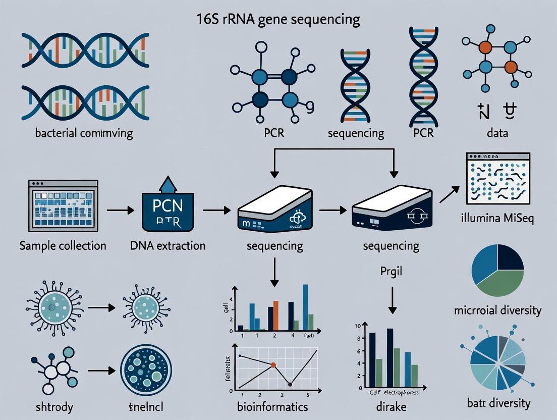
This article provides a detailed framework for applying 16S rRNA gene sequencing to analyze bacterial communities, tailored for researchers and drug development professionals.
16S rRNA Gene Sequencing: A Comprehensive Guide for Microbial Community Analysis in Biomedical Research
Abstract
This article provides a detailed framework for applying 16S rRNA gene sequencing to analyze bacterial communities, tailored for researchers and drug development professionals. It covers foundational principles, step-by-step methodology from sample prep to data analysis, common troubleshooting strategies, and validation against alternative techniques. The guide synthesizes current best practices to ensure robust, reproducible results for studies in microbiome research, infectious disease, and therapeutic development.
The 16S rRNA Gene: Why It's the Gold Standard for Bacterial Phylogeny and Taxonomy
The 16S ribosomal RNA (rRNA) gene serves as the cornerstone of bacterial identification and phylogenetic classification. Its universal presence across the bacterial domain, coupled with conserved regions flanking variable hypervariable regions (V1-V9), makes it an ideal genetic barcode. This Application Note, framed within a thesis on 16S rRNA gene sequencing for microbial ecology and translational research, details the protocols and considerations for employing this principle to profile complex bacterial communities, a critical step in understanding microbiome dynamics in health, disease, and drug development.
Comparative Analysis of 16S rRNA Hypervariable Regions
The choice of hypervariable region(s) for sequencing is critical and influences taxonomic resolution and bias. The table below summarizes key characteristics of commonly targeted regions.
Table 1: Comparative Characteristics of 16S rRNA Gene Hypervariable Regions
| Region | Approx. Length (bp) | Taxonomic Resolution | Common PCR Primers (Examples) | Notes on Bias/Challenges |
|---|---|---|---|---|
| V1-V3 | ~500 | High for many Gram-positives; moderate for others | 27F, 519R | Can be long for some platforms; may under-amplify some Gram-negatives. |
| V3-V4 | ~460 | Good balance; widely used | 341F, 805R | Current Illumina MiSeq standard. Robust performance across samples. |
| V4 | ~290 | Moderate to High | 515F, 806R | Highly conserved primer sites; minimizes amplification bias. |
| V4-V5 | ~390 | Good for environmental samples | 515F, 926R | Good resolution for diverse communities. |
| V6-V8 | ~400 | Variable | 926F, 1392R | Useful for specific phyla. |
| V7-V9 | ~340 | Lower for some groups | 1100F, 1392R | Often used for Archaea; shorter length suits older 454 platforms. |
Detailed Protocol: 16S rRNA Gene Amplicon Sequencing Workflow
Protocol 1: Library Preparation via Two-Step PCR (Illumina MiSeq)
Principle: Amplify target 16S region with gene-specific primers, then add platform-specific adapters and indices via a second PCR.
Materials & Reagents (Research Reagent Solutions):
Table 2: Key Reagents for 16S rRNA Library Preparation
| Item | Function | Example Product/Note |
|---|---|---|
| DNA Polymerase (High-Fidelity) | PCR amplification with low error rate. | KAPA HiFi HotStart, Q5 Hot Start. |
| 16S V3-V4 Primer Mix | First-stage target amplification. | 341F (5'-CCTACGGGNGGCWGCAG-3'), 805R (5'-GACTACHVGGGTATCTAATCC-3'). |
| Nextera XT Index Kit v2 | Provides unique dual indices for sample multiplexing. | Illumina Catalog #FC-131-2001/2002. |
| AMPure XP Beads | Solid-phase reversible immobilization (SPRI) for size selection and purification. | Beckman Coulter #A63881. |
| Qubit dsDNA HS Assay Kit | Accurate quantification of DNA libraries. | Thermo Fisher Scientific #Q32851. |
| Library Quantification Kit | qPCR-based precise molarity for pooling. | KAPA Biosystems #KK4824. |
| Agilent Bioanalyzer HS DNA Kit | Fragment size analysis and QC. | Agilent #5067-4626. |
Procedure:
- Genomic DNA Extraction & QC: Extract using a validated kit (e.g., DNeasy PowerSoil Pro) and quantify via fluorometry.
- First-Stage PCR (Target Amplification):
- Reaction Mix (25 µL): 12.5 µL 2X Master Mix, 1.25 µL each primer (10 µM), 5-20 ng gDNA, nuclease-free water to volume.
- Thermocycling: 95°C 3 min; 25-30 cycles of: 95°C 30s, 55°C 30s, 72°C 30s; final extension 72°C 5 min.
- First-Stage Cleanup: Purify amplicons using 0.8X volume of AMPure XP beads. Elute in 20 µL nuclease-free water.
- Second-Stage PCR (Indexing):
- Reaction Mix (25 µL): 12.5 µL 2X Master Mix, 2.5 µL each Nextera XT index primer (i5 & i7), 2.5 µL purified first-stage product.
- Thermocycling: 95°C 3 min; 8 cycles of: 95°C 30s, 55°C 30s, 72°C 30s; final extension 72°C 5 min.
- Library Cleanup & QC: Purify with 0.9X AMPure beads. Assess concentration (Qubit) and size profile (Bioanalyzer). Precisely quantify via qPCR (KAPA kit).
- Normalization & Pooling: Dilute libraries to 4 nM based on qPCR data, then combine equal volumes into a final sequencing pool. Denature and dilute per Illumina guidelines for loading.
Protocol 2: Bioinformatic Analysis via QIIME 2 (2024.2 Core Workflow)
Principle: Process raw sequence data into Amplicon Sequence Variants (ASVs) and assign taxonomy.
Materials: Demultiplexed paired-end FASTQ files, QIIME 2 environment (https://qiime2.org), reference database (e.g., SILVA 138.99 or Greengenes2 2022.10).
Procedure:
- Import Data: Use
qiime tools importwith appropriate manifest file. - Denoising & ASV Generation: Use DADA2 for quality filtering, denoising, merging, and chimera removal.
- Command example:
qiime dada2 denoise-paired --i-demultiplexed-seqs demux.qza --p-trunc-len-f 280 --p-trunc-len-r 220 --o-table table.qza --o-representative-sequences rep-seqs.qza --o-denoising-stats stats.qza
- Command example:
- Phylogenetic Tree Construction: Generate a tree for diversity metrics with
qiime phylogeny align-to-tree-mafft-fasttree. - Taxonomic Assignment: Use a pre-trained Naïve Bayes classifier.
- Command:
qiime feature-classifier classify-sklearn --i-classifier silva-138-99-nb-classifier.qza --i-reads rep-seqs.qza --o-classification taxonomy.qza
- Command:
- Analysis: Generate core metrics (alpha/beta diversity) with
qiime diversity core-metrics-phylogenetic. Visualize with Emperor for PCoA plots.
Visualization of Workflows and Principles
Title: 16S rRNA Amplicon Sequencing & Analysis Workflow
Title: 16S rRNA Gene Structure & Amplicon Targeting
Within a broader thesis on 16S rRNA gene sequencing for bacterial community analysis, the selection of hypervariable regions (V1-V9) for PCR amplification is a critical foundational decision. The full-length 16S rRNA gene (~1,500 bp) contains nine variable regions (V1-V9) interspersed with conserved sequences. Due to the limitations of current high-throughput sequencing technologies (e.g., Illumina MiSeq, NovaSeq), it is often impractical to sequence the entire gene. Therefore, targeted amplification and sequencing of one or several hypervariable regions is standard. The choice of region(s) directly impacts the depth, accuracy, and biological relevance of taxonomic classification, influencing all downstream analyses and conclusions of the research.
Comparative Analysis of Hypervariable Regions
The discriminatory power and performance of each variable region vary significantly across bacterial taxa and sample types. The following table summarizes key quantitative metrics from recent evaluations.
Table 1: Comparative Performance of 16S rRNA Gene Variable Regions
| Region(s) | Amplicon Length (approx.) | Taxonomic Resolution | Common Primer Pairs (Examples) | Key Strengths | Key Limitations |
|---|---|---|---|---|---|
| V1-V3 | ~500-600 bp | Genus to species-level for some phyla (e.g., Firmicutes). | 27F (8F) / 534R | Good for skin, respiratory microbiota. High discrimination for certain pathogens. | Poor for Bifidobacterium. Length may exceed ideal for some platforms. |
| V3-V4 | ~460 bp | Genus-level. Most common and widely validated. | 341F / 805R | Excellent balance of length and discrimination. Supported by Earth Microbiome Project. | May miss discrimination within Lactobacillus. |
| V4 | ~250-290 bp | Genus to family-level. Highly robust. | 515F / 806R | Short, highly conserved primers. Minimal bias. Best for diverse, unknown communities. | Lower discriminatory power than multi-region spans. |
| V4-V5 | ~390 bp | Genus-level. | 515F / 926R | Good resolution for marine and gut microbiomes. | Less commonly used than V3-V4 or V4 alone. |
| V6-V8 | ~420 bp | Family to genus-level. | 926F / 1392R | Useful for distinguishing cyanobacteria. | Less comprehensive reference database coverage. |
| V7-V9 | ~330-380 bp | Family-level. | 1114F / 1392R | Effective for endolithic and extreme environment microbes. | Generally lower resolution than upstream regions. |
| Full-length | ~1,500 bp | Species to strain-level potential. | 27F / 1492R | Highest possible resolution. Enables rare variant detection. | Requires long-read tech (PacBio, Nanopore). Higher cost, lower throughput. |
Table 2: Region-Specific Bias and Coverage
| Region(s) | PCR Bias | GC Content Bias | Read Length for 2x300bp PE* | Chimera Formation Risk |
|---|---|---|---|---|
| V1-V3 | Moderate-High | Moderate | Excellent overlap (>50bp). | Moderate |
| V3-V4 | Low-Moderate | Low | Good overlap (~140bp). | Low |
| V4 | Lowest | Lowest | Excellent overlap (>200bp). | Lowest |
| V4-V5 | Low | Low | Good overlap (~110bp). | Low |
| V6-V8 | Moderate | Moderate | Limited/no overlap. | Moderate |
| V7-V9 | High | High | Limited/no overlap. | High |
*PE: Paired-End sequencing on Illumina MiSeq.
Experimental Protocols
Protocol A: Library Preparation for V3-V4 Region (Illumina Platform)
Objective: To amplify the bacterial 16S rRNA gene V3-V4 region from genomic DNA extracts for Illumina sequencing.
Materials:
- Template DNA (10-30 ng/µL).
- KAPA HiFi HotStart ReadyMix (or equivalent high-fidelity polymerase).
- Primer Mix: Forward (341F:
5′-CCTACGGGNGGCWGCAG-3′) and Reverse (805R:5′-GACTACHVGGGTATCTAATCC-3′) with overhang adapters. - PCR-grade water.
- Magnetic bead-based purification kit (e.g., AMPure XP).
- Indexing primers (Nextera XT Index Kit v2).
- Thermal cycler.
Procedure:
- First-Stage PCR (Amplification):
- Prepare 25 µL reaction: 12.5 µL 2X KAPA HiFi Mix, 2.5 µL Primer Mix (1 µM each), 5 µL template DNA, 5 µL PCR-grade water.
- Cycling: 95°C for 3 min; 25 cycles of [95°C for 30s, 55°C for 30s, 72°C for 30s]; 72°C for 5 min; hold at 4°C.
- Amplicon Purification:
- Clean PCR products using a 0.8X ratio of AMPure XP beads. Elute in 25 µL of 10mM Tris buffer, pH 8.5.
- Second-Stage PCR (Indexing):
- Prepare 50 µL reaction: 25 µL 2X KAPA HiFi Mix, 5 µL each of unique i5 and i7 indexing primers, 5 µL purified amplicon, 10 µL water.
- Cycling: 95°C for 3 min; 8 cycles of [95°C for 30s, 55°C for 30s, 72°C for 30s]; 72°C for 5 min; hold at 4°C.
- Final Library Purification & Normalization:
- Purify with a 0.9X ratio of AMPure XP beads.
- Quantify library concentration (e.g., via Qubit), check fragment size (e.g., TapeStation), and pool equimolar amounts.
Protocol B: In Silico Evaluation of Region Selection
Objective: To computationally predict the theoretical taxonomic resolution of different variable regions for a specific research question.
Materials:
- Reference database (e.g., SILVA, Greengenes, RDP).
- Bioinformatics tools: QIIME 2, mothur, or the R package
dada2. - In silico PCR tool (e.g.,
EMBOSS: primearchormotifSearchin R).
Procedure:
- Define Target Taxa: List bacterial genera/species of primary interest from your thesis hypothesis.
- Extract Reference Sequences: Download full-length 16S sequences for these taxa from a curated database.
- Perform In Silico PCR: Using the primer sequences for candidate regions (e.g., V4, V3-V4, V1-V3), extract the corresponding sub-sequences from the full-length references.
- Calculate Pairwise Distance: Align the extracted region-specific sequences (e.g., using NAST or MUSCLE). Compute genetic distances (e.g., Kimura-2 parameter) between sequences of different taxa.
- Assess Resolution: A region that yields greater genetic distance between distinct species, while maintaining minimal distance within the same species, has higher discriminatory power for your target taxa.
Visualizations
Title: Decision Workflow for 16S Region Selection
Title: V3-V4 Library Prep and Sequencing Workflow
The Scientist's Toolkit: Research Reagent Solutions
Table 3: Essential Reagents for 16S rRNA Region-Targeted Sequencing
| Item | Function & Rationale | Example Product(s) |
|---|---|---|
| High-Fidelity DNA Polymerase | Minimizes PCR amplification errors and bias, critical for accurate community representation. | KAPA HiFi HotStart, Q5 High-Fidelity DNA Polymerase. |
| Region-Specific Primer Cocktails | Contain degenerate bases to maximize amplification across diverse bacterial phyla. | Illumina 16S Metagenomic Library Prep Kit (targets V3-V4). Custom synthesized oligos. |
| Magnetic Bead Cleanup Kit | For size-selective purification of PCR amplicons, removing primer dimers and non-specific products. | AMPure XP Beads, SPRIselect. |
| Dual-Indexed Adapter Kit | Allows multiplexing of hundreds of samples by attaching unique barcode combinations. | Nextera XT Index Kit v2, IDT for Illumina UD Indexes. |
| Fluorometric DNA Quant Kit | Accurate quantification of library concentration for precise pooling. | Qubit dsDNA HS Assay. |
| Library Quality Control Assay | Assesses library fragment size distribution and detects adapter contamination. | Agilent Bioanalyzer HS DNA Kit, Fragment Analyzer. |
| Phylogenetically Diverse Mock Community | Positive control containing known genomic DNA from multiple bacterial species to assess bias and resolution. | ZymoBIOMICS Microbial Community Standard. |
Within the broader thesis on 16S rRNA gene sequencing for bacterial community analysis, understanding the technological evolution from Sanger to NGS is paramount. This progression has dramatically increased throughput, reduced cost, and enabled high-resolution profiling of complex microbiomes, fundamentally reshaping microbial ecology and drug discovery research.
Technological Evolution & Comparative Data
Table 1: Comparative Analysis of 16S rRNA Gene Sequencing Technologies
| Feature | Sanger Sequencing (Capillary Electrophoresis) | Next-Generation Sequencing (Illumina MiSeq) |
|---|---|---|
| Read Output per Run | 96 - 384 reads | Up to 25 million paired-end reads |
| Read Length | ~900-1000 bp (full-length 16S) | Up to 2x300 bp (targeting V3-V4 hypervariable regions) |
| Approximate Cost per Sample | $5 - $15 (at high throughput) | <$1 - $5 (multiplexed) |
| Primary Application in 16S Analysis | Clonal sequencing, reference database generation | High-throughput community profiling, alpha/beta diversity |
| Key Advantage | Long, accurate reads for definitive classification | Unparalleled depth for rare taxa detection |
| Primary Limitation | Low throughput, not suited for complex communities | Shorter reads may limit species-level resolution |
Table 2: Common 16S Hypervariable Regions Targeted by NGS Platforms
| Platform | Typical Read Type | Commonly Targeted 16S Region(s) | Approximate Amplicon Length |
|---|---|---|---|
| Illumina MiSeq | 2x300 bp | V3-V4 | ~460 bp |
| Illumina iSeq | 2x150 bp | V4 | ~250 bp |
| Ion Torrent PGM | 400-600 bp | V4-V6 or V6-V9 | Variable |
| PacBio Sequel | >1,000 bp (HiFi) | Full-length 16S gene | ~1,500 bp |
Detailed Protocols
Protocol 1: Sanger Sequencing of Cloned 16S rRNA Gene Inserts
Application Note: Used for generating high-quality reference sequences from isolated bacterial colonies or clone libraries.
Materials:
- Purified plasmid DNA from cloned 16S PCR products.
- M13 Forward (-20) or Reverse primer (10 µM).
- BigDye Terminator v3.1 Cycle Sequencing Kit.
- Ethanol/EDTA precipitation solutions.
- Capillary sequencer (e.g., Applied Biosystems 3730xl).
Methodology:
- Cycle Sequencing Reaction: In a 0.2 mL tube, mix: 50-100 ng plasmid DNA, 1 µL primer (10 µM), 2 µL 5X Sequencing Buffer, 0.5 µL BigDye Terminator, and nuclease-free water to 10 µL.
- Thermocycling: 96°C for 1 min, then 25 cycles of: 96°C for 10 s, 50°C for 5 s, 60°C for 4 min. Hold at 4°C.
- Purification: Add 10 µL of nuclease-free water and 30 µL of a 1:5 EDTA:Ethanol (95%) mixture. Incubate at room temperature for 15 min. Centrifuge at 3,000 x g for 30 min. Carefully aspirate supernatant.
- Wash: Add 70 µL of 70% ethanol, vortex gently, and centrifuge at 3,000 x g for 15 min. Aspirate supernatant completely and air-dry pellet.
- Resuspension & Sequencing: Resuspend in 10 µL Hi-Di formamide. Denature at 95°C for 2 min, then snap-cool on ice. Load onto sequencer.
Protocol 2: Illumina MiSeq 16S rRNA Gene Amplicon Sequencing (V3-V4)
Application Note: Standardized protocol for high-throughput bacterial community profiling.
Materials:
- Genomic DNA from microbial community sample.
- 16S V3-V4 primers (341F: 5'-CCTACGGGNGGCWGCAG-3', 805R: 5'-GACTACHVGGGTATCTAATCC-3') with overhang adapters.
- KAPA HiFi HotStart ReadyMix.
- AMPure XP beads.
- Nextera XT Index Kit v2.
- MiSeq Reagent Kit v3 (600 cycles).
Methodology: A. Primary PCR (Amplify Target Region):
- Reaction Setup: For each sample, mix: 12.5 ng genomic DNA, 5 µL each forward and reverse primer (1 µM), 12.5 µL KAPA HiFi mix, and water to 25 µL.
- Thermocycling: 95°C for 3 min; 25 cycles of 95°C for 30 s, 55°C for 30 s, 72°C for 30 s; final extension at 72°C for 5 min.
- Clean-up: Purify amplicons using AMPure XP beads (0.8x ratio). Elute in 25 µL nuclease-free water.
B. Index PCR (Attach Dual Indices & Sequencing Adaptors):
- Reaction Setup: Mix 5 µL purified primary PCR product, 5 µL each Nextera XT index primer (i5 and i7), 25 µL KAPA HiFi mix, and 10 µL water for a 50 µL reaction.
- Thermocycling: 95°C for 3 min; 8 cycles of 95°C for 30 s, 55°C for 30 s, 72°C for 30 s; final extension at 72°C for 5 min.
- Clean-up: Purify with AMPure XP beads (0.8x ratio). Quantify using fluorometry (e.g., Qubit).
- Pooling & Sequencing: Normalize and pool all indexed libraries. Denature with NaOH, dilute to 8 pM in HT1 buffer, and load onto the MiSeq cartridge following manufacturer instructions.
Visualizations
Diagram 1: 16S Sequencing Technology Workflow Comparison
Diagram 2: Evolution of 16S Sequencing Technology Eras
The Scientist's Toolkit: Research Reagent Solutions
Table 3: Essential Reagents for 16S rRNA Gene Sequencing Studies
| Item | Function in 16S Analysis | Example Product(s) |
|---|---|---|
| DNA Extraction Kit | Lyse cells and purify total genomic DNA from complex samples. Critical for bias minimization. | DNeasy PowerSoil Pro Kit (QIAGEN), MagAttract PowerMicrobiome Kit |
| High-Fidelity DNA Polymerase | Amplify 16S region with minimal PCR errors to avoid artificial diversity. | KAPA HiFi HotStart ReadyMix, Q5 High-Fidelity DNA Polymerase |
| 16S rRNA Gene Primers | Target conserved regions flanking hypervariable zones (e.g., V4, V3-V4). | 515F/806R (V4), 341F/805R (V3-V4) with Illumina overhangs. |
| Size-Selective Magnetic Beads | Purify PCR amplicons and perform library normalization by removing primer dimers and large fragments. | AMPure XP Beads, SPRIselect Beads |
| Indexing/Primer Kit | Attach unique dual indices and full sequencing adapters to amplicons for multiplexing. | Illumina Nextera XT Index Kit v2, 16S Metagenomic Sequencing Library Prep Kit |
| Quantification Assay | Accurately measure DNA library concentration for optimal pooling and sequencing loading. | Qubit dsDNA HS Assay, Library Quantification Kit for Illumina (qPCR) |
| Positive Control DNA | Standardized genomic DNA from a mock microbial community to assess run performance and bias. | ZymoBIOMICS Microbial Community Standard, ATCC Mock Microbial Communities |
Within the context of 16S rRNA gene sequencing for bacterial community analysis, the choice of bioinformatic metric for clustering sequences into taxonomic units is fundamental. Historically, Operational Taxonomic Units (OTUs) defined by a 97% similarity threshold were the standard. Recently, Amplicon Sequence Variants (ASVs), exact sequences differentiated by a single nucleotide, have emerged. This application note details these two paradigms, their methodological workflows, and their impact on the interpretation of microbial ecology data in research and drug development.
Operational Taxonomic Unit (OTU): A cluster of sequencing reads grouped based on a user-defined sequence similarity threshold (typically 97%), intended to approximate a species-level grouping. This method assumes that sequences within the cluster are functionally and phylogenetically related.
Amplicon Sequence Variant (ASV): A unique sequence inferred from high-resolution data, representing a single biological sequence without pre-defined clustering. ASVs are resolved to the level of single-nucleotide differences over the sequenced region.
The following table summarizes the key differences:
Table 1: Comparative Analysis of OTU and ASV Methodologies
| Feature | OTU (97% Clustering) | ASV (DADA2, UNOISE3, etc.) |
|---|---|---|
| Definition Basis | Similarity-based clustering (97% identity). | Exact biological sequence inference. |
| Resolution | Lower, groups sequences into bins. | Single-nucleotide resolution. |
| Bioinformatics Tools | QIIME1 (uclust, mothur), VSEARCH. | DADA2, UNOISE3 (deblur), QIIME2 (Deblur plugin). |
| Threshold Dependence | Yes, arbitrary (e.g., 97%, 99%). | No, threshold-free. |
| Cross-Study Comparison | Difficult; clusters are study-dependent. | Straightforward; ASVs are reproducible and portable. |
| Handling of Sequencing Errors | Errors are often clustered with real sequences. | Explicitly models and removes errors. |
| Interpretation | Ecological groups, but may contain multiple strains. | Can represent strain-level variation. |
| Rarefaction Sensitivity | High; clustering is affected by sampling depth. | Low; sequences are identified independently of depth. |
Table 2: Impact on Key Microbial Community Metrics (Representative Data)
| Data Interpretation Metric | OTU-Based Analysis | ASV-Based Analysis | Interpretive Impact |
|---|---|---|---|
| Alpha Diversity (Richness) | Typically lower counts; saturates quickly. | Typically higher counts; more sensitive to rare taxa. | ASVs reveal greater diversity, especially in low-complexity environments. |
| Beta Diversity (Between-Sample) | Can be inflated by technical variation. | More precise; better separation of technical vs. biological variation. | ASV-based ordinations often show tighter sample clusters within groups. |
| Tracking Taxa Across Studies | Low portability; requires re-clustering. | High portability; ASVs are absolute identifiers. | Enables robust meta-analyses and reference database development. |
| Identification of Biomarkers | May group ecologically distinct variants. | Can pinpoint specific sequence variants linked to phenotypes. | Crucial for drug development targeting specific pathogenic strains. |
Detailed Experimental Protocols
Protocol 3.1: Classic OTU Picking Pipeline (QIIME1/mothur-style)
Objective: To process raw 16S rRNA sequencing reads into OTU tables via clustering.
- Demultiplex & Quality Filter: Assign reads to samples based on barcodes. Trim primers and low-quality bases (Q-score <20, no Ns). Merge paired-end reads (e.g., using PEAR or VSEARCH).
- Pick OTUs:
- De Novo: Cluster all quality-filtered sequences at 97% identity using a greedy algorithm (e.g., uclust, CD-HIT). The most abundant sequence in each cluster becomes the representative sequence.
- Closed-Reference: Map all sequences against a reference database (e.g., Greengenes, SILVA) at 97% identity. Sequences failing to match are discarded.
- Assign Taxonomy: Use a classifier (e.g., RDP Classifier, BLAST) against a reference database to taxonomically label each representative sequence.
- Build OTU Table: Generate a sample-by-OTU observation count matrix (BIOM format).
- Downstream Analysis: Apply normalization (e.g., rarefaction) and calculate diversity metrics, ordination (PCoA), and differential abundance.
Protocol 3.2: ASV Inference Pipeline (DADA2 in R)
Objective: To infer exact Amplicon Sequence Variants from raw reads.
- Filter & Trim: Inspect quality profiles (
plotQualityProfile). Trim forward/reverse reads to consistent quality (e.g.,truncLen=c(240,160)). Filter reads with expected errors >2 (maxEE=c(2,2)). - Learn Error Rates: Model the error rates specific to the dataset using a machine-learning algorithm (
learnErrors). - Dereplication: Combine identical reads into unique sequences with abundance counts (
derepFastq). - Core Inference: Apply the DADA algorithm to the dereplicated data to distinguish sequencing errors from true biological variation (
dada). This yields an ASV table. - Merge Paired Reads: Merge the inferred forward and reverse ASVs (
mergePairs). - Construct Sequence Table: Create the final ASV abundance table (
makeSequenceTable). - Remove Chimeras: Identify and remove chimeric sequences (
removeBimeraDenovo). - Assign Taxonomy: Use the
assignTaxonomyfunction with a training database (e.g., SILVA). Optionally add species-level assignment withaddSpecies. - Analysis: Proceed with analysis using the sequence table. Rarefaction is often not required but can be applied for specific comparative metrics.
Visualization of Workflows
Figure 1: OTU vs. ASV Bioinformatics Workflow Comparison
Figure 2: Impact of Metric Choice on Data Interpretation
The Scientist's Toolkit: Research Reagent Solutions
Table 3: Essential Materials and Reagents for 16S rRNA Analysis Workflows
| Item | Function / Role | Example Product / Note |
|---|---|---|
| PCR Primers (V4 Region) | Amplify the hypervariable V4 region of the 16S rRNA gene for sequencing. | 515F (GTGYCAGCMGCCGCGGTAA) / 806R (GGACTACNVGGGTWTCTAAT). |
| High-Fidelity DNA Polymerase | Minimize PCR amplification errors to preserve true sequence variation. | Phusion High-Fidelity DNA Polymerase, KAPA HiFi HotStart. |
| Quantitation Kit (dsDNA) | Accurately measure library concentration for pooling and sequencing. | Qubit dsDNA HS Assay Kit, Fragment Analyzer systems. |
| Sequencing Standards | Control for cross-study comparisons and pipeline validation. | ZymoBIOMICS Microbial Community Standards. |
| Bioinformatics Software | Implement OTU clustering or ASV inference algorithms. | QIIME2 (for ASVs/plugins), mothur, DADA2 (R package), USEARCH. |
| Reference Taxonomy Database | Assign taxonomic labels to OTU/ASV representative sequences. | SILVA, Greengenes, RDP. Must match primer region. |
| Positive Control DNA | Verify the entire wet-lab workflow from extraction to PCR. | Genomic DNA from a known, culturable bacterial strain. |
| Negative Control Reagents | Identify contamination from reagents or the extraction process. | Nuclease-free water carried through extraction and PCR. |
Within a thesis on 16S rRNA gene sequencing for bacterial community analysis, the accurate taxonomic classification of sequence data is a foundational step. This process is entirely dependent on high-quality, curated reference databases. Three major databases—SILVA, Greengenes, and the Ribosomal Database Project (RDP)—are pivotal resources. Each offers unique attributes, curation philosophies, and classification tools that significantly influence downstream ecological interpretations. This application note provides a detailed comparison, protocols for their use, and practical guidance for researchers, scientists, and drug development professionals seeking to identify microbial taxa or discover biomarkers.
The choice of database directly impacts taxonomic assignment accuracy, resolution, and reproducibility. The following table summarizes the core quantitative and qualitative attributes of each database as of current information.
Table 1: Core Comparison of Major 16S rRNA Reference Databases
| Feature | SILVA | Greengenes | RDP |
|---|---|---|---|
| Current Version | SSU r138.1 (2020) | gg138 (2013) | RDP 11. Update 5 (2016) |
| Update Status | Actively curated; periodic releases | Archived; no longer actively updated | Archived; minor updates possible |
| Primary Source | Comprehensive rRNA database (Bacteria, Archaea, Eukarya) | Primarily bacterial and archaeal sequences | Curated bacterial and archaeal sequences |
| # of Quality-aligned Sequences | ~2.7 million (Ref NR) | ~1.3 million (97% OTUs) | ~3.4 million (Bacteria & Archaea) |
| Taxonomy System | Based on LTP, Bergey's, and original publications | Based on NCBI taxonomy, manually curated | RDP's proprietary taxonomy (consistent with Bergey's) |
| Alignment & Tree | Provided (ARB format), based on SSU/LSU alignment | Provided (.fna), based on a profile alignment |
Provided, secondary-structure aware alignment |
| Primary Tool/Classifier | SINA aligner, SILVA Incremental Aligner |
RDP Classifier, QIIME-compatible files |
RDP Classifier (Naïve Bayesian) |
| Strengths | Broad domain coverage, actively updated, high-quality alignment | Stable benchmark, integrated into many pipelines (e.g., QIIME 1) | Fast, accurate classifier with confidence estimates |
| Key Considerations | Larger size requires more computational resources; Eukaryotic rRNA may be irrelevant for some studies. | Outdated; may lack novel taxa discovered post-2013. | Less frequently updated than SILVA; classifier is database-specific. |
Experimental Protocols for Database Utilization
Protocol 3.1: Taxonomic Classification with the RDP Classifier
The RDP Classifier is a widely used tool for assigning taxonomy to 16S rRNA sequences, often employed with all three databases when formatted appropriately.
Materials & Reagents:
- Input Data: Demultiplexed, quality-filtered, and chimera-checked FASTA sequences (e.g., from DADA2 or USEARCH).
- Reference Files: Formatted training set files for the desired database (
trainsetXX_YYXX.rdp.fa&trainsetXX_YYXX.rdp.tax). - Software: RDP Classifier (v2.13) jar file, Java Runtime Environment.
Procedure:
- Prepare Reference Data: Download and place the RDP-formatted training set for your chosen database (e.g., SILVA, Greengenes, or native RDP) in your working directory.
- Execute Classification: Run the classifier from the command line:
- Interpret Output: The output file will list each query sequence ID followed by its taxonomic assignment from domain to genus (or species), with bootstrap confidence scores for each rank.
Protocol 3.2: Alignment and Classification using the SILVA Database and SINA
For maximum alignment accuracy with the SILVA database, the SINA aligner is recommended.
Materials & Reagents:
- Input Data: Quality-controlled FASTA sequences.
- Reference Files: SILVA SSU Ref NR dataset (
.arbor.fasta). - Software: SINA aligner (v1.7.2 or later), ARB (optional for manual curation).
Procedure:
- Download & Prepare SILVA: Download the SILVA SSU Ref NR dataset and extract the
.fastaand.taxfiles. - Perform Alignment: Align your query sequences to the SILVA reference alignment using SINA:
- Taxonomic Assignment: Use the alignment output and the provided taxonomy file to assign taxonomy, often integrated within pipelines like mothur or QIIME2 via feature-classifier plugins.
Protocol 3.3: Integrating Greengenes into a QIIME2 Pipeline
Greengenes, though archived, remains a common reference in legacy or comparative studies. QIIME2 provides tools to import and use it.
Materials & Reagents:
- Input Data: QIIME2 artifact of representative sequences (
rep-seqs.qza). - Reference Files: Greengenes 13_8 99% OTUs reference sequences (
99_otus.fasta) and taxonomy (99_otu_taxonomy.txt). - Software: QIIME2 (2024.5 or later).
Procedure:
- Import Reference Data: Create QIIME2 artifacts from Greengenes files.
Extract Region-Specific Reads: If your sequences target a specific hypervariable region (e.g., V4), extract that region from the reference.
Train a Classifier: Train a naïve Bayes classifier on the prepared references.
Classify Sequences: Apply the classifier to your data.
Visualizing the Database Selection and Classification Workflow
Decision Workflow for 16S rRNA Database Selection
The Scientist's Toolkit: Essential Research Reagents & Materials
Table 2: Key Research Reagent Solutions for 16S rRNA Classification Workflows
Item
Function in Context
Example/Specification
Curated Reference Database
Provides the gold-standard sequences and taxonomy against which unknown sequences are classified.
SILVA SSU Ref NR, Greengenes 13_8 OTUs, RDP training set.
Alignment & Classifier Software
Executes the algorithm for matching query reads to the reference database and assigning taxonomy.
RDP Classifier jar, SINA aligner, QIIME2 feature-classifier plugin.
Pre-formatted Training Files
Database-specific files formatted for immediate use with a chosen classifier, saving preprocessing time.
trainset18_062020.rdp.fa, gg_13_8_99.refseqs.qza.
Primer Sequence Files
Essential for extracting the exact hypervariable region sequenced from full-length references during classifier training.
FASTA file containing the forward and reverse primers used in your study (e.g., 515F/806R for V4).
High-Performance Computing (HPC) Resources
Classification against large databases (>1M sequences) requires significant memory (RAM) and CPU resources.
Access to a cluster or server with ≥16 GB RAM and multiple cores for timely processing.
Taxonomy Table Template
A standardized file format (e.g., TSV) for storing and visualizing classification results across samples.
QIIME2 .qza artifact or a simple tab-separated file with columns: FeatureID, Taxon, Confidence.
From Sample to Insight: A Step-by-Step Protocol for 16S rRNA Sequencing Workflow
This application note, framed within a thesis on 16S rRNA gene sequencing for bacterial community analysis, details the critical first step in the microbial ecology workflow: sample collection and preservation. The integrity of downstream sequencing data and community composition analysis is entirely contingent upon the initial stabilization of the in-situ microbial profile. This protocol provides best practices for diverse sample matrices to minimize bias from post-sampling shifts.
The following table summarizes key findings from current literature on the efficacy of various preservation methods for maintaining bacterial community integrity prior to DNA extraction and 16S sequencing.
Table 1: Comparison of Sample Preservation Methods for 16S rRNA Gene Sequencing
| Matrix | Preservation Method | Maximum Storage Time (at indicated temp) for Minimal Community Shift | Key Metric Impacted (vs. Fresh Processing) | Reported Bias / Notes |
|---|---|---|---|---|
| Stool / Feces | Immediate freezing at -80°C | Gold Standard | N/A (Baseline) | Minimal change over months. |
| Commercial Stabilization Buffer (e.g., OMNIgene•GUT, RNAlater) | 7-60 days at room temp | Alpha Diversity (Shannon Index) | <10% shift vs. -80°C freeze for up to 7 days. Effective for transport. | |
| Soil & Sediment | -80°C freezing | > 4 weeks | Relative Abundance of Taxa | Minor shifts in low-abundance taxa after 4 weeks at -20°C. |
| 95% Ethanol (for DNA) | 24 hours at RT, then -80°C | Community Composition (Bray-Curtis) | Effective short-term; may lyse Gram-positives less efficiently. | |
| Skin & Oral Swabs | Dry Swab in Stabilizing Tube (e.g., with beads) | 1 week at -80°C; 24h at RT | Biomass Yield | Significant DNA degradation after 24h at RT on dry swab. |
| Swab in Liquid Stabilizer (e.g., Zymo DNA/RNA Shield) | 30 days at RT | Bacterial Load (qPCR) | >95% DNA integrity maintained vs. immediate extraction. | |
| Water (Fresh/Marine) | Filtration + Immediate -80°C freeze | Gold Standard | N/A (Baseline) | Filtration captures biomass; freezing halts activity. |
| Filtration + Preservation Buffer (e.g., RNAlater, LifeGuard) | 2 weeks at 4°C | Community Structure | Preserves community better than just 4°C storage for >24h. | |
| Tissue (Mucosal) | Snap-freeze in LN₂, then -80°C | Gold Standard | N/A (Baseline) | Rapid freezing prevents autolysis and microbial growth. |
| Immersion in Stabilization Buffer | 48 hours at 4°C | Ratio of Firmicutes/Bacteroidetes | Potential for selective permeation; for flash-freeze is superior. |
Detailed Experimental Protocols
Protocol 3.1: Fecal Sample Collection for Human Microbiome Studies
Objective: To collect and stabilize fecal samples for 16S rRNA gene sequencing, minimizing changes in microbial community composition. Materials: OMNIgene•GUT stool collection kit (or equivalent), disposable spatula, gloves, cooler with ice packs or -80°C freezer access. Procedure:
- Using the provided spatula, collect approximately 50-100 mg of feces (pea-sized) from multiple locations within the stool specimen.
- Immediately place the sample into the tube containing stabilization buffer. Ensure the sample is fully submerged.
- Securely close the lid and shake vigorously for 30 seconds to homogenize.
- Label the tube with a unique subject ID and collection timestamp.
- Short-term: Store at room temperature (15-25°C) for up to 7 days before transfer to -80°C. Long-term: Place directly at -80°C within 24 hours for optimal preservation.
- For DNA extraction, use a bead-beating step to ensure lysis of tough Gram-positive bacteria.
Protocol 3.2: Environmental Water Filtration & Preservation
Objective: To concentrate microbial biomass from water and preserve it for community analysis. Materials: Peristaltic pump or vacuum manifold, 0.22µm polyethersulfone (PES) membrane filters, sterile filter housings, forceps, sterile scissors, preservation tubes with DNA/RNA Shield or RNAlater. Procedure:
- Assemble the filtration unit under aseptic conditions. Record the volume of water filtered (typically 100mL-1L, depending on turbidity).
- Filter the water sample through the 0.22µm membrane.
- Using sterile forceps, carefully fold the filter (biomass side inward) and cut it into 4-6 pieces with sterile scissors.
- Immediately transfer the filter pieces to a tube containing 1-2 mL of preservation buffer. Ensure all pieces are immersed.
- Invert the tube several times to coat the filter.
- Store at 4°C for up to 2 weeks, or transfer to -80°C for long-term storage.
Protocol 3.3: Skin Swab Collection with Stabilization
Objective: To standardize the collection of skin microbiota while preserving community DNA. Materials: Sterile polyester or nylon-flocked swabs, pre-moistened with sterile 0.15M NaCl + 0.1% Tween 20 (or commercial swab kit), sterile template (e.g., 2cm²), stabilizing tube with bead-beating matrix. Procedure:
- Moisten the swab in the sterile solution and express excess liquid.
- Place the sterile template on the skin site (e.g., volar forearm).
- Firmly rub the swab over the defined area for 30 seconds, rotating the swab to use all surfaces.
- Immediately place the swab head into the stabilizing tube containing a lysis buffer (e.g., PowerBead solution from DNeasy PowerSoil kit).
- Break or cut the swab shaft to seal the tube.
- Vortex for 1 minute to dislodge cells onto the beads. Store at -20°C or -80°C until DNA extraction.
Visualized Workflows
Diagram 1: Universal Sample Integrity Workflow
Diagram 2: Preservation Method Selection Logic
The Scientist's Toolkit: Research Reagent Solutions
Table 2: Essential Materials for Sample Collection & Preservation
| Item / Reagent | Primary Function | Key Considerations for 16S Studies |
|---|---|---|
| OMNIgene•GUT (DNA Genotek) | Stabilizes fecal microbial DNA at room temperature. | Inhibits nuclease activity and bacterial growth. Allows for non-cold-chain transport. Compatible with bead-beating extraction. |
| DNA/RNA Shield (Zymo Research) | Inactivates nucleases and preserves nucleic acids in diverse matrices (swabs, tissue, water). | Broad-spectrum, room-temperature stabilization. Prevents overgrowth and degradation. |
| RNAlater (Thermo Fisher) | Aqueous, non-toxic tissue storage reagent that stabilizes and protects cellular RNA and DNA. | Penetration can be slow for dense tissues; best for small biopsies or filters. May require removal before extraction. |
| PowerBead Tubes (Qiagen) | Tubes containing a mixture of ceramic and silica beads for mechanical lysis. | Critical for homogenizing tough matrices (stool, soil, biofilms) and lysing robust Gram-positive cell walls. |
| Polyethersulfone (PES) Membrane Filters (0.22µm) | For concentrating microbial cells from low-biomass liquid samples (water, saline solutions). | Low protein binding minimizes biomass loss. Compatible with downstream DNA extraction protocols. |
| Flocked Nylon Swabs | Maximize cell collection efficiency from surfaces (skin, mucosa). | Flocked design releases cells more efficiently than wound-fiber swabs during vortexing in lysis buffer. |
| Cryogenic Vials & LN₂ | For snap-freezing tissue and liquid samples to instantly halt all biological activity. | Most effective method to preserve the in-situ community without chemical additives. Requires immediate access. |
Within a thesis focused on 16S rRNA gene sequencing for bacterial community analysis, the DNA extraction step is a critical determinant of data fidelity. Biases introduced during lysis of complex, mixed samples can skew microbial abundance profiles. Gram-positive bacteria, with their thick peptidoglycan layer, and Gram-negative bacteria, with their outer membrane, require distinct optimization strategies to achieve equitable, high-yield, and inhibitor-free DNA extraction for subsequent PCR and sequencing.
Comparative Challenges in Lysis
| Characteristic | Gram-Positive Bacteria | Gram-Negative Bacteria |
|---|---|---|
| Primary Barrier | Thick, multi-layered peptidoglycan (20-80 nm) | Thin peptidoglycan layer (2-7 nm) + Outer Membrane |
| Key Lysis Target | Peptidoglycan cross-links | Outer membrane (LPS) followed by peptidoglycan |
| Common Chemical Agents | Lysozyme, Lysostaphin, Mutanolysin, high-concentration EDTA | Lysozyme, Chelators (EDTA), Detergents (SDS, Sarkosyl) |
| Mechanical Force Required | Generally higher | Generally lower |
| Inhibitor Concern | Teichoic acids can co-precipitate with DNA | Lipopolysaccharides (LPS, endotoxins) can inhibit enzymes |
| Typical Lysis Time | Extended (30-120 min enzymatic pre-treatment common) | Shorter (5-30 min enzymatic pre-treatment often sufficient) |
Optimized Protocols for Mixed Communities
Dual-Mechanism Lysis Protocol for Fecal/Soil Samples
This protocol is designed for maximal community representation.
Reagents & Equipment:
- Bead-beating tubes (0.1 mm silica/zirconia beads)
- Lysis Buffer A (for Gram-negative): 20 mM Tris-Cl (pH 8.0), 2 mM EDTA, 1.2% Triton X-100.
- Lysis Buffer B (for Gram-positive): 20 mM Tris-Cl (pH 8.0), 20 mM EDTA, 200 mM NaCl.
- Lysozyme (50 mg/mL stock)
- Lysostaphin (for Staphylococci; 1 mg/mL stock)
- Mutanolysin (for Streptococci/Lactobacilli; 5 U/µL stock)
- Proteinase K (20 mg/mL)
- SDS (20% w/v)
- Phenol:Chloroform:Isoamyl Alcohol (25:24:1)
- Isopropanol
- 70% Ethanol
- TE Buffer (pH 8.0)
Procedure:
- Sample Preparation: Resuspend 180 mg of pelleted cells or environmental sample in 480 µL of Lysis Buffer A.
- Enzymatic Pre-treatment (Gram-targeted):
- Add 50 µL of Lysozyme stock. Vortex.
- Add 10 µL of Lysostaphin stock if Staphylococci are suspected.
- Add 5 µL of Mutanolysin stock if Lactobacilli/Streptococci are suspected.
- Incubate at 37°C for 45 minutes with gentle agitation.
- Chemical Lysis: Add 60 µL of 20% SDS and 20 µL of Proteinase K stock. Mix by inversion. Incubate at 56°C for 30 minutes.
- Mechanical Disruption: Transfer mixture to a bead-beating tube. Process on a high-speed homogenizer for 90 seconds. Place on ice for 2 minutes.
- Phase Separation: Centrifuge at 12,000 x g for 5 min. Transfer supernatant to a fresh tube. Add an equal volume of Phenol:Chloroform:Isoamyl Alcohol. Vortex vigorously for 1 minute. Centrifuge at 12,000 x g for 10 minutes at 4°C.
- DNA Precipitation: Transfer the upper aqueous phase to a new tube. Add 0.7 volumes of room-temperature isopropanol. Mix by inversion. Incubate at -20°C for 1 hour. Centrifuge at 16,000 x g for 20 minutes at 4°C.
- Wash & Elution: Carefully discard supernatant. Wash pellet with 1 mL of 70% ethanol. Centrifuge at 16,000 x g for 5 minutes. Air-dry pellet for 10 minutes. Resuspend in 100 µL of TE Buffer. Quantify via fluorometry.
Commercial Kit Optimization Table
| Kit Name | Recommended for | Gram-Positive Enhancement | Gram-Negative Enhancement | Yield (approx.) from Mixed Culture |
|---|---|---|---|---|
| DNeasy PowerSoil Pro | Environmental, tough cells | Integrated bead-beating step | Efficient detergent-based lysis | 2-5 µg per 0.25 g soil |
| MasterPure Gram DNA Purification | Pure cultures, differentiation | Separate, tailored protocols for each Gram type in manual | Separate, tailored protocols for each Gram type in manual | 5-15 µg per 10^8 cells |
| QIAamp DNA Stool Mini | Fecal samples | Addition of heat (95°C) step post-lysozyme | Inhibitor Removal Technology column | 1-3 µg per 200 mg stool |
| Optimization Tip | Add 30-min lysozyme (10 mg/mL) pre-treatment at 37°C | Add 10-min proteinase K (1 mg/mL) step at 56°C |
The Scientist's Toolkit: Research Reagent Solutions
| Item | Function & Rationale |
|---|---|
| Lysozyme | Hydrolyzes β-1,4-glycosidic bonds in peptidoglycan of both Gram types, more effective on Gram-negative. |
| Lysostaphin | Zinc-dependent endopeptidase specifically cleaves Staphylococcus peptidoglycan cross-bridges. |
| Mutanolysin | Glycosidase effective against Streptococcus and Lactobacillus cell walls. |
| EDTA (Ethylenediaminetetraacetic acid) | Chelates divalent cations, destabilizing the outer membrane of Gram-negatives and weakening Gram-positive peptidoglycan. |
| SDS (Sodium Dodecyl Sulfate) | Ionic detergent that solubilizes membranes and denatures proteins, aiding in comprehensive lysis. |
| Proteinase K | Broad-spectrum serine protease degrades cellular proteins and nucleases, protecting DNA. |
| Zirconia/Silica Beads (0.1 mm) | Provides mechanical shearing via bead-beating, essential for disrupting tough Gram-positive cells and spores. |
| Inhibitor Removal Technology (IRT) Columns | Specific silica-membrane columns designed to adsorb humic acids, polysaccharides, and bile salts common in environmental/clinical samples. |
| PCR Inhibitor Removal Reagents (e.g., PVPP, BSA) | Polyvinylpolypyrrolidone binds phenolics; Bovine Serum Albumin sequesters inhibitors like heparin, improving downstream PCR. |
Workflow & Pathway Visualizations
Diagram 1 Title: DNA Extraction Optimization Workflow for 16S Sequencing
Diagram 2 Title: Comparative Lysis Pathways for Gram-Positive vs. Gram-Negative Bacteria
Application Notes
This protocol details the critical step of amplifying target hypervariable (V) regions of the 16S rRNA gene for subsequent high-throughput sequencing, enabling taxonomic profiling of complex bacterial communities. The selection of primers, optimization of PCR conditions, and stringent contamination controls are paramount to achieving representative and unbiased amplicon libraries. Within the broader thesis on 16S rRNA gene sequencing for microbial ecology and dysbiosis research, this step directly influences data quality, resolution, and the validity of downstream comparative analyses.
Primer Selection and Design Principles Primers must exhibit broad taxonomic coverage across Bacteria while targeting specific, information-rich V regions. Common target regions include V1-V3, V3-V4, and V4-V5, each offering different trade-offs in length, taxonomic resolution, and compatibility with sequencing platforms. Key design considerations include minimizing primer bias, avoiding primer-dimer formation, and incorporating required sequencing adapter overhangs.
Quantitative Data Summary
Table 1: Common Primer Pairs for 16S rRNA Gene Amplicon Sequencing
| Target Region | Forward Primer (27F) | Reverse Primer (1492R) | Amplicon Size (bp) | Primary Sequencing Platform |
|---|---|---|---|---|
| V1-V3 | 27F: AGAGTTTGATCMTGGCTCAG | 519R: GWATTACCGCGGCKGCTG | ~500-600 | 454, Illumina MiSeq |
| V3-V4 | 341F: CCTACGGGNGGCWGCAG | 785R: GACTACHVGGGTATCTAATCC | ~450-550 | Illumina MiSeq/NextSeq |
| V4 | 515F: GTGCCAGCMGCCGCGGTAA | 806R: GGACTACHVGGGTWTCTAAT | ~250-300 | Illumina MiSeq/NextSeq, Ion Torrent |
| V4-V5 | 515F: GTGCCAGCMGCCGCGGTAA | 926R: CCGYCAATTYMTTTRAGTTT | ~400-420 | Illumina MiSeq |
Table 2: Typical PCR Reaction Setup for 16S rRNA Amplicon Library Preparation
| Component | Volume (µL) for 25µL Rxn | Final Concentration |
|---|---|---|
| Sterile, PCR-grade Water | Variable (to 25 µL) | - |
| 5X High-Fidelity Buffer | 5.0 | 1X |
| dNTP Mix (10 mM each) | 0.5 | 200 µM each |
| Forward Primer (10 µM) | 0.5 | 0.2 µM |
| Reverse Primer (10 µM) | 0.5 | 0.2 µM |
| Template DNA (1-10 ng/µL) | 1.0 | ~1-10 ng |
| High-Fidelity DNA Polymerase | 0.25 | 0.5-1.25 U/µL |
Experimental Protocol
Protocol: 16S rRNA Target Region Amplification for Illumina Sequencing
I. Materials and Equipment
- Purified genomic DNA from environmental or clinical samples.
- High-fidelity, proofreading DNA polymerase (e.g., Q5, KAPA HiFi).
- Target-specific primers with Illumina overhang adapter sequences.
- Thermal cycler with heated lid.
- Agencourt AMPure XP beads or equivalent magnetic beads.
- Qubit fluorometer and dsDNA HS assay kit.
- Electrophoresis equipment for agarose gel verification.
II. Methodology
A. PCR Amplification
- Reaction Setup: Prepare the master mix (excluding template) on ice in a sterile, DNA-free workspace. Include negative (no-template) and positive (known bacterial DNA) controls.
- Thermocycling Conditions:
- Initial Denaturation: 98°C for 30 seconds.
- 25-35 Cycles:
- Denaturation: 98°C for 10 seconds.
- Annealing: 55-65°C (primer-dependent) for 30 seconds.
- Extension: 72°C for 20-30 seconds per kb.
- Final Extension: 72°C for 2 minutes.
- Hold: 4°C.
- Post-PCR Verification: Analyze 5 µL of PCR product via 1.5% agarose gel electrophoresis to confirm amplicon size and specificity.
B. PCR Product Purification
- Clean amplicons using magnetic bead-based purification (0.8X bead-to-sample volume ratio).
- Elute DNA in 20-30 µL of 10 mM Tris-HCl (pH 8.5).
- Quantify purified DNA using the Qubit dsDNA HS assay.
C. Indexing PCR (Adapter Addition)
- Perform a second, limited-cycle (8 cycles) PCR to attach unique dual indices and full Illumina sequencing adapters to the purified amplicons.
- Purify the final library with magnetic beads (0.8X ratio).
- Validate library size distribution using a Bioanalyzer or TapeStation and quantify via qPCR (KAPA Library Quantification Kit) for precise pooling and sequencing.
Visualization
Title: 16S Amplicon Library Prep Workflow
Title: Primer Selection Decision Logic
The Scientist's Toolkit
Table 3: Research Reagent Solutions for 16S rRNA PCR Amplification
| Item | Function & Rationale |
|---|---|
| High-Fidelity DNA Polymerase | Minimizes PCR errors, crucial for accurate sequence representation. |
| Dual-Indexed Primers | Allows multiplexing of hundreds of samples while preventing index hopping artifacts. |
| Magnetic Bead Purification Kit | Removes primers, dimers, and salts; enables size selection and buffer exchange. |
| Fluorometric DNA Quantitation Kit | Accurately measures low-concentration DNA libraries without interferences from RNA. |
| Automated Library Size Analyzer | Precisely assesses amplicon library fragment size distribution and quality. |
| PCR Decontamination Reagent | Degrades contaminating DNA in master mixes and workspaces (e.g., UNG, DTT-based solutions). |
| Standardized Mock Community DNA | Positive control containing defined bacterial genomes to assess primer bias and PCR error. |
This protocol details the library preparation and sequencing steps for 16S rRNA gene amplicon sequencing, a cornerstone methodology in microbial ecology and drug development research. This step follows PCR amplification of hypervariable regions (e.g., V3-V4) and is critical for generating high-throughput sequencing data compatible with major platforms. Consistent and accurate library construction is paramount for comparative analysis of bacterial communities in clinical, environmental, and pharmaceutical samples.
Library Preparation Protocol for Illumina Platforms
Principle: Attach platform-specific adapter sequences and sample-specific dual indices (barcodes) to the purified 16S rRNA gene amplicons via a second, limited-cycle PCR. This enables multiplexed sequencing of hundreds of samples in a single run.
Reagents & Equipment:
- Purified 16S rRNA gene amplicons (e.g., ~550 bp for V3-V4 region).
- Illumina Nextera XT Index Kit v2 (or equivalent).
- KAPA HiFi HotStart ReadyMix PCR Kit.
- AMPure XP Beads.
- Microcentrifuge, thermal cycler, magnetic stand, Qubit fluorometer, Agilent Bioanalyzer or TapeStation.
Detailed Protocol:
- Index PCR Setup: In a clean PCR tube, combine:
- 25 ng purified amplicon DNA (5 µL, measured by Qubit).
- 5 µL Nextera XT Index Primer 1 (N7XX).
- 5 µL Nextera XT Index Primer 2 (S5XX).
- 15 µL PCR-grade water.
- 25 µL KAPA HiFi HotStart ReadyMix.
- Total Volume: 50 µL.
- Index PCR Cycling:
- 95°C for 3 min (initial denaturation).
- 8 cycles of:
- 95°C for 30 sec (denaturation).
- 55°C for 30 sec (annealing).
- 72°C for 30 sec (extension).
- 72°C for 5 min (final extension).
- Hold at 4°C.
- Clean-up 1 (SPRI Beads): Add 50 µL (1.0x) of AMPure XP Beads to each 50 µL reaction. Mix thoroughly. Incubate for 5 min at room temperature. Place on magnetic stand for 2 min. Discard supernatant. Wash beads twice with 200 µL freshly prepared 80% ethanol. Air dry for 5 min. Elute DNA in 27.5 µL 10 mM Tris-HCl (pH 8.5).
- Normalization & Pooling: Quantify each library using Qubit. Pool equal molar amounts (e.g., 4 nM each) of up to 384 uniquely indexed libraries into a single tube.
- Clean-up 2 (Pooled Library): Perform a final 1.0x SPRI bead clean-up on the pooled library as in step 3. Elute in 20-30 µL buffer.
- Quality Control: Assess library concentration (Qubit) and size profile (Bioanalyzer/TapeStation; expect a peak ~630 bp for V3-V4 amplicons with adapters). Validate library molarity by qPCR (KAPA Library Quantification Kit) for accurate loading on sequencer.
Library Preparation Protocol for Ion Torrent Platforms
Principle: Ligation of platform-specific adapters containing barcode sequences (Ion Xpress Barcode Adapters) to the purified amplicons using a ligase-based approach, optimized for semiconductor sequencing chemistry.
Reagents & Equipment:
- Purified 16S rRNA gene amplicons.
- Ion Plus Fragment Library Kit.
- Ion Xpress Barcode Adapters (1-16 or 1-96 Kit).
- Agencourt AMPure XP Beads.
- Microcentrifuge, thermal cycler, magnetic stand, Qubit fluorometer, Agilent 2100 Bioanalyzer.
Detailed Protocol:
- Blunt Ending & Repair: In a PCR tube, combine:
- 100 ng purified amplicon DNA.
- 5 µL 10x End Repair Buffer.
- 4 µL End Repair Enzyme.
- Nuclease-free water to 50 µL.
- Incubate at room temperature for 15 min.
- Ligation: Without cleaning, add:
- 4 µL DNA Ligase.
- 2 µL Ion P1 Adapter (diluted 1:10).
- 2 µL of a unique Ion Xpress Barcode Adapter.
- 60 µL Ligation Buffer.
- Total Volume: 120 µL.
- Incubate at 25°C for 15 min.
- Clean-up 1 (SPRI Beads): Add 108 µL (0.9x) of AMPure XP Beads. Mix and incubate for 5 min. Place on magnetic stand for 2 min. Transfer supernatant (~120 µL) to a new tube. Do not discard. Add 60 µL (0.5x) of beads to the supernatant, mix, and incubate. Place on magnet, discard supernatant. Wash beads twice with 200 µL 70% ethanol. Air dry for 5 min. Elute DNA in 25 µL Low TE buffer.
- Size Selection (Optional but Recommended): Perform a double-SPRI size selection (e.g., 0.6x/0.2x ratios) to remove adapter dimers and retain the target amplicon library.
- Amplification & Final Clean-up: Amplify the library using Platinum PCR SuperMix High Fidelity and Library Amplification Primer Mix for 5-8 cycles. Perform a final 1.0x SPRI bead clean-up.
- Quality Control: Assess library concentration (Qubit) and size profile (Bioanalyzer; expect a peak ~330-380 bp for V4 region amplicons with Ion adapters). The library is now ready for template preparation on the Ion Chef system.
Sequencing Platforms: Comparison & Parameters
Table 1: Platform Comparison for 16S rRNA Gene Sequencing
| Feature | Illumina MiSeq | Illumina iSeq 100 | Ion Torrent PGM/Ion S5 |
|---|---|---|---|
| Core Chemistry | Sequencing-by-Synthesis (Reversible terminators) | Sequencing-by-Synthesis (Reversible terminators) | Semiconductor (pH detection of dNTP incorporation) |
| Read Length | Up to 2x300 bp (PE300) | 2x150 bp (PE150) | Up to 400 bp (single-end) |
| Output/Run | 15-25 Gb (V3 kit) | 1.2-1.6 Gb | 80 Mb - 2 Gb (varies by chip) |
| Run Time | ~56 hours (2x300 cycles) | ~17-19 hours | 2.5 - 7.5 hours (chip dependent) |
| Key Advantages | High accuracy (<0.1% error rate), high multiplexing capacity, gold standard for microbiome studies. | Benchtop, fast, integrated cluster generation. | Fast run time, simple workflow, lower initial instrument cost. |
| Considerations | Longer run time, higher capital cost. | Lower throughput per run. | Higher indel error rates in homopolymer regions (>5bp). |
Table 2: Recommended Sequencing Parameters for 16S Studies
| Parameter | Illumina MiSeq (V3-V4) | Ion Torrent S5 (V4) |
|---|---|---|
| Target Region | 16S V3-V4 (~460 bp amplicon) | 16S V4 (~290 bp amplicon) |
| Read Configuration | Paired-end (2x300 bp) | Single-end (400 bp) |
| Minimum Reads/Sample | 50,000 - 100,000 | 100,000 - 200,000 |
| Loading Concentration | 8-12 pM (with 5-20% PhiX spike-in) | Not a molarity; use Ion Chef pre-set recommendations (e.g., 50-100 pM input library) |
| Primary QC Metric | ≥Q30 score > 70% of bases | ISP loading efficiency; Read length histogram. |
Workflow & Pathway Diagrams
Title: 16S Library Prep & Sequencing Workflow
Title: Sequencing Chemistry Core Principles
The Scientist's Toolkit: Research Reagent Solutions
| Item | Platform | Function in 16S Library Prep |
|---|---|---|
| Nextera XT Index Kit | Illumina | Contains unique dual index primers (i5 & i7) for multiplexing hundreds of samples. |
| KAPA HiFi HotStart ReadyMix | Illumina | High-fidelity polymerase for low-error, limited-cycle index PCR. |
| AMPure/SPRIselect Beads | Both | Magnetic beads for size-selective purification and clean-up of DNA fragments. |
| Ion Xpress Barcode Adapters | Ion Torrent | Set of up to 96 unique barcoded adapters for sample multiplexing via ligation. |
| Ion Plus Fragment Library Kit | Ion Torrent | Provides enzymes and buffers for end-repair, ligation, and purification. |
| Library Quantification Kit (qPCR) | Both | Accurately determines the concentration of adapter-ligated molecules for optimal sequencer loading. |
| Agilent High Sensitivity DNA Kit | Both | Used with Bioanalyzer to assess library fragment size distribution and purity. |
| PhiX Control v3 | Illumina | Sequencing control library spiked into runs to monitor cluster generation, sequencing, and alignment metrics. |
| Ion 520/530/540 Chip | Ion Torrent | Semiconductor chips that host the sequencing reaction; choice dictates scale and output. |
Within the broader thesis on 16S rRNA gene sequencing for bacterial community analysis, the choice of bioinformatic pipeline is critical. It dictates the transformation of raw sequencing data into interpretable ecological insights, influencing downstream conclusions about microbial diversity, taxonomy, and dynamics in drug development contexts. This protocol details the application of three cornerstone platforms: QIIME 2, MOTHUR, and DADA2.
Table 1: Quantitative and Qualitative Comparison of 16S rRNA Analysis Pipelines
| Feature | QIIME 2 (v2024.5) | MOTHUR (v1.48.0) | DADA2 (v1.30.0 in R) |
|---|---|---|---|
| Core Philosophy | End-to-end, reproducible, interactive analysis environment. | Comprehensive, single-command-line toolkit for all steps. | Specialized pipeline for error-correction to infer exact amplicon sequence variants (ASVs). |
| Primary Output | Feature Tables of Amplicon Sequence Variants (ASVs) or OTUs. | Operational Taxonomic Units (OTUs). | Exact Amplicon Sequence Variants (ASVs). |
| Error Model | Can incorporate DADA2 or Deblur for ASV inference. | Uses heuristic clustering (e.g., average-neighbor). | Built-in parametric error model for precise correction. |
| Typical Runtime* | ~2-3 hours (for 10,000 reads/sample, 100 samples). | ~3-4 hours (for same dataset, including clustering). | ~1-2 hours (for same dataset, error learning included). |
| Key Strength | Reproducibility, extensive plugins, interactive visualizations. | Fine-grained control, adherence to classic methodologies. | High-resolution ASVs, reduced spurious sequences. |
| Learning Curve | Moderate (relies on qiime commands and artifacts). |
Steep (requires memorizing many command syntaxes). | Moderate for R users (function-based workflow). |
| Citation Prevalence | >24,000 | >19,000 | >14,000 |
*Runtime is approximate for a standard workflow on a high-performance compute node.
Detailed Experimental Protocols
Protocol 1: Core QIIME 2 Workflow (via DADA2 plugin)
Objective: To process paired-end 16S rRNA reads from demultiplexed FASTQ files into an ASV table and phylogenetic tree.
Reagents & Materials:
- Demultiplexed FASTQ files (e.g.,
sample_1.fastq.gz). - Sample metadata TSV file.
- Reference database (e.g., Silva 138 or Greengenes2 2022.10) for taxonomy assignment.
- QIIME 2 environment (installed via Conda).
Procedure:
- Import Data:
Denoise with DADA2: (Trimming parameters must be determined from quality plots)
Generate Phylogenetic Tree:
Assign Taxonomy:
Protocol 2: Standard MOTHUR SOP for OTU Clustering
Objective: To generate a shared file of OTUs (97% similarity) from multiplexed FASTQ files.
Reagents & Materials:
- Multiplexed FASTQ file and mapping file.
- MOTHUR-compatible reference alignment (e.g., SILVA seed alignment).
- Reference taxonomy file.
Procedure:
- Make contigs from paired ends and screen sequences:
Alignment, filtering, and pre-clustering:
Chimera removal and OTU clustering:
Classify OTUs:
Protocol 3: DADA2 R Workflow for ASV Inference
Objective: To implement the core DADA2 algorithm in R for exact sequence variant inference.
Reagents & Materials:
- R environment (v4.3.0+) with
dada2package installed. - Sorted FASTQ files in a dedicated directory.
Procedure:
- Load library and inspect quality profiles:
Filter and trim, learn error rates, and infer ASVs:
Construct sequence table and remove chimeras:
Assign taxonomy:
Workflow Visualizations
Title: QIIME 2 End-to-End Analysis Workflow
Title: MOTHUR Standard Operating Procedure (SOP)
Title: DADA2 Core ASV Inference Process
The Scientist's Toolkit
Table 2: Essential Research Reagent Solutions for 16S rRNA Bioinformatic Analysis
| Item | Function in Analysis | Example/Note |
|---|---|---|
| Reference Database | Provides taxonomic labels for sequences based on alignment or classification. | SILVA, Greengenes, RDP. Critical for consistent taxonomy. |
| Classifier File (.qza) | Pre-trained machine learning model for fast taxonomic assignment in QIIME 2. | silva-138-99-nb-classifier.qza. Must match primer region. |
| Alignment Template | Multiple sequence alignment for positioning reads prior to filtering and OTU clustering. | silva.seed_v138.align for MOTHUR. |
| Primer Sequences | Required for in-silico primer trimming during preprocessing steps. | E.g., 515F/806R for V4 region. Must be exact. |
| Metadata File (.tsv) | Contains sample-associated variables (e.g., treatment, timepoint) for downstream statistical analysis. | Strict format required by QIIME 2. Essential for group comparisons. |
| Chimera Reference | Database of known non-chimeric sequences for reference-based chimera checking. | Used by uchime_ref in MOTHUR or isBimeraDenovo in DADA2. |
| Positive Control Mock Community DNA | Bioinformatic positive control to assess pipeline accuracy and error rate. | e.g., ZymoBIOMICS Microbial Community Standard. |
| Negative Control Sequences | Identifies and permits removal of contaminant sequences arising from reagents. | Processed alongside samples to define "kitome" background. |
Following the bioinformatic processing of 16S rRNA gene sequencing data (Steps 1-5), downstream statistical and ecological analyses are conducted to derive biological insights. This step transforms amplicon sequence variant (ASV) or operational taxonomic unit (OTU) tables into interpretable results concerning microbial community structure and composition. Key objectives include: (1) Quantifying within-sample (alpha) and between-sample (beta) diversity, (2) Identifying taxa differentially abundant between experimental groups, and (3) Visualizing these patterns for publication and hypothesis generation. This phase is critical in drug development for identifying microbial biomarkers associated with disease states or treatment responses.
Key Quantitative Metrics & Data Presentation
Table 1: Common Alpha Diversity Indices
| Index Name | Formula / Description | Interpretation | Typical Range in Gut Microbiota |
|---|---|---|---|
| Observed Features (Richness) | S = Count of unique ASVs/OTUs | Pure count of taxa. Sensitive to sequencing depth. | 50 - 500 |
| Shannon Index (H') | H' = -Σ (pi * ln(pi)) | Combines richness and evenness. Weighted towards abundant taxa. | 2.0 - 5.0 |
| Faith's Phylogenetic Diversity (PD) | Sum of branch lengths on phylogenetic tree for all taxa in sample | Incorporates evolutionary relationships. Higher PD indicates greater evolutionary divergence. | 10 - 100 |
| Pielou's Evenness (J) | J = H' / ln(S) | Measure of uniformity in taxon abundances. Ranges from 0 (uneven) to 1 (perfectly even). | 0.3 - 0.9 |
Table 2: Common Beta Diversity Distance/Dissimilarity Measures
| Measure | Formula (for samples j & k) | Phylogenetic? | Best Use Case |
|---|---|---|---|
| Bray-Curtis Dissimilarity | BCjk = (Σ|xij - xik|) / (Σ(xij + x_ik)) | No | General-purpose, abundance-weighted. Common for ecological studies. |
| Jaccard Distance | J_jk = 1 - (W / (A + B - W)) where W=shared taxa, A/B=taxa in j/k | No | Presence/absence data. Focuses on taxon turnover. |
| Weighted UniFrac | Σ (bi * |xij - xik|) / Σ (bi * (xij + xik)) where b_i=branch length | Yes | Abundance-weighted, includes phylogeny. Sensitive to abundant lineages. |
| Unweighted UniFrac | Σ (bi * I(xij, xik)) / Σ (bi) where I=indicator (present in one sample only) | Yes | Presence/absence, includes phylogeny. Sensitive to rare lineages. |
Table 3: Common Differential Abundance Test Performance (Simulated Data)
| Method | Model Type | Handles Zero-Inflation? | Controls False Discovery Rate (FDR) | Computation Speed |
|---|---|---|---|---|
| DESeq2 (modified) | Negative Binomial | Yes (via normalization) | Good (with Benjamini-Hochberg) | Moderate |
| ANCOM-BC | Linear Model with Bias Correction | Yes | Conservative | Fast |
| MaAsLin2 | Generalized Linear Mixed Model | Yes | Good | Moderate |
| LEfSe | Kruskal-Wallis + LDA | Yes | Uses LDA effect size cutoff | Fast |
| edgeR | Negative Binomial | Yes | Good (with robust estimation) | Fast |
Experimental Protocols
Protocol 3.1: Alpha Diversity Analysis Using QIIME 2 (2023.5 Distribution)
Objective: Calculate and compare within-sample microbial diversity across experimental groups.
Materials:
- A feature table (ASV/OTU table) in QIIME 2 artifact format (.qza).
- Sample metadata file (.tsv).
- Optional: Phylogenetic tree (.qza) for phylogenetic diversity.
- QIIME 2 core distribution installed via Conda.
Procedure:
- Rarefaction (Subsampling): To correct for uneven sequencing depth, create a rarefied table at a depth that retains most samples (e.g., 10,000 sequences/sample).
Alpha Diversity Statistical Testing: Compare alpha diversity indices between groups (e.g., Control vs. Treated) using non-parametric Kruskal-Wallis or pairwise Wilcoxon tests.
Visualization: Generate boxplots via the QIIME 2 view or export data for plotting in R/Python.
Protocol 3.2: Beta Diversity Ordination and PERMANOVA Using R (phyloseq/vegan)
Objective: Visualize between-sample community differences and test for statistical significance of grouping factors.
Materials:
- R environment (v4.2+) with packages
phyloseq,vegan,ggplot2. - ASV table, taxonomy table, and metadata loaded into a
phyloseqobject.
Procedure:
- Calculate Distance Matrix: From the phyloseq object (
ps), compute a Bray-Curtis dissimilarity matrix.
Ordination - Principal Coordinates Analysis (PCoA): Reduce dimensionality for visualization.
Statistical Testing with PERMANOVA: Use
adonis2fromveganto test if group centroids are significantly different (e.g., by "Treatment").Visualization: Plot the PCoA with ellipses/hulls using
ggplot2.
Protocol 3.3: Differential Abundance Analysis with ANCOM-BC
Objective: Identify taxa whose abundances are significantly different between two or more experimental conditions.
Materials:
- R package
ANCOMBC. - Phyloseq object containing counts, taxonomy, and metadata.
Procedure:
- Run ANCOM-BC Analysis: Specify the fixed effect (e.g., Treatment). The function handles zero-inflation and sample-specific bias.
Extract Results: Obtain tables for log-fold changes, standard errors, p-values, and adjusted p-values (q-values).
Visualization: Create a volcano plot or a bar plot of log-fold changes for significant taxa.
Mandatory Visualizations
Title: Downstream Analysis Workflow for 16S Data
Title: Differential Abundance Analysis Pipeline
The Scientist's Toolkit: Research Reagent Solutions
Table 4: Essential Tools for Downstream 16S rRNA Analysis
| Item / Software | Function / Purpose | Key Feature for Drug Development Research |
|---|---|---|
| QIIME 2 (v2023.5+) | Integrated pipeline for diversity analysis and visualization. | Reproducible workflow via artifacts (.qza/.qzv), crucial for auditable preclinical studies. |
| R phyloseq Package | R object and functions for handling phylogenetic sequencing data. | Seamless integration of OTU table, taxonomy, tree, and sample data for flexible in-house analysis. |
| vegan R Package | Community ecology package for PERMANOVA, ordination, and diversity indices. | Standard, peer-reviewed statistical methods for ecological inference from microbial data. |
| ANCOM-BC R Package | Differential abundance testing with bias correction for compositionality. | Reduces false positives from sparse count data, improving biomarker discovery reliability. |
| PICRUSt2 / BugBase | Inferring metagenome functional potential from 16S data. | Provides hypothetical functional insights (e.g., pathway abundance) when shotgun sequencing is not feasible. |
| ggplot2 (R) / Matplotlib (Python) | Publication-quality graphing libraries. | Enables generation of consistent, high-fidelity visualizations for regulatory documents and publications. |
| FastTree | Efficiently generates phylogenetic trees for phylogenetic diversity metrics. | Allows incorporation of evolutionary relationships into analyses without prohibitive compute time. |
Solving Common 16S Sequencing Challenges: Contamination, Bias, and Data Artifacts
Identifying and Mitigating Laboratory and Reagent Contamination.
Within 16S rRNA gene sequencing for bacterial community analysis, contamination from laboratory reagents and environments poses a significant threat to data integrity. Negative control samples consistently reveal that DNA extraction kits, PCR master mixes, and molecular-grade water contain trace microbial DNA, primarily from Acidovorax, Bradyrhizobium, Delftia, and Pseudomonas genera. This contamination can critically skew results in low-biomass samples, such as those from sterile sites, environmental filters, or minimal microbiome studies, leading to erroneous conclusions about community structure and diversity.
Quantitative Analysis of Common Contaminants
Recent meta-analyses and controlled studies have quantified contamination loads across common reagents. The following table synthesizes key findings.
Table 1: Quantification of Bacterial DNA in Common Molecular Biology Reagents
| Reagent Type | Median DNA Concentration (fg/µL) | Most Frequently Detected Genera (via 16S seq) | Primary Source Implicated |
|---|---|---|---|
| DNA Extraction Kits | 5.2 - 25.8 | Delftia, Bradyrhizobium, Pseudomonas | Silica membrane manufacturing, guanidine thiocyanate |
| PCR Water (Molecular Grade) | 0.8 - 3.1 | Comamonadaceae, Sphingomonas | Water purification systems, packaging |
| PCR Master Mix (10X) | 15.0 - 42.5 | Acidovorax, Ralstonia | Polymer enzyme preparations, bovine serum albumin |
| Taq DNA Polymerase | 50.0 - 150.0 | Thermus (target), Pseudomonas | Recombinant production in E. coli |
| Sterile PBS/Saline | 1.5 - 8.7 | Pelomonas, Cupriavidus | Manufacturing process, plasticware leaching |
Application Notes & Detailed Protocols
Protocol 3.1: Systematic Contamination Tracking via Negative Controls
Objective: To identify and catalog contaminant sequences intrinsic to the laboratory workflow. Materials: Sterile, DNA-free water; unused collection swabs/tubes; full suite of standard reagents. Procedure:
- Process "Kit Blank": Substitute sample with sterile water in the DNA extraction protocol. Include this blank from the first lysis step.
- Process "Extraction Blank": Include a tube containing only lysis buffer processed alongside samples.
- Process "PCR Blank": Set up a PCR reaction using molecular grade water as template.
- Sequencing: Sequence all blanks on the same sequencing run as experimental samples using identical primers (e.g., V4 region of 16S rRNA gene, 515F/806R).
- Bioinformatic Subtraction: Using a pipeline like QIIME 2 or mothur, create a "contaminant profile" from the consensus of blank samples. Apply a stringent threshold (e.g., contaminants must appear in >50% of blanks) and subtract these sequences from experimental samples' feature tables before downstream analysis.
Protocol 3.2: Reagent Decontamination with DNase I and Double-Barrier Filtration
Objective: To reduce contaminating DNA load in liquid reagents prior to use in low-biomass studies. Materials: Reagent (e.g., PCR water, TE buffer); DNase I (RNase-free); 0.22 µm sterilizing-grade PES filter; 0.1 µm ultraclean PES filter; sterile syringes. Procedure:
- Add DNase I to the target reagent at a concentration of 0.1 U/µL.
- Incubate at 37°C for 30 minutes.
- Heat-inactivate the DNase I at 75°C for 10 minutes.
- Dual Filtration: First, pass the reagent through a 0.22 µm filter to remove microbial cells and large debris. Immediately follow by passing it through a 0.1 µm filter to remove smaller particles and potential extracellular DNA.
- Aliquot the treated reagent into single-use volumes using sterile techniques to prevent recontamination.
- Validate efficacy by qPCR targeting the bacterial 16S gene (e.g., with 341F/534R primers) against an untreated aliquot.
Protocol 3.3: Implementation of a Dual-Primer Set for Contaminant Verification
Objective: To distinguish genuine low-abundance signals from co-amplified contamination. Materials: Two distinct primer sets targeting different hypervariable regions (e.g., V1-V3 and V4-V5); validated, contaminant-aware bioinformatics pipeline. Procedure:
- Amplify each sample and its corresponding process controls with two independent primer sets.
- Sequence amplicons from both reactions, maintaining separation.
- Perform independent bioinformatic processing on each dataset.
- Cross-reference results: True, sample-derived taxa should be detected with both primer sets, albeit with potential variation in relative abundance. Taxa appearing strongly in one primer set's data but absent or negligible in the other's—and prevalent in the matched controls—are likely primer-specific contaminants and should be flagged for removal.
Visualizing the Contamination Mitigation Workflow
Workflow for Low-Biomass 16S rRNA Sequencing Contamination Control
The Scientist's Toolkit: Essential Research Reagent Solutions
Table 2: Key Reagents and Materials for Contamination-Aware 16S rRNA Sequencing
| Item | Function & Critical Feature | Contamination-Mitigation Role |
|---|---|---|
| UltraPure DNase/RNase-Free Water | Solvent for all molecular reactions. Certified nuclease-free. | Low baseline microbial DNA; used for preparing all blanks. |
| DNA/RNA Shield | Sample preservation buffer that immediately inactivates nucleases and microbes. | Prevents biomass changes and microbial growth between collection and extraction, stabilizing the true signal. |
| DNase I, RNase-free | Enzyme that degrades single and double-stranded DNA. | Used for pre-treatment of reagents (see Protocol 3.2) to degrade contaminant DNA. |
| 0.1 µm Ultraclean PES Syringe Filter | Sterile membrane for filtration of small-volume reagents. | Removes sub-micron particles and potential extracellular DNA post-DNase treatment. |
| UV-Irradiated PCR Plates/Tubes | Plasticware for PCR setup. Pre-treated with UV light. | UV cross-links any residual surface DNA, reducing carryover contamination. |
| "Microbiome-Grade" Certified Extraction Kits | DNA extraction kits (e.g., Qiagen DNeasy PowerSoil Pro) with documented low bioburden. | Manufactured and packaged under conditions that minimize introduction of contaminant DNA. |
| Carrier RNA (e.g., poly-A) | RNA added to lysis buffer during extraction. | Improves yield from low-biomass samples by enhancing nucleic acid binding to silica, reducing stochastic effects of contaminant DNA. |
| Synthetic Spike-In DNA (e.g., ZymoBIOMICS Spike-in Control) | Known, non-biological DNA sequences added at extraction. | Serves as an internal process control to monitor extraction/PCR efficiency and identify batch effects independent of sample or contaminant DNA. |
Within 16S rRNA gene sequencing for bacterial community analysis, PCR amplification introduces critical biases that distort the perceived microbial composition. This application note details the sources and mitigation strategies for three principal biases: chimera formation, differential amplification efficiency, and primer choice effects. Accurate profiling in clinical, environmental, and drug development research hinges on controlling these variables.
Chimera Formation: Mechanisms and Minimization
Chimeric amplicons are hybrid molecules formed from two or more parent sequences during PCR, primarily in later cycles due to incomplete extension. They result in erroneous Operational Taxonomic Units (OTUs).
Quantitative Impact:
| Factor | Effect on Chimera Rate | Typical Range/Value |
|---|---|---|
| Cycle Number | Positive Correlation | Increases 0.5-5% per cycle after 25 |
| Template Diversity | Positive Correlation | Higher in complex communities (>1000 species) |
| Extension Time | Negative Correlation | <20s vs >30s can double chimera rate |
| Polymerase Type | High-Fidelity reduces | 3-5x lower vs standard Taq |
Protocol: In-Silico Chimera Detection & Removal Objective: Identify and filter chimeric sequences from FASTQ files post-sequencing. Materials: VSEARCH v2.14.1, SILVA reference database (v138), computing cluster/workstation. Steps:
- Dereplicate sequences:
vsearch --derep_fulllength input.fasta --output derep.fasta --sizeout - Sort by abundance:
vsearch --sortbysize derep.fasta --output sorted.fasta --minsize 2 - Chimera detection (reference-based):
vsearch --uchime_ref sorted.fasta --db silva_db.fasta --nonchimeras nonchimeras.fasta --strand plus - Chimera detection (de novo):
vsearch --uchime_denovo sorted.fasta --nonchimeras denovo_nonchimeras.fasta - Merge results and proceed with OTU clustering.
Amplification Efficiency: Tackling Sequence-Dependent Bias
Amplicon yield varies with template GC content, length, and secondary structure, skewing abundance estimates.
Quantitative Data on Bias:
| Template Characteristic | Effect on Amplification Efficiency | Bias Magnitude (Fold-Change) |
|---|---|---|
| High GC (>65%) | Decreased | 0.1x - 0.5x relative yield |
| Low GC (<35%) | Decreased | 0.3x - 0.7x relative yield |
| Secondary Structure (ΔG < -5 kcal/mol) | Severe Decrease | Up to 0.01x relative yield |
| Template Length Disparity | Favors shorter fragments | 2-10x bias for 100bp vs 400bp |
| Additive Bias (Betaine, DMSO) | Can improve High GC | Restores efficiency to ~0.8x |
Protocol: qPCR-Based Efficiency Calibration Objective: Measure amplification efficiency (E) for different 16S primer sets using a mock community. Materials: Synthetic microbial mock community (e.g., ZymoBIOMICS D6300), SYBR Green master mix, chosen primer sets (e.g., 27F/338R, 515F/806R), real-time PCR instrument. Steps:
- Extract genomic DNA from the mock community (known equal cell counts).
- Perform 10-fold serial dilutions (10⁰ to 10⁶ copies/µL).
- Run triplicate qPCR reactions for each dilution and primer set. Cycling: 95°C/3min, then 40 cycles of [95°C/30s, 52-55°C/30s, 72°C/45s].
- Generate standard curve: Plot Cq (quantification cycle) vs log10(starting quantity).
- Calculate efficiency: E = [10⁽⁻¹/slope⁾ - 1] x 100%. Target: 90-105%.
- Use efficiency values to correct abundance estimates in subsequent analyses.
Primer Choice: Specificity, Coverage, and Mismatch Tolerance
Primer selection dictates which taxa are amplified and quantified. Universal primers do not exist.
Comparative Table of Common 16S rRNA Gene Primers:
| Primer Pair (Region) | Sequence (5'->3') | Taxonomic Coverage (Bacteria) | Notable Biases | Best For |
|---|---|---|---|---|
| 27F/338R (V1-V2) | AGAGTTTGATCMTGGCTCAG / TGCTGCCTCCCGTAGGAGT | Broad | Under-rep. Bifidobacterium, Gammaproteobacteria | General profiling |
| 515F/806R (V4) | GTGYCAGCMGCCGCGGTAA / GGACTACNVGGGTWTCTAAT | Very Broad | Low bias, standard for Earth Microbiome Project | Most general studies |
| 341F/785R (V3-V4) | CCTACGGGNGGCWGCAG / GACTACHVGGGTATCTAATCC | Broad | Good for Firmicutes | Gut microbiome |
| 1389R (Universal) | ACGGGCGGTGTGTACAAG | Reverse primer for many | Complementary to forward primer choice | Full-length or near-full-length amplification |
Protocol: In-Silico Primer Coverage Evaluation Objective: Assess theoretical coverage and mismatch profiles of primer candidates. Materials: TestPrime tool in SILVA, or USEARCH v11 with reference database (e.g., Greengenes 13_8). Steps:
- Obtain a curated 16S rRNA gene alignment database (FASTA format).
- Using
usearch -search_oligodbor TestPrime web interface, input candidate primer sequence in forward orientation. - Set parameters: Allow up to 2 mismatches, no gaps. Check reverse-complement matches.
- Execute analysis. Output includes: percentage of sequences matched, list of mismatched taxa.
- Repeat for reverse primer.
- Calculate combined in-silico coverage for the pair. Prioritize sets with >85% coverage across target domain (Bacteria/Archaea).
The Scientist's Toolkit: Research Reagent Solutions
| Item | Function in Bias Mitigation |
|---|---|
| High-Fidelity DNA Polymerase (e.g., Q5, Phusion) | Reduces PCR error rates and chimera formation via robust 3'->5' exonuclease proofreading. |
| Betaine (5M stock) | PCR additive that equalizes amplification efficiency by destabilizing GC-rich secondary structures. |
| DMSO (1-3% v/v) | Additive to improve amplification of templates with high secondary structure or GC content. |
| Mock Microbial Community (Genomic) | Defined mix of known bacterial genomes; essential control for quantifying bias in amplification efficiency and primer coverage. |
| Polymerase with Hot Start | Inhibits polymerase activity at room temp, reducing non-specific priming and primer-dimer formation in early cycles. |
| Uniform Template Standards (e.g., gBlocks) | Synthetic, equimolar DNA fragments spanning primer sites; calibrate primer set performance. |
| Magnetic Bead Cleanup Kits (SPRI) | Size-selective post-PCR cleanup; removes primer dimers and non-target fragments that skew quantification. |
Experimental Workflow Diagrams
Title: 16S rRNA Sequencing Workflow with Bias Controls
Title: PCR Chimera Formation Mechanism and Drivers
Within 16S rRNA gene sequencing for bacterial community analysis, determining optimal sequencing depth is critical to capture true diversity without wasteful oversampling or biased undersampling. This application note provides a structured framework for assessing sequencing saturation and navigating rarefaction choices, ensuring robust, reproducible data for downstream drug development and clinical research.
Sequencing depth directly influences the detection of rare taxa and the accuracy of alpha and beta diversity metrics. Insufficient depth leads to undersampling, missing biologically relevant low-abundance members. Excessive depth yields diminishing returns, increasing cost and computational burden while amplifying sequencing errors. The core challenge is to identify the point of saturation where additional sequences no longer substantially change community profiles.
Key Concepts and Quantitative Benchmarks
Saturation Metrics
Saturation assesses how completely a community has been sampled. Common metrics include:
- Sample Completeness: The proportion of expected species (based on a richness estimator) observed.
- Sequence Saturation: The plateau in the accumulation of new ASVs/OTUs with added sequences.
- Coverage Estimators: Good's Coverage, which estimates the probability that the next read is from a previously observed taxon.
Table 1: Common Saturation Metrics and Target Values
| Metric | Formula/Description | Target Value for Saturation | Interpretation |
|---|---|---|---|
| Good's Coverage | C = 1 - (n/N) where n=singletons, N=total reads | >99% for most communities | Probability a randomly selected read represents a novel taxon is <1%. |
| Rarefaction Curve Slope | Slope of species accumulation curve | <0.10 new ASVs per 1000 reads | Approaching plateau. Community sufficiently sampled. |
| Sample Completeness | Observed Richness / Chao1 Estimated Richness | >95% | Nearly all estimated species have been detected. |
The Rarefaction Pitfall
Rarefaction (subsampling to an equal depth) is standard for diversity comparisons but introduces pitfalls:
- Information Loss: Discarding valid data can reduce power to detect rare taxa differences.
- Arbitrary Depth Choice: Subsample depth is often set to the minimum library size, potentially discarding large amounts of data from well-sequenced samples.
- False Negatives for Rare Biosphere: Differential abundance of low-abundance, but potentially functionally important, taxa may be erased.
Table 2: Comparative Analysis of Data Normalization Strategies
| Strategy | Principle | Advantages | Disadvantages | Best For |
|---|---|---|---|---|
| Rarefaction | Random subsampling to even depth. | Simple, enables direct diversity metric comparison. | Discards data, sensitive to outlier samples with low counts. | Initial alpha/beta diversity analysis on comparable samples. |
| DESeq2/Median of Ratios | Models counts based on variance-mean dependence. | No data loss, robust to compositionality. | Complex, assumes most features not differentially abundant. | Differential abundance testing. |
| CSS (MetagenomeSeq) | Cumulative sum scaling to correct for uneven sampling. | Effective for zero-inflated data. | Can be sensitive to outlier samples. | Microbiome data with high sparsity. |
| GMPR (Geometric Mean of Pairwise Ratios) | Size factor calculation for sparse data. | Designed specifically for microbiome data. | Computationally intensive for large sample numbers. | Normalizing severe case-control sequencing depth disparities. |
Protocols for Determining Optimal Depth & Saturation
Protocol 3.1: Empirical Saturation AnalysisIn Silico
Objective: To determine the sequencing depth at which community profiles stabilize for a specific study type.
Materials: High-depth 16S sequencing data from a pilot or previous study (minimum 100,000 reads/sample recommended).
Software: QIIME 2, R (with vegan, phyloseq, iNEXT packages).
Procedure:
- Data Preparation: Import demultiplexed sequences into QIIME 2. Denoise (DADA2 or Deblur) to generate an Amplicon Sequence Variant (ASV) table.
- Generate Subsampled Data: Using the
rarefyfunction in R (veganpackage), create multiple rarefied subsets of each sample at incrementally increasing depths (e.g., 1000, 5000, 10000, ... up to max depth). - Calculate Metrics: For each subsampled depth, calculate:
- Observed ASV richness.
- Good's Coverage.
- Shannon Diversity Index.
- Plot & Analyze: Plot each metric against sequencing depth. Fit a non-linear asymptotic model (e.g., Michaelis-Menten) to the richness curve. The depth at which the curve reaches 95% of its asymptote is a robust estimate of saturation depth.
- Define Optimal Depth: The optimal depth is the minimum depth beyond which key diversity metrics (richness, Shannon) stabilize and Good's Coverage exceeds 99%.
Title: In Silico Saturation Analysis Workflow
Protocol 3.2: Validating Rarefaction Decisions with Alpha Diversity
Objective: To ensure chosen rarefaction depth does not distort biological conclusions.
Materials: ASV table, sample metadata.
Software: R (phyloseq, vegan, ggpubr).
Procedure:
- Rarefy: Subsample all samples to the proposed depth (D). Retain only samples with read count > D.
- Compute Alpha Diversity: Calculate Observed Richness, Shannon, and Faith's PD for the rarefied table.
- Statistical Correlation: Perform a Spearman correlation test between the pre-rarefaction library size (for samples > D) and each alpha diversity metric calculated from the rarefied data.
- Interpretation: A significant positive correlation (p < 0.05) indicates that diversity estimates are still influenced by original library size, suggesting depth D is too low and may introduce bias. An ideal rarefaction depth removes this correlation.
The Scientist's Toolkit: Research Reagent Solutions
Table 3: Essential Materials for 16S rRNA Gene Sequencing Depth Optimization
| Item | Function & Relevance to Depth Optimization |
|---|---|
| Standardized Mock Community DNA (e.g., ZymoBIOMICS) | Contains known, fixed ratios of bacterial genomes. Critical for validating sequencing saturation and detecting technical bias across low-abundance members. |
| High-Fidelity DNA Polymerase (e.g., Q5, KAPA HiFi) | Minimizes PCR errors during library prep, reducing spurious rare variants that can be misinterpreted as biological rare taxa. |
| Dual-Indexed PCR Primers (Nextera-style) | Enables high-level multiplexing without index crosstalk, allowing sequencing capacity to be focused on deep sampling of fewer samples or broad sampling of many. |
| Library Quantification Kit (qPCR-based, e.g., KAPA Library Quant) | Ensures precise, equimolar pooling of libraries to avoid uneven sequencing depth across samples, which complicates saturation analysis. |
| PhiX Control v3 (Illumina) | Spiked into runs (1-5%) for error rate monitoring and base calling calibration, improving accuracy of low-frequency variant calling. |
| Bioinformatics Pipelines: DADA2, Deblur | Error-correcting algorithms that infer exact ASVs, providing higher resolution for rare biosphere analysis compared to OTU clustering at 97% identity. |
Advanced Strategy: Avoiding Rarefaction with Compositional Data Analysis
For studies where rare taxa are of primary interest, avoid rarefaction and employ compositional methods.
Protocol 5.1: Differential Abundance with ANCOM-BC Objective: Identify differentially abundant taxa without rarefaction, controlling for false discoveries.
- Input: Raw ASV count table. Do not rarefy.
- Normalization: Use Analysis of Compositions of Microbiomes with Bias Correction (ANCOM-BC) in R. It models the observed abundances using a linear regression framework that includes a sample-specific bias term.
- Testing: The method outputs log-fold changes and p-values for each taxon, adjusted for the compositional nature of the data and sampling fraction differences.
Title: Compositional Analysis with ANCOM-BC
Optimal sequencing depth is study-specific. Pilot studies are non-negotiable. For standard community profiling, use Protocol 3.1 to define a saturation depth and apply cautious rarefaction for core diversity analyses, while acknowledging the loss of rare taxa information. For studies focusing on low-abundance members or requiring maximal data use, adopt compositional data analysis pipelines (Protocol 5.1) and forgo rarefaction altogether. Always validate conclusions with mock communities and correlation checks to avoid the pitfalls of both undersampling and inappropriate normalization.
Within the broader thesis on 16S rRNA gene sequencing for bacterial community analysis, the accurate profiling of low-biomass samples (e.g., tissue biopsies, sterile body fluids, air filters, and cleanroom swabs) presents a paramount challenge. The microbial signal in such samples is often dwarfed by contaminating DNA introduced during sampling, DNA extraction kits, and laboratory reagents. Without stringent controls and validation, these contaminants can be erroneously reported as genuine biological findings, fundamentally compromising research conclusions and downstream applications in drug development and diagnostics.
Contamination in low-biomass 16S rRNA studies originates from multiple vectors:
- Reagents and Kits: DNA extraction kits, PCR master mixes, and water are well-documented sources of bacterial DNA, often from Pseudomonas, Delftia, Sphingomonas, and Bradyrhizobium.
- Laboratory Environment: Airborne particles, surfaces, and personnel.
- Cross-Contamination: From high-biomass samples during processing. The low target-to-contaminant ratio means standard sequencing outputs are dominated by non-sample-derived sequences.
Critical Controls: Experimental Design
A robust experimental design for low-biomass analysis must incorporate the following controls, processed identically to biological samples.
Table 1: Essential Negative Controls for Low-Biomass 16S rRNA Sequencing
| Control Type | Description | Purpose | Acceptable Outcome |
|---|---|---|---|
| Extraction Blank | Sterile water or buffer processed through DNA extraction. | Identifies contamination from extraction kits and associated labware. | Minimal to no amplification; if sequenced, yields very low library concentration (<0.1 nM). |
| Template-Free PCR Blank | PCR reaction containing all reagents but no template DNA. | Detects contamination from PCR reagents (polymerase, buffers, water). | No visible amplicon on gel; qPCR Cq > 35. |
| Equipment/Process Blank | A sterile swab wiped on a sterile surface, processed fully. | Captures contamination from sampling equipment and in-lab handling. | Sequencing results should be dominated by kit contaminants, not environmental taxa. |
| Biological Replicate | Multiple independent samples from same source. | Assesses technical variability vs. biological signal. | High inter-replicate correlation for abundant taxa. |
Validation Techniques and Data Analysis
Raw sequencing data must be rigorously validated before biological interpretation.
Protocol 4.1: In Silico Decontamination Using Negative Controls
- Sequence & Combine Data: Sequence all biological samples and negative controls in the same run. Merge datasets into a single feature table (e.g., ASV or OTU).
- Quantify Contaminant Prevalence: Using a tool like
decontam(R package), apply the prevalence method. Identify features (ASVs) significantly more prevalent in negative controls than in true samples (p < 0.1, Fisher's Exact Test). - Apply Threshold: Remove all identified contaminant features from the entire dataset.
- Validation: Post-decontamination, negative control samples should contain negligible reads (< 0.01% of total study reads).
Protocol 4.2: Quantitative PCR (qPCR) for Biomass Assessment
- Purpose: To objectively determine if template DNA is above the limit of detection (LOD) of the assay.
- Method:
- Perform qPCR targeting the V4 region of the 16S rRNA gene on all samples and extraction blanks.
- Use a standardized DNA (e.g., from E. coli) to generate a standard curve (10^1 to 10^8 copies/µL).
- Calculate the 16S rRNA gene copy number per sample.
- Interpretation: Samples with copy numbers within 1 log of the extraction blanks should be considered potentially compromised and interpreted with extreme caution or excluded.
Table 2: Validation Metrics and Thresholds
| Metric | Method/Software | Recommended Threshold for Data Inclusion |
|---|---|---|
| Library Concentration | Fluorometry (Qubit, Bioanalyzer) | Sample > 10x concentration of extraction blank. |
| qPCR Cq Value | SYBR Green qPCR on 16S V4 region | Sample Cq < (Extraction Blank Cq - 2). |
| Post-Decontamination Read Count | decontam (prevalence method) |
Negative controls contain < 0.01% of total study reads. |
| Sample Purity | 260/280 & 260/230 Nanodrop ratios | 260/280 ~1.8, 260/230 > 2.0 (indicates low organics/salt carryover). |
Optimized Wet-Lab Protocol for Low-Biomass 16S rRNA Sequencing
Protocol 5.1: Low-Biomass DNA Extraction and Library Prep
- Principle: Minimize handling, use dedicated equipment, and include controls from the start.
- Materials: See "The Scientist's Toolkit" below.
- Workflow:
- Pre-Clean: Wipe all surfaces, pipettes, and equipment with 10% bleach followed by 70% ethanol. Use UV-irradiated biosafety cabinet if possible.
- Sample Lysis: Use a bead-beating protocol in a single, closed tube to maximize yield and minimize aerosol contamination.
- DNA Extraction: Perform using a kit validated for low-biomass (e.g., with carrier RNA). Include one extraction blank per extraction batch (max 12 samples).
- qPCR Biomass Check: Quantify 16S copy number as per Protocol 4.2.
- Amplification: If biomass passes threshold, perform a limited-cycle (25-30 cycles) PCR for the 16S target region. Include a template-free PCR blank per plate.
- Library Clean-up: Use size-selection beads to remove primer dimers.
- Quantification & Pooling: Quantify libraries via fluorometry. Do not pool samples with concentrations within 2x of the extraction blank library.
Low-Biomass 16S rRNA Sequencing Workflow
Primary Sources of Contaminating DNA
The Scientist's Toolkit
Table 3: Essential Research Reagent Solutions for Low-Biomass Studies
| Item | Function | Example/Note |
|---|---|---|
| Carrier RNA | Added during lysis to bind silica membranes, improving recovery of low-concentration nucleic acids. | Essential for extraction kits when input biomass is very low. |
| DNA/RNA-Free Water | Used for all reagent preparation and blanks. Must be certified nuclease and nucleic-acid free. | Purchased in small, single-use aliquots to prevent contamination. |
| UV-Irradiated Tips & Tubes | Pre-sterilized consumables exposed to UV-C light to degrade any contaminating DNA. | Critical for PCR setup and library preparation steps. |
| Bleach (10%) & Ethanol (70%) | For decontaminating surfaces and equipment. Bleach degrades DNA; ethanol cleans. | Wipe sequentially; allow to evaporate before use. |
| Negative Control Kits | Dedicated, pre-qualified lots of extraction kits with known, low contaminant profile. | Some suppliers now provide "low-biomass" certified kits. |
| Mock Microbial Community | A defined mix of genomic DNA from known organisms at low concentration. | Used as a positive control to assess sensitivity and bias. |
| Decontamination Software | Computational tool to statistically identify and remove contaminant sequences. | decontam (R) is the current standard; requires negative controls. |
Application Notes
Within the framework of a thesis on 16S rRNA gene sequencing for bacterial community analysis, a primary limitation is the reliable classification of sequences beyond the genus level. The ~500 bp reads from hypervariable regions (e.g., V3-V4) often lack sufficient discriminatory power for species- or strain-level identification due to high sequence conservation among closely related organisms and database inaccuracies. This ambiguity hinders precise microbial profiling in critical applications such as tracking antibiotic resistance gene carriers, identifying probiotic strains, or discerning pathogens in clinical samples during drug development. The following protocols and solutions address these challenges by integrating advanced bioinformatics tools, curated databases, and complementary experimental validations.
Protocol 1: In Silico Pipeline for High-Resolution Taxonomic Classification
Objective: To assign 16S rRNA gene sequences to the lowest possible taxonomic rank with improved confidence using a multi-database, consensus-based bioinformatics approach.
Materials & Software:
- Demultiplexed FASTQ files from Illumina MiSeq (2x300 bp) targeting the V3-V4 region.
- Computational Environment: Unix/Linux server with minimum 16 GB RAM.
- Bioinformatics Tools: QIIME 2 (2024.5 distribution), DADA2,
taxmachineplugin. - Reference Databases: SILVA 138.1, RDP 18, GTDB R220, and a custom-curated database of type strains.
Procedure:
- Sequence Quality Control & ASV Inference:
- Import paired-end reads into QIIME 2 using the
q2-demuxplugin. - Denoise and infer Amplicon Sequence Variants (ASVs) using DADA2 (
q2-dada2). Use truncation lengths determined from interactive quality plots (e.g.,trunc-len-f 280,trunc-len-r 220). - Generate a feature table of ASVs and a representative sequences file (
rep-seqs.qza).
- Import paired-end reads into QIIME 2 using the
Multi-Database Taxonomic Assignment:
Classify ASVs against each reference database separately using a
sklearnnaïve Bayes classifier pre-trained on the respective database.Repeat for RDP, GTDB, and the custom database.
Consensus Calling & Ambiguity Flagging:
- Use the
q2-taxmachineplugin to apply a consensus rule. An ASV is assigned to a species rank only if ≥3 out of 4 databases agree, and the assigned species is present in the custom type-strain database. - ASVs with conflicting assignments are flagged as "ambiguous" and subjected to further analysis (see Protocol 2).
- Use the
Confidence Metric Calculation:
- For each assignment, compute a weighted confidence score based on bootstrap values from each classifier and database completeness metrics.
Table 1: Performance Comparison of Taxonomic Classifiers on a Mock Community (ZymoBIOMICS D6300)
| Classification Method | Database | Genus-Level Accuracy (%) | Species-Level Accuracy (%) | Avg. Confidence at Species Rank |
|---|---|---|---|---|
| Naïve Bayes (single) | SILVA 138 | 99.8 | 72.3 | 0.81 |
| Naïve Bayes (single) | GTDB R220 | 99.7 | 85.1 | 0.88 |
| Consensus (This Protocol) | Multi-DB | 99.8 | 96.4 | 0.95 |
| BLAST+ (megablast) | NCBI 16S rRNA | 98.9 | 78.5 | N/A |
Diagram Title: Multi-Database Consensus Taxonomy Workflow
Protocol 2: Resolution of Ambiguous ASVs via Targeted Sequence Analysis
Objective: To resolve the taxonomic identity of ASVs flagged as ambiguous by Protocol 1 through analysis of hypervariable sub-regions and phylogenetic inference.
Procedure:
- Hypervariable Sub-region Extraction:
- Align ambiguous ASVs to the full-length 16S rRNA gene model using
mafftwithin QIIME 2. - Extract the sequence corresponding to the V1, V2, V5, and V6 hypervariable regions based on E. coli position indices.
- Align ambiguous ASVs to the full-length 16S rRNA gene model using
- Phylogenetic Placement:
- Construct a reference tree from full-length 16S sequences of candidate genera/species using
FastTree. - Place the ambiguous ASV sequences onto the reference tree using the
pplacertool to infer evolutionary relationships.
- Construct a reference tree from full-length 16S sequences of candidate genera/species using
- Discriminatory Nucleotide Position Check:
- Identify single nucleotide polymorphisms (SNPs) at known discriminatory positions (e.g., E. coli positions 500-510, 980-1000) that differentiate closely related species.
Table 2: Resolution Success Rate for Ambiguous ASVs from a Gut Microbiome Dataset
| Source of Ambiguity | Number of ASVs Flagged | Resolved to Species | Resolved to Genus Only | Remain Unresolved |
|---|---|---|---|---|
| Inter-Database Conflict | 145 | 110 (75.9%) | 30 (20.7%) | 5 (3.4%) |
| Low Bootstrap Support (<80%) | 89 | 45 (50.6%) | 40 (44.9%) | 4 (4.5%) |
| Total | 234 | 155 (66.2%) | 70 (29.9%) | 9 (3.8%) |
Diagram Title: Ambiguous ASV Resolution Pathway
The Scientist's Toolkit: Key Research Reagent Solutions
| Item | Function in Taxonomy Resolution |
|---|---|
| ZymoBIOMICS Microbial Community Standards | Validated mock communities with known strain composition for benchmarking classifier accuracy and precision at species level. |
| DNeasy PowerSoil Pro Kits | Standardized, high-yield DNA extraction critical for avoiding bias and ensuring representative template for 16S amplification. |
| KAPA HiFi HotStart ReadyMix | High-fidelity PCR enzyme mix to minimize amplification errors that can create spurious ASVs, complicating classification. |
| Illumina 16S Metagenomic Sequencing Library Prep Reagents | Optimized, standardized protocol for preparing amplicon libraries from the V3-V4 regions, ensuring data consistency. |
| Custom Curated Type-Strain 16S Database | An in-house or commercially sourced database containing only sequences from type strains, reducing misclassification from non-type references. |
| Phylogenetic Marker Gene Panels | Multiplex PCR panels for housekeeping genes (rpoB, gyrB, dnaK) to use as orthogonal validation for critical ambiguous identifications. |
Beyond 16S: Comparing Methodologies and Validating Findings for Robust Research
Application Notes
In the context of a thesis on 16S rRNA gene sequencing for bacterial community analysis, understanding its complementary role with shotgun metagenomics is crucial. 16S rRNA sequencing provides a cost-effective, high-throughput method for profiling microbial taxonomy and diversity, particularly valuable for exploratory studies and large cohort analyses. However, its resolution is often limited to the genus level, and it cannot directly infer the functional potential of a community. Shotgun metagenomics, by sequencing all genomic DNA, enables simultaneous taxonomic profiling at species or strain resolution and reveals the functional gene repertoire, metabolic pathways, and antimicrobial resistance genes. The choice between these techniques hinges on the research question: 16S for "who is there?" in a broad survey, and shotgun for "what are they capable of doing?" with greater taxonomic precision.
Quantitative Comparison of Key Parameters
Table 1: Technical and Analytical Comparison of 16S rRNA and Shotgun Metagenomics
| Parameter | 16S rRNA Gene Sequencing | Shotgun Metagenomics |
|---|---|---|
| Target Region | Hypervariable regions (e.g., V1-V9) of the 16S rRNA gene | All genomic DNA in sample |
| Typical Sequencing Depth | 10,000 - 100,000 reads/sample | 5 - 20 million reads/sample |
| Approximate Cost per Sample | $20 - $100 | $150 - $500+ |
| Primary Output | Amplicon Sequence Variants (ASVs) or Operational Taxonomic Units (OTUs) | Metagenomic-Assembled Genomes (MAGs) & gene catalogs |
| Taxonomic Resolution | Genus to species (limited) | Species to strain level |
| Functional Insight | Indirect, via predictive tools (PICRUSt2, Tax4Fun2) | Direct, via alignment to functional databases (KEGG, COG, Pfam) |
| Host DNA Interference | Low (specific amplification) | High, requires depletion or deep sequencing |
| Bioinformatics Complexity | Moderate (e.g., QIIME 2, mothur) | High (e.g., KneadData, MetaPhlAn, HUMAnN) |
| Key Databases | SILVA, Greengenes, RDP | NCBI nr, GTDB, UniRef, MGnify |
Detailed Protocols
Protocol A: 16S rRNA Gene Amplicon Sequencing for Community Profiling
Objective: To characterize the taxonomic composition of a bacterial community from a complex sample (e.g., stool, soil).
Workflow:
- DNA Extraction: Use a bead-beating mechanical lysis kit (e.g., Qiagen DNeasy PowerSoil Pro) to ensure robust cell wall disruption of diverse bacteria.
- PCR Amplification: Amplify the target hypervariable region (e.g., V3-V4) using barcoded universal primers (e.g., 341F/806R). Use a high-fidelity polymerase and minimal PCR cycles to reduce bias.
- Library Preparation & Sequencing: Pool purified amplicons in equimolar ratios. Sequence on an Illumina MiSeq platform with paired-end 300bp chemistry.
- Bioinformatics Analysis (QIIME 2 Pipeline):
- Import demultiplexed data (
qiime tools import). - Denoise with DADA2 to correct errors and infer exact Amplicon Sequence Variants (ASVs) (
qiime dada2 denoise-paired). - Assign taxonomy using a pretrained classifier (e.g., SILVA 138) (
qiime feature-classifier classify-sklearn). - Build a phylogenetic tree (
qiime phylogeny align-to-tree-mafft-fasttree). - Perform diversity analysis (alpha & beta diversity) (
qiime diversity core-metrics-phylogenetic).
- Import demultiplexed data (
Title: 16S rRNA Amplicon Sequencing Workflow
Protocol B: Shotgun Metagenomic Sequencing for Functional Profiling
Objective: To obtain taxonomic and functional profiles of a microbial community at high resolution.
Workflow:
- High-Input DNA Extraction: Use a kit designed for high molecular weight DNA (e.g., MagAttract PowerSoil DNA KF Kit). Quantify via Qubit.
- Host DNA Depletion (if required): Use probe-based kits (e.g., NEBNext Microbiome DNA Enrichment Kit) for human-associated samples.
- Library Preparation: Fragment DNA (~550bp), perform end-repair, adapter ligation, and PCR amplification using a kit like Illumina DNA Prep.
- Deep Sequencing: Sequence on Illumina NovaSeq for >5M paired-end 150bp reads per sample.
- Bioinformatics Analysis (Standard Pipeline):
- Quality Control & Host Read Removal: Use FastQC, Trimmomatic, and KneadData (Bowtie2 vs. host genome).
- Taxonomic Profiling: Align reads to a marker database using MetaPhlAn 4 for species-level abundance.
- Functional Profiling: Use HUMAnN 3.0: align reads to pangenome databases (ChocoPhlAn) and pathway databases (MetaCyc) to quantify gene families and metabolic pathways.
- Assembly & Binning: For deeper analysis, assemble reads (MEGAHIT) and bin contigs into Metagenome-Assembled Genomes (MAGs) using MetaBAT2.
Title: Shotgun Metagenomics Sequencing Workflow
The Scientist's Toolkit
Table 2: Essential Research Reagent Solutions
| Item | Function in 16S Protocol | Function in Shotgun Protocol |
|---|---|---|
| Bead-Beating Lysis Kit (e.g., Qiagen PowerSoil) | Standardized mechanical and chemical lysis for diverse bacteria from complex matrices. | Foundation for obtaining high-yield, high-molecular-weight DNA suitable for fragmentation. |
| Universal 16S Primers (e.g., 341F/806R) | Targets conserved regions flanking hypervariable zones for specific amplification of prokaryotic 16S genes. | Not used. |
| High-Fidelity DNA Polymerase (e.g., KAPA HiFi) | Reduces PCR amplification errors and bias during amplicon generation. | Used in library amplification post-adapter ligation to minimize artifacts. |
| Shotgun Library Prep Kit (e.g., Illumina DNA Prep) | Not used. | Standardized workflow for fragmenting, repairing ends, ligating adapters, and amplifying whole-genome DNA. |
| Host Depletion Kit (e.g., NEBNext Microbiome) | Rarely used. | Critical for host-dominated samples (e.g., biopsies, blood) to enrich microbial reads and reduce sequencing cost waste. |
| Size Selection Beads (e.g., SPRIselect) | Used for post-PCR amplicon clean-up. | Used twice: post-fragmentation for target size selection and post-amplification for final library clean-up. |
| Metagenomic Standard (e.g., ZymoBIOMICS Microbial Community Standard) | Validates extraction, amplification, and bioinformatics pipeline for taxonomic accuracy. | Validates entire workflow for both taxonomic and functional analysis accuracy. |
Within the broader thesis on 16S rRNA gene sequencing for bacterial community analysis, a critical limitation is its primary focus on taxonomic presence based on conserved genomic DNA. It infers function and activity only indirectly from taxonomy. Metatranscriptomics, the sequencing of total RNA (primarily mRNA) from a community, directly profiles gene expression and activity. This Application Note details the comparative use of these tools.
Table 1: Core Comparison of 16S rRNA Sequencing and Metatranscriptomics
| Feature | 16S rRNA Gene Sequencing | Metatranscriptomics |
|---|---|---|
| Target Molecule | Genomic DNA (specific gene region) | Total RNA (converted to cDNA) |
| Primary Output | Taxonomic profile (who is there) | Gene expression profile (what functions are active) |
| Resolution | Typically genus/species, sometimes strain | Species/Strain + functional pathways |
| Identifies Activity? | No (infers potential from taxonomy) | Yes (direct measure of expression) |
| Technical Challenge | Moderate (PCR bias, copy number variation) | High (RNA instability, host/bacterial rRNA depletion, high dynamic range) |
| Cost per Sample | Low to Moderate | High |
| Bioinformatics Complexity | Moderate (ASV/OTU clustering, taxonomy assignment) | High (assembly, annotation, differential expression) |
| Best For | Census-taking, diversity studies, cheaply profiling many samples | Mechanistic insights, functional response to perturbation, active community roles |
Detailed Experimental Protocols
Protocol A: Standard 16S rRNA Gene Amplicon Sequencing (Illumina MiSeq)
- Sample Lysis & DNA Extraction: Use a bead-beating mechanical lysis kit (e.g., DNeasy PowerSoil Pro) to ensure Gram-positive cell breakage. Include negative extraction controls.
- PCR Amplification: Amplify the V3-V4 hypervariable region using primers 341F (5’-CCTACGGGNGGCWGCAG-3’) and 805R (5’-GACTACHVGGGTATCTAATCC-3’). Use a high-fidelity polymerase (e.g., Q5 Hot Start) and a minimal number of cycles (25-30) to reduce chimeras. Include PCR controls.
- Library Preparation & Sequencing: Clean amplicons, attach dual-index barcodes and Illumina adapters via a second limited-cycle PCR. Pool libraries at equimolar concentrations. Sequence on a MiSeq platform using a 2x300 bp v3 kit.
- Bioinformatics (QIIME 2 workflow):
- Demultiplex and quality filter (demux plugin).
- Denoise (DADA2 or Deblur) to generate Amplicon Sequence Variants (ASVs).
- Assign taxonomy using a pre-trained classifier (e.g., Silva 138 or Greengenes) against the 16S rRNA database.
- Generate diversity metrics (alpha/beta) and visualizations.
Protocol B: Metatranscriptomic Profiling of Microbial Communities
- Sample Stabilization: Immediately preserve samples in RNAlater or flash-freeze in liquid N₂. Store at -80°C.
- Total RNA Extraction: Use a robust, inhibitor-removing kit (e.g., RNeasy PowerMicrobiome). Include DNase I treatment on-column. Quantify with Qubit RNA HS Assay; check integrity via Bioanalyzer (RIN >7 desired).
- rRNA Depletion: Deplete host and bacterial rRNA using a pan-prokaryotic/microbial rRNA depletion kit (e.g., Illumina Ribo-Zero Plus). Critical: Do not use poly-A selection.
- Library Construction: Convert depleted RNA to cDNA using random hexamer priming (e.g., NEBNext Ultra II Directional RNA Library Prep). Fragment, end-repair, adapter ligate, and amplify (∼12 cycles). Validate library size (∼350 bp insert) on a Bioanalyzer.
- Sequencing: Pool libraries and sequence on an Illumina NovaSeq or HiSeq platform to achieve a minimum of 20-40 million paired-end (2x150 bp) reads per complex sample.
- Bioinformatics (Snakemake/Nextflow workflow):
- Quality and adapter trim (Trim Galore!).
- Remove residual host reads (Bowtie2 against host genome).
- Optional: De novo assembly of all reads (MEGAHIT) or map directly to reference genomes/protein databases.
- Quantify gene expression by mapping reads (Bowtie2/Salmon) to a curated genomic database (e.g., MG-RAST, IMG/M, or a custom Genomes-based database).
- Annotate genes for function (KEGG, COG, CAZy) and taxonomy (using lowest common ancestor algorithms).
- Perform differential expression analysis (DESeq2/edgeR).
Visualizations
Diagram 1: Comparative Workflow: 16S vs Metatranscriptomics (78 chars)
Diagram 2: Decision Framework for Microbial Study Design (99 chars)
The Scientist's Toolkit: Research Reagent Solutions
Table 2: Essential Materials for Comparative Microbial Profiling
| Item (Example Product) | Application | Critical Function |
|---|---|---|
| RNAlater Stabilization Solution | Metatranscriptomics | Immediately preserves RNA integrity in situ by inhibiting RNases. |
| Bead-Beating Lysis Kit (DNeasy PowerSoil Pro / RNeasy PowerMicrobiome) | Both (DNA/RNA) | Mechanical disruption of tough microbial cell walls for complete nucleic acid recovery. |
| High-Fidelity DNA Polymerase (Q5 Hot Start) | 16S rRNA | Reduces PCR errors and chimeric sequence formation during amplicon generation. |
| Broad-Spectrum rRNA Depletion Kit (Ribo-Zero Plus) | Metatranscriptomics | Removes abundant host and bacterial ribosomal RNA to enrich for informative mRNA. |
| RNA-seq Library Prep Kit (NEBNext Ultra II) | Metatranscriptomics | Converts fragile, depleted RNA into stable, sequencing-ready cDNA libraries. |
| Indexed Adapter Primers (Nextera XT / IDT for Illumina) | Both | Allows multiplexing of many samples in a single sequencing run, reducing cost. |
| Quantitation Assay (Qubit dsDNA HS / RNA HS) | Both | Accurate, dye-based quantification of nucleic acids, insensitive to contaminants. |
| Bioanalyzer / TapeStation RNA Kit | Metatranscriptomics | Assesses RNA and final library quality/integrity (RIN and fragment size). |
| Positive Control Mock Community (ZymoBIOMICS) | Both | Validates entire workflow, from extraction to sequencing, for accuracy and bias. |
| Negative Extraction Control (Molecular Grade Water) | Both | Deters contamination introduced during sample processing. |
Integrating 16S Data with Culturomics and Targeted qPCR for Validation
Within the broader thesis of 16S rRNA gene sequencing for bacterial community analysis, a central limitation is its inherent taxonomic and functional inference. 16S data provides a profile of relative abundance but cannot distinguish between viable and non-viable cells, often misses rare taxa due to sequencing depth, and offers limited functional insight. This application note details a robust integrative validation framework. The proposed tripartite approach uses 16S sequencing for community-wide discovery, culturomics to isolate and expand viable taxa of interest, and targeted qPCR for absolute quantification of specific taxa across original samples, thereby transforming relative compositional data into validated, quantitative biological insights.
Core Experimental Workflow
The following diagram illustrates the integrative validation pipeline.
Diagram Title: Tripartite Validation Workflow for 16S Data
Detailed Methodologies and Protocols
Protocol 3.1: Culturomics for Targeted Taxon Isolation
Objective: To isolate viable bacterial taxa identified in 16S data.
- Sample Preparation: Homogenize original sample in anaerobic PBS. Perform serial dilutions (10⁻¹ to 10⁻⁶).
- Multi-Media Plating: Plate each dilution on a panel of media:
- General-purpose: Tryptic Soy Agar (TSA), Brain Heart Infusion (BHI) agar.
- Selective: Based on 16S predictions (e.g., Bacteroides Bile Esculin agar, Clostridioides difficile Cycloserine-Cefoxitin-Fructose agar).
- Enrichment Broths: Use for fastidious taxa, followed by sub-culturing.
- Incubation: Incubate plates under multiple conditions: 37°C aerobic, anaerobic (80% N₂, 10% H₂, 10% CO₂), and microaerophilic. Monitor for 48h to 7 days.
- Colony Picking & Purity: Pick morphologically distinct colonies. Re-streak for purity.
- Identification: Perform colony PCR (16S rRNA gene, universal primers 27F/1492R) and Sanger sequencing, or analyze using MALDI-TOF MS. Cross-reference with 16S-derived OTU/ASV sequences.
Protocol 3.2: Design and Validation of Taxon-Specific qPCR Assays
Objective: To develop qPCR assays for absolute quantification of target taxa.
- Target Sequence Alignment: Align full-length 16S sequences from cultured isolates and public databases (e.g., SILVA, RDP) for the taxon of interest (e.g., a specific Faecalibacterium species).
- Primer/Probe Design: Use Primer-BLAST or ARB software to design:
- Specificity: Ensure 1-2 mismatches in the 3' end against non-target sequences.
- Amplicon Size: 80-150 bp for optimal qPCR efficiency.
- Probe (if TaqMan): Design with a higher Tm than primers, labeled with FAM/BHQ1.
- In Silico Validation: Check specificity via ProbeMatch against 16S databases.
- In Vitro Validation:
- Specificity Test: qPCR against DNA from a panel of non-target cultures.
- Standard Curve: Use serial dilutions (e.g., 10⁸ to 10¹ gene copies/µL) of a gBlock gene fragment or quantified PCR product. Acceptable efficiency: 90-110%, R² > 0.99.
- Limit of Detection (LOD): Determine with dilute target DNA.
Protocol 3.3: Integrated qPCR Validation on Original Samples
Objective: To quantify absolute abundance of targets in the original community.
- qPCR Reaction Setup: Use a master mix (e.g., TaqMan Environmental or SYBR Green). Include no-template controls and standard curve on every plate.
- Run Conditions: Standard cycling: 95°C for 3 min, then 40 cycles of 95°C for 15s and 60°C for 1 min (acquire fluorescence).
- Data Analysis: Calculate gene copy number (16S copies/g of sample) from the standard curve.
- Normalization (Optional): Co-amplify a universal bacterial 16S target to determine total bacterial load. Express target abundance as both absolute copies and percentage of total bacterial 16S.
Data Presentation: Comparative Analysis of Methods
Table 1: Comparative Analysis of 16S Sequencing, Culturomics, and Targeted qPCR
| Feature | 16S rRNA Gene Sequencing | Culturomics | Targeted qPCR |
|---|---|---|---|
| Primary Output | Relative taxonomic profile (ASVs/OTUs) | Live bacterial isolates | Absolute gene copy number |
| Viability Assessed | No | Yes | No |
| Throughput | High (1000s of sequences) | Low-Moderate (100s of colonies) | High (96/384-well plates) |
| Quantification | Relative abundance (%) | Semi-quantitative (CFU/g) | Absolute (copies/g) |
| Functional Potential | Inferred only | Direct (phenotypic & genomic) | None |
| Key Advantage | Unbiased community census | Provides live strains for experimentation | Sensitive, specific, and quantitative |
| Key Limitation | PCR/sequencing biases, relative data | Cultivation bias, labor-intensive | Requires a priori target knowledge |
Table 2: Example qPCR Validation Data for a Hypothetical Faecalibacterium prausnitzii Target
| Sample ID | 16S Rel. Abundance (%) | Culturomics (CFU/g) | qPCR (16S gene copies/g) | qPCR as % of Total Bacterial Load* |
|---|---|---|---|---|
| Healthy_1 | 8.5 | 2.1 x 10⁷ | 3.4 x 10⁸ (± 0.2 x 10⁸) | 7.1% |
| Healthy_2 | 7.2 | 1.8 x 10⁷ | 2.9 x 10⁸ (± 0.3 x 10⁸) | 6.5% |
| Disease_1 | 0.5 | 5.0 x 10⁴ | 1.2 x 10⁶ (± 0.1 x 10⁶) | 0.3% |
| Disease_2 | 0.3 | Below Detection | 4.5 x 10⁵ (± 0.05 x 10⁵) | 0.1% |
*Total bacterial load determined by universal 16S qPCR.
The Scientist's Toolkit: Research Reagent Solutions
Table 3: Essential Materials for Integrated Validation
| Item | Function & Rationale |
|---|---|
| ZymoBIOMICS DNA Miniprep Kit | Consistent co-extraction of DNA from Gram-positive and Gram-negative bacteria for downstream 16S and qPCR. |
| DNeasy PowerSoil Pro Kit | Optimal for environmental/fecal samples with high inhibitor content. |
| Anaerobic Chamber (Coy Labs) | Essential for cultivating obligate anaerobic gut microbiota. |
| Pre-reduced Media (e.g., YCFA, BHI+supplements) | Supports growth of fastidious anaerobes by maintaining redox potential. |
| gBlocks Gene Fragments (IDT) | Synthetic, quantifiable standards for qPCR assay development and absolute standard curves. |
| TaqMan Environmental Master Mix 2.0 | Resistant to common PCR inhibitors found in complex samples. |
| MALDI-TOF MS System (Bruker) | Rapid, high-throughput identification of cultured isolates to species level. |
| Nucleotide BLAST (NCBI) | Critical in silico tool for checking primer specificity and identifying cultured isolates. |
Within the broader thesis on 16S rRNA gene sequencing for bacterial community analysis, a critical assessment of its limitations is paramount. This application note details the inherent constraints of 16S rRNA gene sequencing in resolving bacterial identity to the strain level and predicting the functional potential of microbial communities. These limitations have direct implications for microbiome research in drug development, where precise taxonomic resolution and functional understanding are often required.
Table 1: Comparative Resolution of Microbial Genomics Methods
| Method | Target Region | Approx. Taxonomic Resolution | Functional Prediction Capability | Key Limitation |
|---|---|---|---|---|
| Full-Length 16S Sequencing | V1-V9 (∼1,500 bp) | Species-level (for some taxa) | Indirect (via reference databases) | Cannot reliably differentiate strains; conserved gene. |
| Hypervariable Region Sequencing | V3-V4, V4, etc. (∼250-500 bp) | Genus-level (sometimes species) | Indirect (limited accuracy) | Shorter read length reduces resolution further. |
| Shotgun Metagenomics | Whole-genome shotgun | Strain-level (with sufficient depth) | Direct (via gene annotation) | High cost, host DNA contamination, complex analysis. |
| Metatranscriptomics | Expressed RNA | Strain-level (context-dependent) | Direct functional activity | Technically challenging; captures only expressed functions. |
Table 2: Impact of 16S rRNA Gene Conservation on Strain Discrimination
| Genetic Element | Average Nucleotide Identity (ANI) for Strain Differentiation | 16S rRNA Gene Sequence Identity Between Strains |
|---|---|---|
| Core Genome | < 99.0 - 99.5% | Not Applicable |
| Pan Genome (Accessory Genes) | Highly Variable | Not Applicable |
| 16S rRNA Gene | > 99.5% (Often 99.8-100%) | > 99.5% (Often 99.8-100%) |
| Implication | Strains often show >99.5% ANI but differ in virulence/drug resistance. | 16S is too conserved to capture these critical strain-level differences. |
Experimental Protocols for Validating Limitations
Protocol 3.1:In SilicoAnalysis of Strain Discrimination Failure
Objective: To computationally demonstrate that different strains of the same species share identical or near-identical 16S rRNA gene sequences.
Materials:
- High-performance computing cluster or workstation.
- NCBI GenBank database access (via command-line tools or web interface).
- Bioinformatics software: BLAST+, MUSCLE, or MAFFT for alignment.
Procedure:
- Strain Selection: Identify a well-characterized bacterial species with multiple sequenced strains exhibiting known phenotypic differences (e.g., Escherichia coli strains K-12, O157:H7, and CFTO73).
- Data Retrieval: Download the complete genome assemblies (FASTA format) for at least three distinct strains from the NCBI Assembly database.
- 16S rRNA Gene Extraction:
a. Use a hidden Markov model (HMM) search (e.g., with
barrnaporRNAmmer) to identify and extract all 16S rRNA gene copies from each genome assembly. b. Consolidate identical sequences from within a single genome. - Sequence Alignment and Comparison: a. Perform a multiple sequence alignment of all extracted 16S rRNA gene sequences using MUSCLE or MAFFT. b. Calculate pairwise sequence identity percentages from the alignment.
- Analysis: Confirm that the 16S rRNA gene sequences from phenotypically distinct strains are ≥99.5% identical, while whole-genome comparisons (using tools like FastANI) will show ANI values consistent with strain-level variation (e.g., 98.5-99.9%).
Protocol 3.2: Wet-Lab Validation via Parallel Sequencing
Objective: To empirically show that 16S rRNA gene sequencing fails to distinguish strains detected by culture-based or strain-specific PCR methods.
Materials:
- Microbial community sample (e.g., stool, soil).
- DNA extraction kit (e.g., DNeasy PowerSoil Pro Kit).
- 16S rRNA gene PCR primers (e.g., 515F/806R for V4 region).
- Next-generation sequencing platform (e.g., Illumina MiSeq).
- Strain-specific PCR primers or selective culture media.
Procedure:
- Sample Processing: Homogenize the sample and divide into three aliquots.
- Parallel Analysis: a. Aliquot 1 (16S Sequencing): Extract total genomic DNA. Amplify the 16S V4 region, prepare libraries, and sequence on the MiSeq platform (2x250 bp). Process data through a standard pipeline (QIIME 2, DADA2) to generate Amplicon Sequence Variants (ASVs). b. Aliquot 2 (Strain-Specific PCR): Use the same DNA extract or a new one. Perform PCR with primers designed to target a unique genetic marker (e.g., a virulence gene or a polymorphic locus) of a specific strain of interest. c. Aliquot 3 (Culture): Perform serial dilutions and plate on selective and differential media designed to isolate the target strain based on phenotypic traits (e.g., antibiotic resistance, colony morphology).
- Comparative Data Interpretation: a. From the 16S data, note the highest possible taxonomic classification for the target species (likely genus or species). b. Correlate with results from strain-specific PCR (presence/absence) and culture (colony count of the specific strain). Document the instance where culture/PCR confirms a specific strain, but 16S data only resolves to the species level or higher.
Visualizing the Limitations and Workarounds
Title: 16S Limitations & Complementary Method Pathways
Title: Strain Discrimination: 16S vs. Whole-Genome Resolution
The Scientist's Toolkit: Research Reagent Solutions
Table 3: Essential Materials for Investigating Strain-Level Variation
| Item & Example Product | Function in Context of Limitation Assessment |
|---|---|
| High-Fidelity DNA Polymerase (e.g., Q5, KAPA HiFi) | Critical for generating accurate amplicons for full-length 16S sequencing, minimizing PCR errors that could be mistaken for real variation. |
| Strain-Specific PCR Primers (Custom Designed) | Used in Protocol 3.2 to directly target and confirm the presence of a strain that 16S sequencing cannot resolve. Targets can be virulence genes (eaeA for E. coli O157), antibiotic resistance genes (mecA), or strain-specific SNPs. |
| Selective & Differential Culture Media (e.g., CHROMagar, MacConkey with antibiotics) | Enables isolation of specific strains based on phenotypic traits (metabolism, resistance), providing biological validation for genomic predictions and a source for downstream validation. |
| Metagenomic DNA Library Prep Kit (e.g., Illumina DNA Prep) | Required for transitioning from 16S amplicon sequencing to shotgun metagenomics (Alternative 1) to directly assess functional potential and strain-level variation. |
| Bioinformatics Pipeline Software (QIIME 2, mothur, MetaPhlAn, HUMAnN) | QIIME 2 for standard 16S analysis. MetaPhlAn (for taxonomy) and HUMAnN (for function) are used with shotgun data to demonstrate superior resolution compared to 16S-based inference. |
| Reference Database (Greengenes, SILVA, GTDB, KEGG, COG) | SILVA/GTDB for 16S taxonomy. KEGG/COG for functional annotation of shotgun data. Highlighting differences in outputs from the same sample underscores 16S inference limitations. |
Within the broader thesis on 16S rRNA gene sequencing for bacterial community analysis, this case study emphasizes that sequencing data alone is insufficient for causal inference in drug development. Multi-method validation, integrating sequencing with complementary biochemical and phenotypic assays, is critical to deconvolute drug effects, distinguish microbiome-mediated mechanisms from direct host effects, and establish robust biomarkers for clinical trials.
Application Notes: A Multi-Method Validation Framework
Key validation pillars move from correlation to causation:
- Pillar 1: Taxonomic & Functional Profiling: 16S rRNA gene sequencing (V3-V4 region) identifies taxonomical shifts. Concurrently, shotgun metagenomic sequencing or targeted genomic DNA qPCR arrays (e.g., for butyrate producers) assess functional potential.
- Pillar 2: Metabolite Verification: Quantification of predicted functional outputs (e.g., SCFAs, bile acids, tryptophan metabolites) via LC-MS/MS validates microbial metabolic activity.
- Pillar 3: Phenotypic Confirmation: Ex vivo assays, such as culturing patient-derived microbes and measuring drug metabolism or immunomodulatory molecule production, confirm microbial community function.
- Pillar 4: In Vivo Causal Testing: Utilizing gnotobiotic mouse models colonized with human-derived microbiota to test drug efficacy and mechanism in a controlled system.
Experimental Protocols
Protocol 3.1: Integrated 16S rRNA Sequencing and Metabolomics from Fecal Samples
A. Sample Processing & DNA Extraction (for 16S)
- Homogenize 100 mg of frozen fecal sample in 1 mL of PBS.
- Extract genomic DNA using a bead-beating kit optimized for hard-to-lyse bacteria.
- Quantify DNA using a fluorometric assay; verify integrity by gel electrophoresis.
- For metabolomics, extract metabolites from a separate 50 mg aliquot using 80% methanol, vortex, centrifuge (14,000 x g, 15 min, 4°C), and collect supernatant for LC-MS.
B. 16S rRNA Gene Amplicon Library Preparation
- Amplify the V3-V4 hypervariable region using primers 341F (5'-CCTAYGGGRBGCASCAG-3') and 806R (5'-GGACTACNNGGGTATCTAAT-3').
- Use a 2-step PCR protocol: First PCR with 25 cycles to amplify the region; second PCR with 8 cycles to attach unique dual indices and sequencing adapters.
- Pool libraries equimolarly, clean up using magnetic beads, and quantify via qPCR.
- Sequence on an Illumina MiSeq platform using a 2x300 bp paired-end kit.
C. LC-MS/MS for Short-Chain Fatty Acid (SCFA) Quantification
- Chromatography: Use a HILIC column. Mobile Phase A: 10 mM ammonium acetate in water (pH 9.0); B: acetonitrile. Gradient: 90% B to 60% B over 10 min.
- Mass Spectrometry: Operate in negative ionization mode (ESI-). Use Multiple Reaction Monitoring (MRM) for acetate (m/z 59→59), propionate (73→73), and butyrate (87→87).
- Quantification: Quantify against a standard curve of pure SCFAs (0.1-100 µM).
Protocol 3.2:Ex VivoMicrobial Culture & Drug Metabolism Assay
- Anaerobically prepare a modified Gut Microbiota Medium (GMM).
- Inoculate medium with 2% (w/v) homogenized fecal sample from control or drug-treated subjects.
- Add the investigational drug (at physiologically relevant concentration) or vehicle control.
- Incubate anaerobically at 37°C for 24-48 hours.
- Centrifuge culture; analyze supernatant for drug metabolites via HPLC or LC-MS/MS and for immunomodulatory cytokines (e.g., IL-10, IL-6) via multiplex ELISA.
Data Presentation
Table 1: Multi-Method Data from a Hypothetical Drug D Study
| Method | Parameter Measured | Vehicle Group (Mean ± SEM) | Drug D Group (Mean ± SEM) | p-value | Inference |
|---|---|---|---|---|---|
| 16S Sequencing | Faecalibacterium Relative Abundance | 8.2% ± 0.9% | 12.5% ± 1.1% | 0.007 | Increase in putative beneficial taxa |
| qPCR Array | F. prausnitzii Gene Copies/g feces | 4.3e8 ± 0.9e8 | 1.1e9 ± 0.2e9 | 0.002 | Confirms absolute increase |
| LC-MS/MS (SCFAs) | Fecal Butyrate (µM/g) | 45.3 ± 6.7 | 89.4 ± 10.2 | 0.003 | Functional validation of increased butyrate production |
| Ex Vivo Culture | Drug D Metabolism (%) | 15% ± 4% | N/A | N/A | Direct microbial biotransformation confirmed |
| Ex Vivo Culture | IL-10 in Supernatant (pg/mL) | 120 ± 20 | 350 ± 45 | 0.001 | Immunomodulatory functional output |
Table 2: Key Research Reagent Solutions
| Reagent/Material | Function/Application | Example Product/Catalog |
|---|---|---|
| Bead-Beating DNA Extraction Kit | Mechanical and chemical lysis of diverse bacterial cell walls for unbiased DNA recovery. | ZymoBIOMICS DNA Miniprep Kit |
| 16S rRNA PCR Primers (341F/806R) | Amplify the V3-V4 region for Illumina sequencing, providing genus-level taxonomic resolution. | Illumina 16S Metagenomic Sequencing Library Prep |
| Gut Microbiota Medium (GMM) | A complex, anaerobic culture medium designed to support the growth of a wide diversity of gut bacteria. | Custom formulation or commercial anaerobic broth systems. |
| Anaerobic Chamber | Maintains a nitrogen/hydrogen/carbon dioxide atmosphere for processing and culturing obligate anaerobes. | Coy Laboratory Products Vinyl Glove Box |
| SCFA Standard Mix | Quantitative calibration standard for LC-MS/MS analysis of acetate, propionate, butyrate, etc. | Sigma-Aldrish SCFA Mix |
| Multiplex Cytokine ELISA Panel | Simultaneously measure multiple cytokines (e.g., IL-6, IL-10, TNF-α) from limited sample volumes. | Bio-Plex Pro Human Cytokine Assay |
Visualizations
Multi-Method Validation Workflow in Microbiome Drug Studies
Proposed Microbiome-Mediated Drug Mechanism
Conclusion
16S rRNA gene sequencing remains an indispensable, cost-effective tool for profiling bacterial community composition and diversity. Mastery of its workflow—from informed experimental design and rigorous contamination control to appropriate bioinformatic analysis—is critical for generating reliable data. While it provides robust taxonomic profiles, researchers must acknowledge its limitations in resolving strain-level variation and functional capacity. The future lies in strategically integrating 16S sequencing with shotgun metagenomics, metabolomics, and culturomics to move from correlation to causation. This multi-omics approach will be pivotal in unlocking the translational potential of the microbiome for novel diagnostics, therapeutics, and personalized medicine, ultimately driving innovation in clinical and pharmaceutical research.