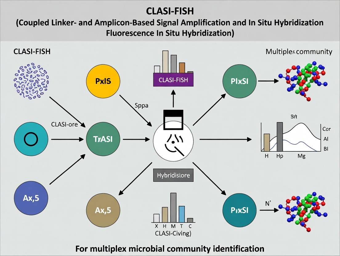
CLASI-FISH: The Complete Guide to High-Plex Microbial Community Profiling for Research and Drug Development
This comprehensive guide explores Combinatorial Labeling and Spectral Imaging - Fluorescence In Situ Hybridization (CLASI-FISH), a revolutionary technique for multiplex microbial identification.
CLASI-FISH: The Complete Guide to High-Plex Microbial Community Profiling for Research and Drug Development
Abstract
This comprehensive guide explores Combinatorial Labeling and Spectral Imaging - Fluorescence In Situ Hybridization (CLASI-FISH), a revolutionary technique for multiplex microbial identification. Designed for researchers, scientists, and drug development professionals, the article covers foundational principles, detailed methodological workflows, troubleshooting protocols, and comparative validation against other omics techniques. We examine how CLASI-FISH enables the spatial, taxonomic, and functional profiling of complex microbiomes with unprecedented multiplexing capability, offering critical insights for biomedical research, therapeutic discovery, and clinical diagnostics.
What is CLASI-FISH? Unlocking the Principles of High-Plex Microbiome Imaging
Within the broader thesis on advancing multiplex microbial community identification, CLASI-FISH (Combinatorial Labeling and Spectral Imaging - Fluorescence In Situ Hybridization) represents a paradigm shift. It overcomes the spectral limitation of standard FISH, enabling the simultaneous identification of dozens to hundreds of microbial taxa in a single sample. This application note details its core principles, evolution, and protocols to empower research in complex microbiomes, a critical frontier for drug development and microbial ecology.
Core Principles and Evolution from Standard FISH
The evolution from standard FISH to CLASI-FISH is marked by a move from direct, spectrally distinct labeling to combinatorial encoding.
| Feature | Standard FISH | CLASI-FISH |
|---|---|---|
| Primary Limitation | Spectral overlap limits multiplexity (~3-8 targets). | Spectral overlap is circumvented by encoding. |
| Labeling Principle | One fluorophore (or mix) per target rRNA sequence. | Targets assigned unique binary codes from a fluorophore panel. |
| Encoding Strategy | Direct, spectral differentiation. | Combinatorial (binary) encoding. |
| Max Targets (Typical) | 3-8 with spectral imaging. | Dozens to hundreds theoretically (e.g., 7 fluorophores = 2⁷-1=127 codes). |
| Key Enabling Tech | Epifluorescence/Confocal microscopy. | Spectral imaging, computational decoding. |
| Data Analysis | Direct channel observation. | Spectral unmixing and code validation. |
| Application Scope | Low-complexity communities, abundance quantification. | High-complexity spatial mapping, network analysis. |
Core Principle: In CLASI-FISH, each microbial taxon is targeted by a unique set of oligonucleotide probes, each labeled with a different fluorophore from a small panel (e.g., Cy3, Cy5, FITC). A taxon is identified not by a single color, but by a unique combination of presence/absence signals from the fluorophore panel—a binary barcode. Spectral imaging and unmixing deconvolve the overlapping emission signals to read these barcodes.
Title: Evolution from Standard FISH to CLASI-FISH
Detailed Application Notes & Protocols
Protocol 1: CLASI-FISH Probe Design and Validation
Objective: Design and validate taxon-specific oligonucleotide probes for combinatorial labeling.
Methodology:
- Target Retrieval: Retrieve 16S/23S rRNA gene sequences for target taxa from databases (SILVA, RDP).
- Probe Design: Use ARB or probeDesigner to find hypervariable regions. Check specificity in silico against a database.
- Combinatorial Code Assignment: Assign each target a unique binary code from the fluorophore panel (e.g., Taxon1: FluorA+FluorC; Taxon2: FluorB+FluorC).
- Probe Synthesis: Order oligonucleotides with a 5'-amino modifier (C6) for later fluorophore conjugation.
- Fluorophore Labeling: Conjugate amino-linked probes with NHS-ester dyes (e.g., Cy3, Cy5, FITC, Texas Red) following manufacturer protocol. Purify via HPLC.
- Specificity Validation: Perform standard FISH on pure cultures or defined synthetic communities. Confirm signal and lack of off-target binding.
Protocol 2: Sample Hybridization and Spectral Imaging
Objective: Hybridize CLASI-FISH probes to a fixed microbial sample and acquire spectral image cubes.
Reagents & Materials:
- Formalin-fixed sample (biofilm, tissue, soil smear).
- Ethanol series (50%, 80%, 96%).
- Hybridization buffer (0.9 M NaCl, 20 mM Tris/HCl pH 7.2-8.0, 0.01% SDS, Formamide concentration probe-dependent).
- Washing buffer (various NaCl concentrations based on formamide %).
- DAPI counterstain.
- Antifading mounting medium.
Methodology:
- Sample Preparation: Apply fixed sample to slide. Dehydrate through ethanol series (3 min each).
- Hybridization: Apply hybridization buffer containing the pooled, labeled CLASI-FISH probe set (typically 2-8 ng/µL each). Incubate at 46°C for 2-3 hours in a dark, humid chamber.
- Washing: Immerse slide in pre-warmed washing buffer at 48°C for 15-20 minutes. Rinse briefly with ice-cold dH₂O. Air dry in dark.
- Counterstaining & Mounting: Apply DAPI stain (1 µg/mL) for 5 min. Rinse, air dry, and mount with antifading medium.
- Spectral Imaging: Acquire images using a confocal microscope with a spectral detector or a widefield system with tunable filters. Parameters:
- For each field of view, collect an emission spectrum (e.g., 500-750 nm) at each pixel at multiple (e.g., 5-10 nm) intervals.
- Use consistent laser power, exposure time, and a high numerical aperture objective (63x or 100x oil).
- Generate a spectral library from control samples hybridized with single probes.
Protocol 3: Spectral Unmixing and Microbial Identification
Objective: Deconvolve spectral image cubes to assign binary codes and identify taxa.
Methodology:
- Spectral Library Creation: Image control samples (pure cultures or spots) labeled with each individual fluorophore used. Extract reference emission spectra.
- Linear Unmixing: For each pixel in the experimental image cube, model the measured spectrum as a linear combination of the reference spectra. Use algorithms (e.g., least squares) to calculate the contribution (weight) of each reference fluorophore.
Pixel_spectrum = (a * Spectrum_Cy3) + (b * Spectrum_Cy5) + (c * Spectrum_FITC) + ... - Thresholding & Binarization: Apply a signal-to-noise ratio threshold (e.g., 5-10) to each fluorophore channel. Weights above threshold are scored as "1" (present), below as "0" (absent).
- Code Assignment & Visualization: Assign the binary code for each pixel to a specific taxon based on the probe code table. Generate false-color identification maps.
Title: CLASI-FISH Experimental and Computational Workflow
The Scientist's Toolkit: Key Research Reagent Solutions
| Item | Function / Rationale |
|---|---|
| Amino-Modified Oligonucleotides | Probe backbone for covalent, stable attachment of NHS-ester fluorophores. |
| NHS-Ester Fluorophores (Cy3, Cy5, etc.) | Reactive dyes for amine coupling; provide bright, photostable signals for the encoding panel. |
| Formamide (Molecular Biology Grade) | Critical component of hybridization buffer; lowers melting temperature to allow stringent, sequence-specific binding. |
| Spectral Imaging Microscope | Equipped with spectral detector or tunable filters to capture full emission spectra for unmixing. |
| Spectral Unmixing Software | (e.g., Zeiss Zen, CytoSpectre, in-house scripts) to perform linear unmixing and decode fluorescence signals. |
| Antifading Mounting Medium | (e.g., Vectashield, Citifluor) Preserves fluorescence signal during imaging by reducing photobleaching. |
| Stringent Washing Buffer (NaCl/EDTA/Tris) | Removes non-specifically bound probes after hybridization; concentration is calculated based on formamide % for stringency. |
Combinatorial Labeling and Spectral Imaging - Fluorescence In Situ Hybridization (CLASI-FISH) represents a paradigm shift for multiplex microbial community analysis. The core thesis posits that by using combinatorial binary labeling schemes with a limited set of fluorophores, one can exponentially increase the number of distinguishable targets, thereby bypassing the fundamental spectral limits imposed by conventional fluorescence microscopy. This application note details the protocols and reagents enabling this breakthrough, translating theoretical multiplexing capacity into practical workflows for microbial ecology, host-microbiome interaction studies, and drug discovery targeting microbial consortia.
Quantitative Comparison: Spectral vs. Combinatorial Multiplexing
Table 1: Multiplexing Capacity Comparison
| Parameter | Conventional Spectral FISH | Combinatorial CLASI-FISH | Fold Increase |
|---|---|---|---|
| Number of Fluorophores (n) | 5 | 5 | 1x |
| Distinct Targets (Spectral) | 5 | - | - |
| Distinct Targets (Combinatorial) | - | 2^n - 1 = 31 | 6.2x |
| Practical Achieved Targets (Published) | 5-8 | 30+ | 4-6x |
| Required Detection Channels | 5 | 5 | 1x |
| Spatial Co-localization Analysis | Limited | High-plex, network mapping | N/A |
| Reference | (Valm et al., 2011) | (Shi et al., 2020; Moffitt et al., 2022) |
Table 2: Key Performance Metrics for CLASI-FISH
| Metric | Typical Value/Range | Protocol Section | Impact on Data Quality |
|---|---|---|---|
| Hybridization Efficiency | >85% for abundant rRNA | 3.2 | Defines detection limit |
| False Positive Rate (Binary Code) | <1% per bit | 3.4, 3.5 | Limits maximum multiplex |
| False Negative Rate (Binary Code) | 2-5% per bit | 3.4 | Affects code accuracy |
| Signal-to-Noise Ratio (Post-Processing) | 10-30 dB | 3.6 | Critical for decoding |
| Spatial Resolution Maintained | ~200-300 nm (diffraction-limited) | 3.3 | Enables single-cell mapping |
| Experiment Duration (for 30 targets) | 2-3 days | 3.0 | Throughput consideration |
Detailed Experimental Protocols
Diagram Title: CLASI-FISH End-to-End Workflow
Probe Design and Binary Code Assignment
- Objective: Assign a unique binary barcode to each target microbial taxon.
- Materials: rRNA sequence database (e.g., SILVA, Greengenes), probe design software (e.g., ARB, mathFISH), oligonucleotide synthesis service.
- Protocol:
- Retrieve 16S/23S rRNA target sequences for organisms of interest.
- Design ~15-20mer oligonucleotide probes with matched Tm (∼55°C). Label 5’ end with a primary amine for later dye conjugation.
- For N fluorophores, assign each target a unique N-bit binary code. Reserve code '00000' (no signal) as null.
- Synthesize probe pools: For each taxonomic target, synthesize a mixture of probes targeting multiple sites on its rRNA, all associated with the same binary code.
- Conjugate fluorophores (Cy3, Cy5, Alexa Fluor dyes) to amine-labeled probes via NHS-ester chemistry. Purify via HPLC.
Sample Preparation and Pre-Hybridization
- Objective: Prepare microbial biofilm or tissue sections for FISH.
- Materials: Multi-well chamber slides, paraformaldehyde (4%), ethanol, lysozyme (for Gram-positives), permeabilization buffers.
- Protocol:
- Fix samples in 4% PFA for 2-3 hours at 4°C.
- Wash in 1x PBS. Apply to charged microscope slides. Dehydrate in 50%, 80%, 98% ethanol series (3 min each).
- (Optional for Gram-positives) Apply lysozyme solution (10 mg/mL in 0.1M Tris, 0.05M EDTA) for 10-60 min at 37°C.
- Dehydrate again through ethanol series. Air dry.
Combinatorial Hybridization Cycle
- Objective: Hybridize probes corresponding to one "bit" position of the binary code.
- Materials: Hybridization buffer (0.9M NaCl, 20mM Tris-HCl pH 7.5, 0.01% SDS, 20% Formamide), humidified chamber, hybridization oven.
- Protocol:
- For hybridization round
i(where i=1 to N, for N fluorophores):- Prepare a master hybridization mix containing ALL probe pools whose binary code has a '1' in the i-th bit position, each labeled with fluorophore
i. - Apply 30-50 µL of mix to sample area, add coverslip.
- Prepare a master hybridization mix containing ALL probe pools whose binary code has a '1' in the i-th bit position, each labeled with fluorophore
- Incubate in dark, humidified chamber at 46°C for 2-4 hours.
- Remove coverslip and wash in pre-warmed wash buffer (according to formamide concentration) at 48°C for 15 min.
- Rinse briefly with ice-cold dH2O. Air dry in dark.
- Mount with anti-fade mounting medium.
- For hybridization round
Sequential Imaging and Signal Stripping
- Objective: Acquire image for the current bit and remove signal to prepare for next round.
- Materials: Epifluorescence or confocal microscope with motorized stage, stable LED or laser light sources, appropriate filter sets.
- Protocol:
- Image Acquisition: Using the filter set for fluorophore
i, acquire images for all fields of view (FOVs). Use identical exposure times across rounds. - Image Registration: Include fiducial markers (fluorescent beads) in a control channel to enable precise image alignment in post-processing.
- Chemical Stripping: Immerse slide in stripping solution (e.g., 30-50% formamide in 2x SSC at 48°C for 30 min, or 20mM NaOH for 1-2 min).
- Efficiency Check: Re-image the same FOV with the same settings to confirm >95% signal loss.
- Repeat Sections 3.3 & 3.4 for all N hybridization rounds.
- Image Acquisition: Using the filter set for fluorophore
Image Processing and Barcode Decoding
- Objective: Generate a decoded map identifying each cell by its taxonomic assignment.
- Materials: Image analysis software (e.g., MATLAB, Python with scikit-image, Ilastik, custom scripts).
- Protocol:
- Image Registration: Align all N image stacks from each FOV using the fiducial marker channel. Use sub-pixel registration algorithms.
- Cell Segmentation: Use a DAPI or general nucleic acid stain image from a final round to create a mask identifying individual cells.
- Signal Extraction: For each segmented cell, measure the mean intensity in each channel (bit) from each registered round.
- Thresholding & Barcode Assignment: Apply a threshold (typically 5-6x standard deviation of background) to each bit to convert intensities to binary '1' or '0'.
- Error Correction: Compare each cell's measured binary code against a predefined library of target codes. Assign identity to the closest valid code using Hamming distance, allowing for 1-bit errors.
Diagram Title: Image Decoding Logic Flow
The Scientist's Toolkit: Research Reagent Solutions
Table 3: Essential Materials for CLASI-FISH
| Item Name / Category | Example Product / Specification | Function in Protocol |
|---|---|---|
| Fluorophore-Conjugated Oligos | Alexa Fluor 488/546/647, Cy3, Cy5 NHS esters | Provides the signal for each "bit"; spectral separation is key. |
| Probe Design Software | ARB, mathFISH, probeBase | Ensures probe specificity and matched hybridization conditions. |
| Chambered Slides | Lab-Tek II, 8-well removable chamber | Holds samples for sequential hybridization and washing. |
| Hybridization Buffer | 0.9M NaCl, 20mM Tris-HCl, 0.01% SDS, variable formamide (0-40%) | Creates optimal stringency for specific probe binding. |
| Chemical Stripping Solution | 50% Formamide / 2x SSC or 20mM NaOH | Removes hybridized probes without damaging sample morphology for subsequent rounds. |
| Fiducial Markers | TetraSpeck or FluoSpheres multicolor beads (0.1-0.2 µm) | Provides invariant reference points for perfect image registration across rounds. |
| Anti-Fade Mountant | ProLong Diamond, Vectashield | Preserves fluorescence signal during imaging; some are compatible with stripping. |
| Automated Fluidics System | Optional: Microfluidic pump/manifold (e.g., BioTek) | Standardizes and automates hybridization/wash/stripping steps, improving reproducibility. |
| Spectral Imaging Microscope | Confocal or widefield with motorized stage, stable light source, and filter sets for all fluorophores. | Acquires high-quality, comparable images across multiple experimental rounds. |
Application Notes
Context within CLASI-FISH for Microbial Community Identification
Combinatorial Labeling and Spectral Imaging - Fluorescence In Situ Hybridization (CLASI-FISH) is a transformative approach for the spatial identification of dozens of microbial taxa within complex communities. Its power hinges on the precise integration of three core technological pillars: (1) specifically designed oligonucleotide probes, (2) spectrally distinct fluorophores, and (3) advanced spectral imaging systems. This synergy allows researchers to transcend the "spectral limit" of traditional fluorescence microscopy, enabling highly multiplexed analysis crucial for understanding microbiomes in health, disease, and biotechnological applications.
Probes: The Targeting Mechanism
Probes are typically 15-25 nucleotide DNA oligonucleotides complementary to unique ribosomal RNA (rRNA) sequences of target microorganisms. For high-plex CLASI-FISH, probes are designed with computational tools to ensure target specificity and are synthesized with a reactive moiety (e.g., an amino linker) for subsequent fluorophore conjugation.
Key Design Considerations:
- Specificity: Must bind exclusively to the target sequence. Tools like ARB, Silva, and probeBase are used for in silico validation.
- Accessibility: Target site on the rRNA must be accessible for hybridization. Empirical testing is often required.
- Melting Temperature (Tm): Probes within a multiplex set should have similar Tms (~55-65°C) to allow simultaneous hybridization under uniform stringency conditions.
Fluorophores: The Spectral Palette
Fluorophores provide the detectable signal. CLASI-FISH employs a combinatorial labeling scheme where each taxonomic target is identified by a unique combination of fluorophores, not a single color.
Principle: If n spectrally separable fluorophores are available, they can be used in binary combinations (present/absent for each fluorophore on a probe) to theoretically label 2ⁿ - 1 distinct targets. For example, 7 fluorophores can encode 127 unique combinations.
Critical Fluorophore Properties:
- Brightness & Photostability: Essential for detecting low-abundance targets and during spectral scanning.
- Spectral Separability: Emission spectra must be distinct enough for unambiguous unmixing.
- Chemical Compatibility: Must withstand FISH protocols and conjugate efficiently to probes.
Table 1: Common Fluorophore Pairs for CLASI-FISH (Example Panel)
| Fluorophore | Excitation Max (nm) | Emission Max (nm) | Conjugate To |
|---|---|---|---|
| Cy2 | 489 | 506 | Probe Set A |
| Cy3 | 550 | 570 | Probe Set A |
| Cy3.5 | 581 | 596 | Probe Set B |
| Cy5 | 649 | 670 | Probe Set B |
| Cy5.5 | 675 | 694 | Probe Set C |
| Cy7 | 743 | 767 | Probe Set C |
| Alexa Fluor 488 | 495 | 519 | Probe Set D |
| Alexa Fluor 594 | 590 | 617 | Probe Set D |
Spectral Imaging Systems: The Decoding Engine
Spectral imaging captures the full emission spectrum at every pixel in an image. This data is then "unmixed" using reference spectra (single-fluorophore controls) to determine the contribution of each fluorophore at each location, thereby decoding the combinatorial label.
Core Components:
- Light Source: A laser-based or LED-based system capable of exciting the full range of fluorophores.
- Spectral Detector: Typically a spectrometer coupled to a CCD camera, or a filter-based system with a large number of narrow bandpass filters.
- Unmixing Software: Algorithms (e.g., linear unmixing) that decompose the measured mixed signal into its constituent fluorophore contributions.
Table 2: Comparison of Spectral Imaging Approaches
| Approach | Mechanism | Spectral Resolution | Speed | Cost |
|---|---|---|---|---|
| Laser Scanning Confocal + Spectral PMT | Prism disperses light onto a 32-channel PMT array. | High (5-10 nm bins) | Medium | High |
| Filter-based (Liquid Crystal Tunable Filter) | Electronically tunable filter transmits narrow wavelength bands sequentially. | Medium-High | Slow | High |
| Filter-based (Multi-band Pass + Emission Filter Array) | Uses a predefined set of 10-20 emission filters. | Medium | Fast | Medium |
| Widefield + Hyperspectral Camera | Grating projects spectrum directly onto a 2D sensor. | Very High (2-5 nm) | Slow | Very High |
Detailed Protocols
Protocol 1: Synthesis of Fluorophore-Labeled FISH Probes
Objective: Covalently conjugate NHS-ester modified fluorophores to amino-modified oligonucleotides.
Materials:
- Amino-modified oligonucleotide (100 µM in nuclease-free water)
- NHS-ester fluorophore (e.g., Cy3, Cy5) in anhydrous DMSO
- 0.1 M Sodium bicarbonate buffer, pH 8.5
- Sephadex G-25 spin column (for dye removal) or reverse-phase HPLC system
- Microcentrifuge
Procedure:
- Dissolve the amino-linked oligonucleotide to 100 µM in 0.1 M sodium bicarbonate buffer (pH 8.5).
- Prepare a 10 mM stock of the NHS-ester fluorophore in anhydrous DMSO immediately before use.
- Mix the oligonucleotide solution with the fluorophore solution at a 1:10 molar ratio (oligo:dye). Incubate in the dark at room temperature for 2 hours.
- Purification: Remove unconjugated fluorophore using a Sephadex G-25 spin column according to the manufacturer's instructions. Collect the flow-through (labeled probe).
- Verify labeling efficiency by measuring absorbance at 260 nm (DNA) and the fluorophore's peak absorbance (e.g., 550 nm for Cy3). Calculate the degree of labeling (DOL, dyes per oligo). A DOL of 0.8-1.2 is typically targeted.
- Adjust probe concentration to 50 ng/µL, aliquot, and store at -20°C in the dark.
Protocol 2: Multiplex CLASI-FISH Hybridization and Spectral Imaging
Objective: Hybridize a complex microbial sample with a combinatorially labeled probe set and acquire spectral image data.
Materials:
- Fixed microbial samples on glass slides (e.g., biofilm sections)
- Combinatorial probe set (each at 50 ng/µL in hybridization buffer)
- Hybridization buffer: 0.9 M NaCl, 20 mM Tris-HCl (pH 7.4), 0.01% SDS, 30% formamide (stringency adjusted as needed).
- Washing buffer: 20 mM Tris-HCl (pH 7.4), 5 mM EDTA, 0.01% SDS, 80-900 mM NaCl (matched to formamide concentration).
- Ethanol dehydration series (80%, 90%, 100%)
- Spectral imaging microscope system (e.g., confocal with spectral detector).
Procedure: Part A: Hybridization
- Apply 20-50 µL of the probe mix (containing all combinatorially labeled probes in hybridization buffer) to the sample area on the slide. Cover with a coverslip.
- Place the slide in a pre-warmed, humidified hybridization chamber. Incubate at 46°C for 2-4 hours in the dark.
- Carefully remove the coverslip by immersing the slide in pre-warmed washing buffer.
- Wash the slide in washing buffer at 48°C for 20 minutes.
- Briefly rinse the slide in ice-cold deionized water.
- Dehydrate the sample by dipping sequentially in 80%, 90%, and 100% ethanol (3 min each). Air dry in the dark.
- Mount with an anti-fading mounting medium (e.g., Vectashield) and seal.
Part B: Spectral Image Acquisition
- Define Spectral Library: Prior to sample imaging, acquire reference images from control samples labeled with single fluorophores (one per fluorophore used in the combinatorial scheme). Use identical acquisition settings.
- Acquire Experimental Spectral Stack: On the sample, define regions of interest. Set the spectral detector to collect emission across the full relevant range (e.g., 500-800 nm) in 5-10 nm increments. Acquire z-stacks if 3D information is needed. Keep laser power and gain settings consistent with library acquisition.
- Spectral Unmixing: Use the microscope's software (e.g., Zeiss ZEN, Leica LAS X) to perform linear unmixing. Input the single-fluorophore reference spectra from Step 1 as the basis set. The software will generate a separate channel image for each fluorophore, showing its relative contribution at every pixel.
- Decoding & Visualization: Using a lookup table that maps fluorophore combinations to target identities (e.g., "Cy3+Cy5 = Target Species X"), assign colors and identities to the unmixed images for composite visualization and analysis.
Diagrams
Title: CLASI-FISH Experimental Workflow
Title: Spectral Imaging and Linear Unmixing Principle
The Scientist's Toolkit: Research Reagent Solutions
| Item | Function in CLASI-FISH | Key Considerations |
|---|---|---|
| Amino-Modified Oligonucleotides | Probe backbone with reactive -NH2 group for fluorophore coupling. | Position (5' or 3'), linker length, purity (HPLC-grade). |
| NHS-Ester Fluorophores | Reactive dye form for stable amide bond formation with amino-linked probes. | Spectral profile, brightness, solubility in DMSO, matching to imaging system lasers. |
| Formamide (Molecular Biology Grade) | Denaturant in hybridization buffer to control stringency and probe specificity. | Concentration must be optimized for each probe set (typically 30-50%). |
| Sephadex G-25 Spin Columns | Size-exclusion chromatography for rapid purification of labeled probes from free dye. | Fast, effective for removing small molecule dyes; does not separate unlabeled oligo. |
| Anti-Fading Mounting Medium (e.g., Vectashield) | Preserves fluorescence signal during imaging by reducing photobleaching. | Refractive index, hardness (for potential re-imaging), compatibility with fluorophores. |
| Multispectral Calibration Beads | Beads coated with multiple fluorophores, used to validate spectral unmixing accuracy. | Essential for quality control of the spectral imaging and unmixing pipeline. |
| Stringency Wash Buffer (SSC or Tris-EDTA based) | Removes nonspecifically bound probes post-hybridization. | Salt concentration is precisely calculated based on formamide % and desired Tm. |
1. Introduction and Context Within the broader thesis framework on CLASI-FISH (Combinatorial Labeling and Spectral Imaging - Fluorescence In Situ Hybridization) for multiplex microbial community identification, targeting ribosomal RNA (rRNA) remains the cornerstone for phylogenetic identification. The vast majority of environmental microbes resist cultivation, constituting the microbial 'dark matter.' This application note details protocols leveraging the high copy number and genetic conservation of rRNA to identify and visualize these uncultivated organisms in complex samples, enabling their integration into a multiplex CLASI-FISH analytical pipeline.
2. Application Notes: The Role of rRNA-Targeted FISH
- Specificity and Universality: The 16S rRNA gene contains nine hypervariable regions (V1-V9) flanked by conserved sequences. Probes are designed with a conserved 'anchor' for binding and a hypervariable 'fingerprint' for specificity.
- Sensitivity: The high intracellular rRNA copy number (10³–10⁵ copies per cell) provides natural signal amplification.
- Quantitative Data: Key performance metrics for rRNA-FISH are summarized below.
Table 1: Performance Metrics of rRNA-Targeted FISH Probes
| Metric | Typical Range/Value | Notes |
|---|---|---|
| Probe Length | 15-25 nucleotides | Optimizes specificity and binding kinetics. |
| Hybridization Temperature | 46-48°C (±5°C formamide) | Critical for stringency; varies with probe GC%. |
| Formamide Concentration | 0-60% (v/v) in buffer | Used to adjust stringency; higher % lowers effective Tm. |
| Detection Limit (Cell Count) | >10³ cells/mL (direct) | Can detect single cells microscopically. |
| Label Incorporation (Fluorophores per probe) | 1-5 | Higher labeling can reduce hybridization efficiency. |
| Phylogenetic Resolution | Species to Domain level | Depends on probe target region design. |
Table 2: Common rRNA Target Regions and Specificity
| Target Region (16S rRNA) | Phylogenetic Resolution | Common Probe Examples |
|---|---|---|
| V1-V3 Region | High (Genus/Species) | EUB338 (Bacteria), ARCH915 (Archaea) |
| V3-V4 Region | Medium-High (Genus) | Used extensively in NGS, good for FISH. |
| V4-V5 Region | Medium (Family/Genus) | Balanced between conservation and variability. |
| V6-V8 Region | Medium (Phylum/Class) | Suitable for broader group identification. |
3. Experimental Protocols
Protocol 1: Design and Validation of rRNA-Targeted Oligonucleotide Probes
- In Silico Design:
- Retrieve 16S/23S rRNA sequences from databases (SILVA, RDP, Greengenes).
- Align sequences to identify taxon-specific target sites.
- Design 18-22 nt oligonucleotide probes with a Tm of ~55°C. Check specificity against non-target sequences using tools like ARB or probeCheck.
- Probe Labeling:
- Synthesize probes with a primary amine or thiol modification at the 5’-end.
- Conjugate NHS-ester or maleimide-activated fluorophores (e.g., Cy3, Cy5, FAM) following manufacturer protocols.
- Purify labeled probes via HPLC or column purification.
- In Vitro Validation:
- Perform dot blot hybridization against target and non-target rRNA extracted from pure cultures.
- Quantify signal-to-noise ratio. A successful probe shows >10-fold higher signal for target rRNA.
Protocol 2: rRNA-FISH for Fixed Environmental Samples (Pre-CLASI)
- Sample Fixation: Fix sample (biofilm, sediment) in 4% paraformaldehyde (PFA) for 2-4h at 4°C. Wash with 1x PBS.
- Permeabilization: Apply lysozyme solution (10 mg/mL in 0.1M Tris-HCl, 0.05M EDTA, pH 8.0) for 30-60 min at 37°C.
- Hybridization:
- Prepare hybridization buffer: 0.9M NaCl, 20mM Tris/HCl (pH 7.4), 0.01% SDS, and a defined concentration of formamide (see Table 1). Pre-warm to 46°C.
- Add labeled probe(s) to buffer (final conc. 2-10 ng/μL).
- Apply buffer to sample and incubate in a dark, humidified chamber at 46°C for 2-4 hours.
- Washing:
- Prepare pre-warmed wash buffer: 20mM Tris/HCl (pH 7.4), 5mM EDTA, 0.01% SDS, and NaCl concentration adjusted based on formamide % used.
- Immerse slide in wash buffer at 48°C for 15-30 minutes.
- Rinsing & Mounting: Rinse briefly with ice-cold distilled water. Air dry and mount with anti-fading mounting medium (e.g., Vectashield with DAPI).
Protocol 3: Integration with CLASI-FISH for Multiplexing
- Probe Set Design: Assign a unique binary fluorescent color code (e.g., Cy3 = 1, Cy5 = 0) to each phylogenetic probe.
- Sequential Hybridization: Perform Protocol 2 iteratively. After each FISH round, document signals via epifluorescence/confocal microscopy, then strip probes using a low-pH buffer (e.g., 0.1% HCl in 70% EtOH for 10 min).
- Image Registration & Decoding: Use computational tools to align images from all hybridization rounds. Decode the fluorescent 'barcode' for each cell to assign phylogenetic identity.
4. Visualizations
Workflow for rRNA-CLASI-FISH Identification
rRNA Gene Target Site Selection
5. The Scientist's Toolkit: Research Reagent Solutions
Table 3: Essential Reagents for rRNA-Targeted CLASI-FISH
| Reagent / Material | Function / Role | Example / Note |
|---|---|---|
| Paraformaldehyde (PFA) | Chemical fixative. Cross-links proteins to preserve cellular morphology and retain rRNA. | 4% solution in PBS. Handle in fume hood. |
| Lysozyme | Enzymatic permeabilization agent. Digests peptidoglycan in bacterial cell walls for probe entry. | From Gallus gallus; 10-50 mg/mL working concentration. |
| Formamide | Denaturant in hybridization buffer. Lowers effective melting temperature (Tm) for stringent binding. | Molecular biology grade. Concentration is probe-specific (0-60%). |
| SSC Buffer | Provides ionic strength (Saline-Sodium Citrate) for hybridization and washing. | 20x stock solution. Dilute to appropriate stringency (e.g., 0.2x-2x). |
| Labeled Oligonucleotide Probes | rRNA-targeted, fluorophore-conjugated DNA strands. Provide specificity and detection signal. | HPLC-purified, 5'-labeled with Cy3, Cy5, Alexa Fluor dyes. |
| Anti-fading Mountant | Preserves fluorescence signal during microscopy by reducing photobleaching. | Vectashield, ProLong Diamond. Often includes DAPI for counterstain. |
| Probe Stripping Buffer | Removes hybridized probes between CLASI-FISH rounds without damaging sample. | Low-pH buffer (e.g., 0.1% HCl/70% EtOH) or chaotropic salt solutions. |
Application Notes
The application of Combinatorial Labeling and Spectral Imaging - Fluorescence In Situ Hybridization (CLASI-FISH) represents a paradigm shift in microbial ecology and systems biology. This multiplex imaging technique enables the simultaneous identification of dozens of microbial taxa within their native spatial context, moving beyond compositional lists to architectural mapping. For drug development professionals, this spatial intelligence is critical for understanding polymicrobial infection sites, biofilm resilience, and host-microbiome interfaces that influence therapeutic outcomes. The following notes detail its primary applications and quantitative benchmarks.
Key Quantitative Performance Metrics
The efficacy of CLASI-FISH is quantified by several parameters, as summarized in the table below.
Table 1: CLASI-FISH Performance Metrics
| Metric | Typical Performance Range | Technical Notes |
|---|---|---|
| Multiplexing Capacity | 20 - 100+ distinct taxa | Dependent on fluorophore spectral separation and combinatorial labeling scheme. |
| Spatial Resolution | ~200 nm (xy), ~500 nm (z) | Limited by optical diffraction; can be enhanced with super-resolution modalities. |
| Sample Throughput | 1 - 4 samples per imaging run | Bottleneck is often high-resolution, multi-channel spectral imaging time. |
| Taxonomic Resolution | Species to strain-level | Dictated by probe design specificity and stringency of hybridization. |
| Signal-to-Noise Ratio | 10:1 to 50:1 | Improved via tyramide signal amplification (TSA) or hybridization chain reaction (HCR). |
| Tissue Penetration Depth | 30 - 100 µm | Thicker samples require tissue clearing protocols (e.g., CLARITY, CUBIC). |
Core Applications in Research and Drug Development
- Polymicrobial Infection Biofilms: Mapping the consortia architecture in chronic wounds, cystic fibrosis lungs, and medical device-related infections to identify keystone species and spatial niches.
- Gut Microbiome-Mucosa Interface: Visualizing the structured organization of taxa relative to crypts, mucus layers, and immune cells in health, inflammatory bowel disease (IBD), and cancer.
- Microbial Biogeography in Soils and Plants: Deciphering nutrient gradients and symbiotic interactions at the micrometer scale relevant to agriculture and biotech.
- Drug Efficacy Testing: Assessing how antimicrobials or biologics disrupt the spatial organization of a community, a metric often more informative than bulk biomass reduction.
Experimental Protocols
Protocol 1: CLASI-FISH Probe Design and Validation
Objective: To create a panel of species-specific rRNA-targeted oligonucleotide probes for multiplexed identification.
Materials:
- Research Reagent Solutions:
- ARB Silva Database: Repository of aligned rRNA sequences for target and non-target specificity checks.
- MathFISH Probe Design Tool: Algorithm for calculating probe binding efficiency and specificity.
- Oligonucleotide Probes: Synthesized with a 5'-amine or azide modification for subsequent fluorophore conjugation.
- NHS-Ester Fluorophores: Set of spectrally distinct dyes (e.g., Cy3, Cy5, Alexa Fluor 488, 594, 750).
- Purification Columns: For removing unconjugated fluorophore from labeled probes (e.g., NAP-10 columns).
Methodology:
- Target Selection: Retrieve full-length 16S/23S rRNA sequences for all target microorganisms from NCBI or RDP.
- Specificity Check: Align target sequences against a database using BLAST or the DECIPHER probeMatch function. Ensure a minimum of 2 mismatches to non-target sequences.
- Probe Design: Use software (e.g., MathFISH, probeBase) to select ~15-25 nt regions with predicted high accessibility and uniform melting temperature (Tm ~55-60°C).
- Fluorophore Conjugation: Resuspend amine-modified oligonucleotide in 0.1M carbonate buffer (pH 9.0). Incubate with 50-fold molar excess of NHS-ester fluorophore for 6 hours at room temperature in the dark.
- Purification: Purify the reaction mixture using size-exclusion chromatography. Verify labeling efficiency by measuring absorbance at 260 nm (DNA) and the fluorophore's peak wavelength.
Protocol 2: Sample Preparation, Hybridization, and Imaging
Objective: To process a complex microbial sample (e.g., a gut biopsy or biofilm) for multiplex CLASI-FISH imaging.
Materials:
- Research Reagent Solutions:
- 4% Paraformaldehyde (PFA): Fixative for preserving spatial structure and cell morphology.
- Lysozyme or Proteinase K: Enzymes for permeabilizing cell walls of Gram-positive or fixed tissues.
- Hybridization Buffer: Contains formamide, salts, detergents, and blocking agents (e.g., dextran sulfate).
- Tyramide Signal Amplification (TSA) Kit: For amplifying weak signals via HRP-catalyzed deposition of fluorophores.
- Antifade Mounting Medium with DAPI: To preserve fluorescence and counterstain DNA.
- Spectral Confocal or Epifluorescence Microscope: Equipped with a spectral detector or multiple filter sets.
Methodology:
- Fixation & Sectioning: Fix sample in 4% PFA for 4-24 hours at 4°C. For tissues, embed in optimal cutting temperature (OCT) compound and cryo-section at 10-20 µm thickness.
- Permeabilization: Treat slides with lysozyme (1-10 mg/mL) for Gram-positives or proteinase K (optional, titrated) for tissues, for 10-30 minutes at 37°C.
- Combinatorial Hybridization:
- Divide probe set into pools. Each pool contains a unique subset of probes, each labeled with a single fluorophore.
- Apply first probe pool in hybridization buffer (e.g., 30-55% formamide, based on probe Tm) to the sample. Incubate at 46°C for 2-4 hours in a humidified chamber.
- Wash in pre-warmed wash buffer to remove unbound probes.
- Signal Amplification (Optional but Recommended): If using horseradish peroxidase (HRP)-labeled probes, incubate with the corresponding fluorophore-tyramide from the TSA kit per manufacturer instructions. Inactivate HRP with H₂O₂ treatment before the next hybridization round.
- Iterative Hybridization & Imaging: Repeat Steps 3-4 for each probe pool. After each round, acquire a multi-channel image of the sample without moving the slide. Precise registration is critical.
- Final Imaging & Spectral Unmixing: After all hybridization rounds, perform a final high-resolution spectral scan. Use software (e.g., Zen, ImageJ plugins) to unmix overlapping fluorescence spectra and generate a pure signal channel for each fluorophore.
Protocol 3: Image Analysis and Spatial Statistics
Objective: To decode combinatorial fluorescence patterns into taxonomic identities and calculate spatial metrics.
Methodology:
- Image Registration & Decoding: Align image stacks from all hybridization rounds using rigid/affine transformation algorithms. Assign a taxonomic identity to each cell based on its unique binary fluorescence code across all imaging rounds.
- Cell Segmentation: Use machine learning tools (e.g., Ilastik, Cellpose) or intensity thresholding to identify individual cell boundaries from DAPI or general FISH signals.
- Spatial Analysis:
- Co-localization: Calculate metrics like Mander's overlap coefficients for specific taxon pairs.
- Neighborhood Analysis: Determine the frequency of specific microbial neighbors within a defined radius (e.g., 5 µm).
- Global Metrics: Compute community spatial organization indices, such as the Pair Correlation Function (PCF) or Morisita-Horn index for aggregation and dispersion.
Diagrams
Title: CLASI-FISH Experimental Workflow
Title: Combinatorial Probe Decoding
The Scientist's Toolkit: Research Reagent Solutions
Table 2: Essential Materials for CLASI-FISH Experiments
| Item Name | Category | Function & Rationale |
|---|---|---|
| Spectrally Distinct Fluorophores (e.g., Alexa Fluor series, Cy dyes) | Fluorescent Dye | Provide the optical signals for multiplexing. Must have minimal spectral overlap for clean unmixing. |
| NHS-Ester or Click Chemistry Modification Kits | Conjugation Chemistry | Enable covalent, stable attachment of fluorophores to oligonucleotide probes. |
| Formamide (Molecular Biology Grade) | Hybridization Reagent | Component of hybridization buffer; lowers probe Tm to allow stringent temperature control. |
| Tyramide Signal Amplification (TSA) Kit | Signal Amplification | Enzymatically deposits numerous fluorophores per probe, dramatically increasing detection sensitivity. |
| Antifade Mounting Medium (e.g., ProLong Gold, Vectashield) | Imaging Reagent | Reduces photobleaching during extended microscopy and contains counterstains like DAPI. |
| Tissue Clearing Reagents (e.g., CUBIC, ClearT2) | Sample Processing | Renders thick tissues transparent for deep imaging by homogenizing refractive indices. |
| Spectral Imaging Software (e.g., Zeiss Zen, Leica LAS X) | Analysis Software | Performs critical spectral unmixing to separate fluorophore signals and eliminate autofluorescence. |
| Image Registration Software (e.g., ImageJ with StackReg) | Analysis Software | Aligns images from sequential hybridization rounds with sub-pixel accuracy for correct decoding. |
Step-by-Step CLASI-FISH Protocol: From Probe Design to Data Analysis
Probe Design and Validation for Specific, High-Sensitivity Targeting
Within the context of a thesis on Combinatorial Labeling and Spectral Imaging Fluorescence In Situ Hybridization (CLASI-FISH) for multiplex microbial community identification, the design and validation of nucleic acid probes are paramount. This application note details protocols for creating probes that achieve species-level differentiation in complex consortia, a cornerstone for accurate spatial mapping and functional analysis in drug development and microbiome research.
Probe Design Principles for CLASI-FISH
High-performance CLASI-FISH probes must satisfy dual constraints: 1) high binding affinity to target rRNA sequences, and 2) exquisite specificity to avoid cross-hybridization with non-target microbes. The process involves:
- Target Sequence Selection: Identification of unique 16S/23S rRNA regions via comprehensive database mining (e.g., SILVA, RDP). A minimum of two mismatches to non-target sequences is recommended.
- In Silico Specificity Check: Use of tools like ARB, ProbeCheck, and DECIPHER.
- Thermodynamic Parameters: Calculation of melting temperature (Tm) and Gibbs free energy (ΔG) to ensure uniform hybridization conditions across multiplex probe sets.
Table 1: Key Parameters for CLASI-FISH Probe Design
| Parameter | Optimal Target Range | Rationale |
|---|---|---|
| Length | 15-25 nucleotides | Balances specificity and accessibility to structured rRNA. |
| GC Content | 40-60% | Ensures stable hybridization; avoids extreme Tm. |
| Tm | 50-65°C (Formamide-adjusted) | Allows for stringent, uniform wash conditions. |
| Minimum Mismatches | ≥2 (central position preferred) | Maximizes discriminatory power against non-targets. |
| BLAST E-value | < 0.01 | Confirms target uniqueness in public databases. |
Experimental Validation Protocols
Protocol 3.1:In VitroSpecificity Testing using Dot Blot Hybridization
Purpose: To confirm probe binding to target sequences and absence of binding to non-target sequences. Materials:
- Nylon Membrane with spotted genomic DNA from pure culture targets and non-targets.
- DIG-labeled Probe synthesized with target-specific sequence.
- Hybridization Buffer (e.g., 5x SSC, 0.1% N-Lauroylsarcosine, 0.02% SDS, 1% Blocking Reagent).
- Stringency Wash Buffer (e.g., 0.2x SSC, 0.1% SDS).
- Anti-DIG-AP Antibody and CDP-Star Chemiluminescent Substrate.
Procedure:
- Spot 100 ng of genomic DNA from each reference strain onto a positively charged nylon membrane. Denature and crosslink.
- Pre-hybridize membrane at the calculated hybridization temperature (e.g., 46°C) for 30 min.
- Add DIG-labeled probe (10-50 ng/mL) to fresh buffer and hybridize for 2-16 hours.
- Perform two 5-minute stringency washes at the predetermined temperature.
- Detect hybridization signal using anti-DIG-AP and chemiluminescent substrate. Image with a CCD camera.
Protocol 3.2: CLASI-FISH Validation on Complex Communities
Purpose: To validate probe specificity and sensitivity within a structured, multi-species sample. Materials:
- Microbial Community Sample (e.g., biofilm, gut microbiome model).
- CLASI-FISH Probe Set: Multiple, differentially fluorophore-labeled probes designed per Table 1.
- Fixative: 4% paraformaldehyde (PFA) in PBS.
- Hybridization Buffer: 0.9 M NaCl, 20 mM Tris/HCl (pH 7.4), 0.01% SDS, variable formamide (concentration probe-dependent).
- Fluorophores: e.g., Cy3, Cy5, Alexa Fluor 488, CF dyes for combinatorial labeling.
Procedure:
- Fixation: Fix sample in 4% PFA for 2-4 hours at 4°C. Wash with PBS.
- Hybridization: Apply probe cocktail in hybridization buffer. Incubate at 46°C for 2-3 hours in a dark, humidified chamber.
- Washing: Immerse slide in pre-warmed wash buffer (based on probe Tm) for 15-20 minutes at 48°C.
- CLASI Imaging: Rinse briefly with ice-cold dH₂O, air dry, and mount. Image using a spectral confocal or epifluorescence microscope equipped with a spectral detector.
- Analysis: Use linear unmixing software to deconvolve overlapping fluorescence spectra and assign specific spectral signatures to each probe-targeted taxon.
Diagrams
Probe Design and Validation Workflow
CLASI-FISH Multiplex Detection Logic
The Scientist's Toolkit
Table 2: Essential Research Reagent Solutions for CLASI-FISH Probe Validation
| Item | Function | Example/Notes |
|---|---|---|
| High-Quality rRNA Databases | Source for in silico probe design and specificity checks. | SILVA, RDP, Greengenes. Regularly updated. |
| Oligonucleotide Synthesis Service | Production of probes with 5'-end reactive groups for labeling. | Must provide HPLC purification and quality control. |
| Fluorophore Succinimidyl Ester (NHS) | Conjugates amine-modified probes to bright, photostable dyes. | Cy3, Cy5, Alexa Fluor dyes, CF dyes. |
| Stringent Hybridization Buffer | Creates optimal conditions for specific binding; formamide lowers effective Tm. | Standard saline citrate (SSC) buffer with formamide and detergent. |
| Spectral Microscope & Unmixing Software | Captures and deconvolves emission spectra for multiplex detection. | Confocal systems with spectral detectors; software like Zen or ImageJ plugins. |
| Positive & Negative Control Strains | Essential for validating probe specificity in both dot blot and FISH. | Cultured target and closely related non-target organisms. |
Within the broader thesis on combinatorial labeling and spectral imaging fluorescence in situ hybridization (CLASI-FISH) for multiplex microbial community identification, sample preparation is the critical determinant of success. CLASI-FISH demands the simultaneous preservation of cellular morphology, accessibility of numerous diverse rRNA targets, and retention of fluorescent signal integrity across multiple hybridization rounds. This document details optimized Application Notes and Protocols for fixation, permeabilization, and hybridization, validated for complex microbiomes.
The following tables summarize key quantitative findings from recent optimization studies relevant to CLASI-FISH workflows.
Table 1: Fixation Agent Efficacy on Gram-positive vs. Gram-negative Bacteria in Biofilms
| Fixative | Concentration | Fixation Time | Gram-negative Signal (AU) | Gram-positive Signal (AU) | Morphology Preservation (1-5 scale) |
|---|---|---|---|---|---|
| Paraformaldehyde (PFA) | 4% | 2h, 4°C | 1550 ± 120 | 980 ± 95 | 5 |
| Ethanol:Phosphate Buffered Saline (PBS) (1:1) | 50% | 1h, -20°C | 870 ± 110 | 1450 ± 130 | 3 |
| PFA + Glutaraldehyde | 4% + 0.1% | 1h, 4°C | 1620 ± 105 | 750 ± 85 | 4 |
| Methanol | 100% | 10 min, -20°C | 920 ± 75 | 1560 ± 115 | 2 |
Table 2: Permeabilization Treatments for Multiplex FISH on Diverse Taxa
| Treatment | Target Group | Recommended Time | Relative Permeabilization Score* | Notes for CLASI-FISH |
|---|---|---|---|---|
| Lysozyme (10 mg/mL) | Gram-positive | 30 min, 37°C | 4.5 | Essential for Firmicutes; precede with mild detergent. |
| Proteinase K (1 µg/mL) | General/Archaea | 5 min, RT | 3.0 | Use with caution; can degrade morphology. |
| Tris-EDTA Buffer (pH 8.0) | General | 10 min, 95°C | 4.0 | Heat-mediated; effective for many environmental samples. |
| SDS (0.01%) | Biofilm EPS | 5 min, RT | 2.5 | Clears extracellular polymers; can be combined. |
| * Score: 1 (poor) to 5 (excellent) based on post-hybridization signal intensity. |
Table 3: Hybridization Buffer Optimization for High-Stringency Multiplexing
| Buffer Component | Standard Concentration | Optimized CLASI Range | Function & Rationale |
|---|---|---|---|
| Formamide | 0-80% (v/v) | 35-55% | Denatures rRNA; primary stringency control. |
| NaCl | 0-900 mM | 56-250 mM | Ionic strength; inversely related to formamide concentration. |
| SDS | 0-0.2% | 0.01-0.05% | Reduces non-specific probe binding. |
| Blocking Reagent (e.g., RNA) | 0-2 mg/mL | 0.5-1 mg/mL | Competes for non-specific sites, reduces background. |
| pH | ~7.2 | 7.0-7.4 | Maintains probe stability and hybridization kinetics. |
Detailed Experimental Protocols
Protocol 1: Optimized Fixation for Heterotrophic Biofilm Communities Objective: To preserve cellular integrity while maximizing rRNA target accessibility for >20-plex CLASI-FISH. Materials: Filtered sample (e.g., on 0.22 µm polycarbonate filter), 4% PFA in PBS (freshly prepared or aliquoted at -20°C), PBS (pH 7.4), 50% Ethanol in PBS. Procedure:
- Fixation: Transfer filter to a Petri dish. Overlay with 3 mL of ice-cold 4% PFA. Incubate at 4°C for 2 hours.
- Washing: Aspirate PFA. Wash filter three times for 5 minutes each with 3 mL of ice-cold PBS.
- Dehydration: Immerse filter in 50% Ethanol/PBS for 5 minutes at room temperature. This step aids in permeabilization and preserves the sample for storage.
- Storage: Store filter in 50% Ethanol/PBS at -20°C for up to 6 months.
Protocol 2: Tiered Permeabilization for Diverse Microbial Consortia Objective: To achieve uniform probe penetration across phylogenetically diverse cells in a single sample. Materials: Fixed samples on filters, Lysozyme stock (100 mg/mL in 10 mM Tris-HCl, pH 8.0), TE Buffer (10 mM Tris-HCl, 1 mM EDTA, pH 8.0), 0.01% SDS in PBS. Procedure:
- Enzymatic Treatment: Apply 100 µL of Lysozyme working solution (10 mg/mL in TE) onto the sample area of the filter placed on a glass slide. Cover with a coverslip. Incubate in a humid chamber at 37°C for 30 minutes.
- Rinse: Gently remove coverslip and rinse filter by submerging in 50 mL of nuclease-free water for 1 minute.
- Detergent Treatment: Submerge filter in 0.01% SDS solution for 5 minutes at room temperature with gentle agitation.
- Final Rinse: Rinse filter thoroughly with 50 mL of nuclease-free water. Air-dry completely before hybridization.
Protocol 3: High-Stringency Hybridization for Multiplex CLASI-FISH Objective: To enable specific binding of multiple oligonucleotide probes (with varying %GC) simultaneously. Materials: Dried, permeabilized sample, hybridization buffer (see Table 3), fluorescently labeled FISH probes (e.g., Cy3, Cy5, Alexa Fluor derivatives), hybridization oven, humid chamber. Procedure:
- Buffer Preparation: Prepare hybridization buffer with formamide concentration set to the calculated mean for your probe set (e.g., 45%). Add SDS to 0.05% and blocking RNA to 1 mg/mL.
- Hybridization Mix: For each sample, mix 8 µL of hybridization buffer with 1 µL of each probe (50 ng/µL) and 1 µL of nuclease-free water per 10 µL total.
- Application: Apply 10 µL of probe mix to the sample on the filter on a slide. Seal with a coverslip.
- Incubation: Place slide in a pre-warmed, humid chamber. Incubate at 46°C for 2-4 hours in a dark hybridization oven.
- Washing: Prepare pre-warmed wash buffer (appropriate NaCl concentration corresponding to formamide %). Remove coverslip and immediately transfer the filter to 50 mL of wash buffer. Incubate at 48°C for 20 minutes in a water bath. Rinse briefly with ice-cold nuclease-free water and air-dry in the dark.
Visualization: Workflows & Pathways
CLASI-FISH Sample Prep & Imaging Workflow
Optimization Logic for CLASI-FISH Prep
The Scientist's Toolkit: Key Research Reagent Solutions
| Item & Example Product | Function in CLASI-FISH Context |
|---|---|
| Paraformaldehyde (PFA), 16% Aqueous, EM Grade | Primary fixative. Cross-links proteins, preserving 3D structure while retaining nucleic acids for probe access. High purity minimizes autofluorescence. |
| Lysozyme, Molecular Biology Grade | Enzymatic permeabilization agent. Hydrolyzes peptidoglycan in Gram-positive cell walls, crucial for probe entry into diverse community members. |
| Formamide, Ultra Pure | Primary denaturant in hybridization buffer. Concentration fine-tunes stringency to ensure specific binding of multiple probes with different melting temperatures. |
| Blocking Reagent (e.g., yeast total RNA) | Competes with non-specific binding sites on cellular components and the filter substrate, critical for reducing background in multiplex assays. |
| FISH Probes (HRP- or Fluoro-labeled) | Oligonucleotides targeting 16S/23S rRNA. For CLASI, probes are designed for sequential or combinatorial labeling; fluorophore choice is key for spectral separation. |
| Dextran Sulfate (in Hybridization Buffers) | A volume excluder that increases the effective probe concentration, accelerating hybridization kinetics, which is beneficial for complex samples. |
| SlowFade or ProLong Antifade Mountants | Preserves fluorescence photostability during prolonged spectral imaging required for deconvolving multiple signals in CLASI-FISH. |
Within the broader thesis of advancing CLASI-FISH (Combinatorial Labeling and Spectral Imaging - Fluorescence In Situ Hybridization) for multiplex microbial community identification, this application note details the principle and protocol of combinatorial labeling. This method enables the generation of a large number of unique taxonomic identifiers from a limited palette of fluorophores, dramatically expanding the multiplexing capacity for complex environmental or clinical samples.
The combinatorial labeling scheme is based on a simple binary principle. Instead of assigning one fluorophore to one rRNA-targeted oligonucleotide probe, multiple fluorophores are assigned to a single probe. A microbial taxon is then identified by a unique combination of fluorophore signals from multiple probes. With n fluorophores used in k combinations per probe, the number of unique spectral codes scales according to the formula: 2n - 1. This allows for the differentiation of dozens to hundreds of microbes with standard epifluorescence microscopes equipped with 4-7 filter sets.
Table 1: Multiplexing Capacity of Combinatorial Labeling Schemes
| Number of Fluorophores (n) | Probes per Organism (k) | Unique Binary Codes (2n - 1) | Practical Number of Identifiable Taxa* |
|---|---|---|---|
| 3 | 1 | 7 | 7 |
| 4 | 1 | 15 | 15 |
| 4 | 2 | 15 | 105 |
| 5 | 2 | 31 | 465 |
| 6 | 2 | 63 | 1,953 |
| 7 | 2 | 127 | 8,001 |
*Calculated as combinations C(2n-1, k). Assumes perfect spectral separation and no cross-talk.
Table 2: Common Fluorophores for CLASI-FISH (Excitation/Emission Max in nm)
| Fluorophore | Common Excitation (nm) | Common Emission (nm) | Color Group | Notes |
|---|---|---|---|---|
| FITC | 490 | 525 | Green | Bright, but can photobleach. |
| Cy3 | 550 | 570 | Orange | Very bright and photostable. |
| Texas Red | 589 | 615 | Red | Good for spectral separation. |
| Cy5 | 649 | 670 | Far-Red | Requires specific filter sets. |
| Cy3.5 | 581 | 596 | Orange/Red | Good alternative to Cy3. |
| Cy7 | 743 | 767 | NIR | For highly multiplexed setups. |
| ATTO 488 | 501 | 523 | Green | More photostable alternative to FITC. |
Core Protocol: Combinatorial Probe Labeling and CLASI-FISH
A. Probe Design and Combinatorial Code Assignment
- Identify Target Sequences: Using tools like ARB or SILVA, design 16S/23S rRNA-targeted oligonucleotide probes (typically 15-25 nucleotides) with high specificity for your target microbial taxa.
- Assign Combinatorial Codes: Create a spreadsheet mapping each target taxon to a unique binary code using your fluorophore palette (e.g., for fluorophores A, B, C: Taxon1 = A+B, Taxon2 = A+C, Taxon3 = B+C, Taxon4 = A+B+C).
- Order Probes: Synthesize each required probe with the appropriate fluorophore(s) conjugated to the 5'-end. For probes requiring multiple fluorophores, order them conjugated to all required fluorophores on the same oligonucleotide.
B. Sample Fixation and Hybridization
Materials: Phosphate-buffered saline (PBS), Paraformaldehyde (PFA, 4% w/v in PBS), Ethanol, Hybridization oven, Humidity chamber.
- Fix environmental or clinical samples in 4% PFA for 2-4 hours at 4°C.
- Wash with PBS and apply successive ethanol dehydration steps (50%, 80%, 96% ethanol; 3 min each).
- Hybridization Buffer (per 1 mL): 360 µL 5M NaCl, 40 µL 1M Tris/HCl (pH 8.0), 2 µL 10% SDS, 598 µL deionized formamide (concentration varies by probe), 0.5-5 µL of each labeled probe (final concentration ~2-10 ng/µL).
- Apply 30-50 µL of hybridization buffer to dried samples on a slide, cover with a coverslip, and incubate at 46°C for 2-3 hours in a humidity chamber.
C. Stringency Wash and Imaging
Materials: Wash buffer, DAPI stain, Antifade mounting medium.
- Prepare pre-warmed wash buffer: 56mM NaCl, 20mM Tris/HCl (pH 8.0), 5mM EDTA, 0.01% SDS. The EDTA concentration and temperature (typically 48°C) are critical for stringency.
- Remove coverslip and incubate slides in wash buffer for 10-15 minutes.
- Rinse briefly with ice-cold deionized water and air dry in the dark.
- Counterstain with DAPI (1 µg/mL) for 5 min if needed.
- Mount with an antifade reagent (e.g., Vectashield).
- Spectral Imaging: Acquire images using an epifluorescence microscope equipped with motorized filter wheels for each fluorophore channel. Acquire a DIC or phase contrast image for reference. For each field of view, capture a stack of images (one per fluorophore channel + DAPI).
D. Image Analysis and Decoding
- Align all fluorescence channel images using registration software.
- Apply a segmentation algorithm (e.g., based on DAPI or general DNA stain, or signal auto-detection) to identify individual microbial cells.
- Measure the mean fluorescence intensity for each cell across all spectral channels.
- Apply a threshold to binarize the signal (1 = signal present, 0 = absent) for each channel.
- Decode the combinatorial label for each cell by matching its binary code to the predefined taxonomic code table.
Visualizations
Combinatorial Labeling & Decoding Workflow
4-Fluorophore Combinatorial Encoding
The Scientist's Toolkit
Table 3: Essential Research Reagent Solutions for CLASI-FISH
| Item | Function/Benefit | Example/Note |
|---|---|---|
| Fluorophore-Conjugated Oligonucleotides | Core detection reagent. Multiple fluorophores can be conjugated to a single probe. | Ordered from IDT, Sigma, or Biomers. HPLC purification is essential. |
| High-Quality Deionized Formamide | Key component of hybridization buffer; lowers melting temperature (Tm) for stringent hybridization. | Use molecular biology grade to reduce background fluorescence. |
| Antifade Mounting Medium | Prevents photobleaching during prolonged microscopy. | ProLong Gold, Vectashield. Some contain DAPI for counterstaining. |
| Stringent Wash Buffer (with EDTA) | Removes nonspecifically bound probes; EDTA chelates Mg2+, increasing stringency. | Precise temperature and salt concentration are critical for specificity. |
| Spectral Imaging Microscope System | Equipped with motorized filter wheels, sensitive camera (sCMOS/EMCCD), and software for image capture and alignment. | Key for automated, multi-channel acquisition. |
| Image Analysis Software | For image registration, cell segmentation, fluorescence quantification, and binary code assignment. | Fiji/ImageJ with plugins, CellProfiler, or commercial solutions like Arivis. |
| Custom Code Lookup Table (Spreadsheet) | Maps each unique binary fluorescence signature to a specific microbial taxon. | Essential for accurate and rapid decoding of complex samples. |
Within the context of CLASI-FISH (Combinatorial Labeling and Spectral Imaging - Fluorescence In Situ Hybridization) for multiplex microbial community identification, precise image acquisition is paramount. Spectral microscopy enables the discrimination of dozens of fluorescent probes simultaneously, but its effectiveness hinges on minimizing crosstalk—the erroneous detection of one fluorophore's signal in another's detection channel. This application note details protocols and best practices for achieving high-fidelity, multiplexed images essential for robust phylogenetic identification and spatial mapping in complex samples like biofilms, gut microbiomes, and environmental consortia.
Core Principles of Spectral Imaging and Crosstalk
Spectral imaging captures the full emission spectrum at each pixel, allowing for computational linear unmixing to distinguish fluorophores with overlapping spectra. Crosstalk arises from:
- Spectral Overlap: Inherent overlap in emission spectra of different fluorophores.
- Excitation Bleed-Through: Excitation of a fluorophore by a laser line intended for another.
- Optical Aberrations & Detector Non-Linearity: Imperfections in the optical path and camera response.
- Autofluorescence: Native sample fluorescence, a critical concern in environmental and tissue samples.
Best practices focus on minimizing these effects at acquisition to ensure reliable unmixing.
Quantitative Parameters for Optimal Acquisition
Key quantitative parameters must be optimized and recorded. The following table summarizes target values and considerations:
Table 1: Key Acquisition Parameters for CLASI-FISH Spectral Imaging
| Parameter | Recommended Target / Range | Rationale & Impact on Crosstalk |
|---|---|---|
| Spectral Sampling (Bandwidth) | 8-10 nm per detection channel | Finer sampling improves unmixing accuracy but increases acquisition time and photobleaching. |
| Signal-to-Noise Ratio (SNR) | > 20:1 for primary signal | Low SNR increases unmixing errors and perceived crosstalk. |
| Spectral Library Purity | Reference spectra R² > 0.95 to pure signal | Imperfect reference spectra are the primary source of computational crosstalk. |
| Laser Power | Lowest possible to achieve target SNR | Minimizes photobleaching and non-linear effects like excited state absorption. |
| Detector Gain | Set to utilize full dynamic range without saturation (e.g., 70-80% max) | Saturation causes non-linear signal loss and unmixing artifacts. |
| Pixel Dwell Time / Integration Time | Optimized for SNR; balance with sample health | Longer times improve SNR but increase photodamage and total scan time. |
| Spatial Resolution (Pixel Size) | 2-3x smaller than optical resolution (e.g., ~100 nm for confocal) | Prevents undersampling, which can alias signal into adjacent spectral channels. |
| Z-stack Interval | ≤ 0.5 x optical section thickness (e.g., 0.3 μm) | Ensures complete 3D spectral data capture without gaps. |
Protocol 1: Acquisition of Pure Reference Spectra for Unmixing
This protocol is critical for building an accurate spectral library.
Materials:
- Single-stained control samples for each fluorophore used in the multiplex set.
- The same spectral microscope and identical settings (lasers, filters, detector) as for experimental samples.
- Immersion oil (type matched to lens specification).
Procedure:
- Prepare Controls: Use biological or synthetic samples (e.g., FISH-labeled pure cultures, fluorescent beads) stained with a single fluorophore from your panel. Include a sample for autofluorescence (unstained/unhybridized).
- Calibrate the System: Perform all standard system calibrations (laser alignment, spectral detector calibration).
- Define Acquisition Settings: Using experimental settings (laser power, detector range, spectral resolution), acquire an image of a single-stained control.
- Extract Reference Spectrum: Select a Region of Interest (ROI) with high SNR and uniform staining. Use the microscope software to extract the average emission spectrum across all pixels in the ROI.
- Verify Purity: Visually inspect the spectrum for shape anomalies. Ensure no other fluorophore's peak is present.
- Document and Save: Save the spectrum to the library, labeling it with fluorophore, laser excitation, and sample details. Repeat for all fluorophores and the autofluorescence control.
Protocol 2: Optimized Image Acquisition for Experimental CLASI-FISH Samples
A step-by-step guide for acquiring multiplexed spectral image stacks.
Procedure:
- Sample Preparation: Perform CLASI-FISH hybridization and washing according to your established protocol. Mount with an anti-fading, non-fluorescent mounting medium.
- System Warm-up: Turn on lasers and microscope system at least 30 minutes prior to acquisition for light source stability.
- Load Spectral Library: Import the pre-acquired reference spectral library (Protocol 1) into the acquisition software.
- Region Selection: Navigate to a representative field of view. Acquire a low-resolution, non-spectral preview image to check sample integrity and labeling.
- Define Spectral Acquisition Window: Set the detection range to cover the full emission range of all fluorophores used (e.g., 500-750 nm).
- Set Spectral Resolution: Configure the spectral detector or tunable filter to match the bandwidth used for the reference library (e.g., 8 nm steps).
- Optimize Detector Settings (Live):
- On a dim structure, increase detector gain until the signal for the brightest fluorophore is near 70-80% of the detector's maximum count (avoid saturation).
- Adjust laser power sequentially for each line to achieve a similar high SNR without saturating.
- Set Spatial Parameters:
- Set pixel size to meet Nyquist sampling criteria (see Table 1).
- Define the z-stack range based on sample thickness and set step size.
- Acquire Check Image & Unmix Preview: Acquire a single spectral plane. Perform a live linear unmixing using the loaded library. Visually inspect the unmixed channels for obvious crosstalk (e.g., signal from one probe appearing in another's channel).
- Final Acquisition: If preview is acceptable, initiate the full x-y-z-λ scan. Save data in a non-proprietary, spectral-enabled format (e.g., OME-TIFF).
Visualization of Workflows
Spectral Image Acquisition & Unmixing Workflow
The Scientist's Toolkit: Key Research Reagent Solutions
Table 2: Essential Materials for CLASI-FISH Spectral Imaging
| Item | Function & Importance in Crosstalk Avoidance |
|---|---|
| Spectrally Distinct Fluorophores (e.g., Cy dyes, Alexa Fluor, ATTO dyes) | Fluorophores with narrow, well-separated emission peaks minimize inherent spectral overlap, the root of crosstalk. Essential for large multiplex panels. |
| Anti-Fading Mounting Medium (e.g., Vectashield, ProLong Diamond) | Preserves fluorescence intensity during long spectral scans, allowing use of lower laser power to maintain SNR and reduce photobleaching artifacts. |
| Single-Stained Control Samples (Pure cultures, labeled beads) | Critical. Provides pure reference spectra for unmixing. Using the same sample matrix as experiments accounts for mounting medium and sample effects. |
| High-Precision Immersion Oil (Laser-rated, correct dispersion) | Maintains optimal point spread function (PSF) across all emission wavelengths, preventing chromatic aberrations that distort spectral signatures. |
| Calibration Slides (Fluorescent beads, spectral standards) | Verifies spectral detector accuracy and aligns laser lines. Ensures day-to-day reproducibility of the acquisition system. |
| OME-TIFF Compatible Software (e.g., Fiji/ImageJ with Bio-Formats) | Enables open, standardized handling of multidimensional spectral data, ensuring unmixing algorithms are applied consistently post-acquisition. |
Advanced Protocol: Validating and Correcting for Crosstalk Post-Acquisition
Even with optimal acquisition, residual crosstalk requires validation.
Procedure:
- Generate a Crosstalk Matrix: Acquire a multiplex control slide where individual fluorophores are spatially separated (e.g., different colored beads mixed together).
- Acquire and Unmix: Acquire a spectral image and unmix it using your standard library.
- Quantify Signal Leakage: For each unmixed channel, measure the mean intensity in the ROI known to contain ONLY one specific fluorophore.
- Construct Matrix: Create an n x n matrix (where n = number of fluorophores). Diagonal values are the signal in its correct channel. Off-diagonal values represent crosstalk into other channels.
- Apply Correction (if necessary): If crosstalk exceeds a threshold (e.g., >3%), use the inverse of the crosstalk matrix as a post-unmixing correction factor in subsequent experimental image analyses.
Post-Acquisition Crosstalk Validation Pathway
Adherence to these best practices in spectral image acquisition forms the foundation for reliable, high-plex CLASI-FISH data. By meticulously acquiring pure reference spectra, optimizing imaging parameters to maximize SNR while minimizing phototoxicity, and validating system performance, researchers can significantly reduce crosstalk at its source. This rigor ensures the accuracy of downstream microbial identification and spatial analysis, enabling robust insights into the structure and function of complex microbial communities in drug development, microbiome research, and environmental studies.
1. Application Notes
This protocol details the computational workflow for analyzing multiplexed CLASI-FISH (Combinatorial Labeling and Spectral Imaging - Fluorescence In Situ Hybridization) images, a cornerstone technique for spatially resolving complex microbial communities. The pipeline transforms raw, spectrally mixed image cubes into quantitative, single-cell data, enabling the taxonomic identification and morphological quantification of dozens of microbial taxa simultaneously within their native spatial context.
- Spectral Unmixing disambiguates the fluorescence signals of multiple fluorophores present in each pixel. Given the broad emission spectra and potential overlap of dyes used in high-plex CLASI-FISH, linear unmixing is critical.
- Image Segmentation isolates individual bacterial cells or defined regions of interest (ROIs) from the background and from each other in the unmixed images.
- Quantification extracts meaningful data from the segmented objects, including cell abundance, biomass, spatial distributions, and intercellular associations.
The successful execution of this pipeline is essential for testing hypotheses regarding microbial community structure, function, and dynamics in applications ranging from human microbiome research to environmental bioremediation and antibiotic discovery.
2. Experimental Protocols
2.1. Protocol: Linear Spectral Unmixing of CLASI-FISH Image Stacks
- Objective: To decompose the measured fluorescence spectrum at each pixel into the contributions of individual reference fluorophores (pure spectra).
- Materials:
- Raw multi-channel image stack (e.g., .czi, .nd2, .tif).
- Reference emission spectrum for each fluorophore used in the experiment.
- Software: Fiji/ImageJ with appropriate plugins (e.g., "Linear Unmixing" plugin), Python (NumPy, SciPy, scikit-image), or commercial software (e.g., Zeiss ZEN, Leica LAS X).
- Methodology:
- Load Image Stack: Import the hyperspectral or multi-channel image cube where dimensions are X, Y, and λ (wavelength).
- Provide Reference Spectra: Input the reference emission spectrum for each fluorophore, ideally acquired from single-stained control samples under identical imaging conditions. Format as a vector of intensities across the same wavelength bands as the image stack.
- Apply Linear Unmixing Model: Solve the linear equation for each pixel: M = R * C + ε, where:
- M is the measured spectrum vector at a pixel.
- R is the reference matrix (columns = reference spectra).
- C is the vector of unknown fluorophore contributions (coefficients) to be solved.
- ε is the residual error.
- Compute Coefficient Maps: Perform a non-negative least squares (NNLS) regression for each pixel to estimate C. This generates a series of new images, one per fluorophore, where pixel intensity represents the relative abundance of that specific signal.
- Output: Save the set of unmixed coefficient images for further analysis.
2.2. Protocol: Microbial Cell Segmentation using StarDist
- Objective: To accurately identify and delineate individual bacterial cells in unmixed 2D or 3D images.
- Materials:
- Unmixed fluorescence image (single channel, often a general nucleic acid stain like DAPI or SYBR Green).
- Software: Fiji/ImageJ with StarDist plugin, or Python using the stardist library.
- Pre-trained model for bacterial cells (e.g., 'Versatile (fluorescent nuclei)' model can be adapted, or a custom-trained model is ideal).
- Methodology:
- Preprocessing: Apply mild Gaussian blur (σ=0.5-1) to the input channel to reduce noise. Normalize image intensity.
- Model Application: Run the StarDist algorithm. The model predicts a star-convex polygon distance map and object probabilities for each pixel.
- Post-processing: The algorithm uses non-maximum suppression to resolve overlapping polygons, generating distinct instance segmentations for each detected cell.
- Output: A labeled mask image where each segmented cell is assigned a unique integer ID. This mask serves as the ROI map for downstream quantification.
2.3. Protocol: Quantification of CLASI-FISH Signals per Cell
- Objective: To extract single-cell fluorescence intensity data from unmixed spectral channels based on the segmentation mask.
- Materials:
- Unmixed fluorophore coefficient images (from Protocol 2.1).
- Cell segmentation label mask (from Protocol 2.2).
- Software: Python (pandas, scikit-image, numpy), R, or Fiji/ImageJ.
- Methodology:
- ROI Registration: Ensure all unmixed images and the label mask are pixel-aligned.
- Intensity Extraction: For each label (cell) in the mask, measure the following from each unmixed channel:
- Mean Intensity: Average pixel intensity within the cell ROI.
- Integrated Intensity (Volume): Sum of all pixel intensities within the ROI.
- Area/Pixel Count: Cell size in pixels or μm².
- Thresholding & Classification: Apply a per-channel intensity threshold (determined from negative controls) to assign the presence/absence of a specific FISH probe signal to each cell.
- Data Collation: Compile measurements into a table where each row represents one cell and columns represent measurements (CellID, Area, IntensityChannel1, IntensityChannel2, ..., TaxonomyBinaryCode).
- Spatial Analysis: Extract centroid coordinates (X, Y) for each cell to enable nearest-neighbor and spatial cluster analysis (e.g., using Ripley's K-function).
3. Quantitative Data Summary
Table 1: Typical Output Metrics from a CLASI-FISH Analysis Pipeline
| Metric | Description | Typical Range/Value | Interpretation |
|---|---|---|---|
| Taxonomic Richness | Number of distinct taxa identified in a FOV. | 5 - 50+ | Community complexity. |
| Cell Abundance | Total number of segmented cells per FOV. | 10² - 10⁵ | Absolute load in sample. |
| Relative Abundance | % of total cells assigned to a specific taxon. | 0.1% - 99% | Population dominance. |
| Cell Area | Cross-sectional area of a segmented cell (px² or μm²). | 0.2 - 5 μm² | Morphological estimate. |
| Mean Fluorescence Intensity (MFI) | Avg. unmixed signal intensity per cell per channel (AU). | 0 (neg) to 65,535 | Probe binding efficiency. |
| Co-localization Index | % of cells where signals from 2+ probes co-occur. | 0% - 100% | Potential syntrophy or shared phylogeny. |
| Nearest Neighbor Distance (NND) | Avg. distance between cells of the same taxon (μm). | 0.5 - 10 μm | Spatial aggregation/repulsion. |
4. Diagrams
Title: CLASI-FISH Data Analysis Workflow
Title: Linear Spectral Unmixing Principle
5. The Scientist's Toolkit
Table 2: Essential Research Reagent Solutions & Materials for CLASI-FISH Analysis
| Item | Function/Description | Example/Note |
|---|---|---|
| Fluorophore-conjugated Oligonucleotide Probes | Hybridize to target rRNA sequences for taxon-specific labeling. | Cy3, Cy5, Alexa Fluor dyes, FITC. Used in combinatorial schemes. |
| Nucleic Acid Stain (Counterstain) | Labels all microbial cells for segmentation. | DAPI, SYBR Green I, Hoechst. Provides cell boundary definition. |
| Mounting Medium with Antifade | Preserves fluorescence signal during imaging. | ProLong Diamond, VECTASHIELD. Reduces photobleaching. |
| Reference Spectral Control Slides | Provide pure emission spectra for each fluorophore. | Single-stained microbial smears or bead slides. Critical for unmixing. |
| High-Performance Workstation | Processes large hyperspectral image datasets. | ≥32 GB RAM, multi-core CPU, GPU (e.g., NVIDIA RTX). |
| Spectral Imaging Microscope | Acquires image stacks across wavelength dimension. | Confocal or widefield with spectral detector or filter sets. |
| Analysis Software Suite | Implements the core pipeline steps. | Python (scikit-image, stardist), Fiji/ImageJ, or commercial solutions. |
| Positive/Negative Control Samples | Validate probe specificity and unmixing accuracy. | Defined microbial cultures or synthetic communities. |
The analysis of complex, spatially organized microbial consortia—such as those found in the gut microbiome, in biofilms, and at host-pathogen interfaces—presents a significant challenge. Traditional sequencing methods lose critical spatial context, while conventional fluorescence in situ hybridization (FISH) is limited by spectral overlap. This application note frames the investigation of these ecosystems within the broader thesis of leveraging Combinatorial Labeling and Spectral Imaging Fluorescence In Situ Hybridization (CLASI-FISH) for high-resolution, multiplex microbial community identification. CLASI-FISH enables the simultaneous visualization of dozens of microbial taxa in a single sample, preserving their spatial relationships, which is paramount for understanding community structure, function, and interaction with the host.
Key Applications & Quantitative Data
Table 1: Applications of CLASI-FISH in Microbial Ecology and Pathogenesis
| Application Area | Key Research Question | Typical Sample Types | Maximum Taxa Resolved (in a single pass) | Spatial Resolution Achievable |
|---|---|---|---|---|
| Gut Microbiome | Spatial organization along crypt-villus axis; consortia surrounding mucus layer. | Colonic/ileal tissue sections, mucosal scrapes, fecal aggregates. | 20-30+ phyla/species | ~200 nm (diffraction-limited) |
| Polymicrobial Biofilms | Metabolic cooperation/competition; structural stratification in chronic infections. | In vitro biofilm models, explanted medical devices, cystic fibrosis sputum. | 15-20+ species/strains | ~200 nm |
| Host-Pathogen Interface | Microbial localization within immune cells; microcolonies at epithelial barriers. | Infected tissue sections (e.g., intestinal, pulmonary), host cell monolayers. | 10-15+ pathogens & commensals | ~200 nm |
Table 2: Quantitative Performance Metrics of CLASI-FISH
| Parameter | Typical Range/Value | Comparison to NGS | Comparison to Standard FISH |
|---|---|---|---|
| Multiplexing Capacity | 10 - 30+ distinct taxa | NGS identifies all but loses spatial data. | Standard FISH is limited to 3-5 taxa. |
| Sample Processing Time | 2-3 days (hybridization + imaging) | Faster sequencing, but sample prep is separate. | Similar processing time. |
| Taxonomic Resolution | Species to genus level (depends on probe design) | Can achieve strain level. | Similar (species/genus level). |
| Signal-to-Noise Ratio | High (via spectral deconvolution) | Not applicable. | Lower due to channel crosstalk. |
| Throughput (Imaging) | Moderate (spectral imaging is slower than widefield) | Very high. | High (widefield/confocal). |
Experimental Protocols
Protocol 1: CLASI-FISH for Formalin-Fixed Paraffin-Embedded (FFPE) Gut Tissue Sections
Objective: To visualize the spatial distribution of 20+ bacterial taxa within intact intestinal mucosal architecture.
Key Research Reagent Solutions:
- CLASI-FISH Probe Sets: A library of ~8-10 oligonucleotide probes, each labeled with a unique fluorophore, used in combinatorial patterns to encode each target taxon.
- Hybridization Buffer (with Formamide): Contains formamide to control stringency; concentration is probe-specific to ensure specific binding.
- Cyclical Hybridization & Stripping Buffers: For sequential rounds of hybridization, imaging, and gentle probe removal to reuse the sample.
- Spectrally Distinct Fluorophores (e.g., Alexa Fluor dyes): Must have separable emission spectra for accurate spectral unmixing.
- ProLong Diamond Antifade Mountant: Preserves fluorescence signal for long-term imaging.
- Proteinase K Solution: For antigen retrieval and permeabilization of FFPE tissue.
Procedure:
- Sample Preparation: Cut 5 µm FFPE sections onto charged slides. Deparaffinize in xylene and rehydrate through an ethanol series.
- Permeabilization: Treat slides with 1 mg/mL Proteinase K in TE buffer (pH 8.0) for 10-30 min at 37°C. Rinse and dehydrate.
- Combinatorial Probe Design & Hybridization:
- Assign a unique binary code (combination of 2-4 fluorophores from your 8-10 channel palette) to each target microbial taxon.
- Round 1: Apply the first subset of probes in hybridization buffer (46°C, 2-4 hours). Wash in pre-warmed buffer.
- Spectral Imaging: Image the entire slide or ROI using a spectral confocal or widefield microscope. Capture the full emission spectrum per pixel.
- Probe Stripping: Immerse slides in a gentle stripping buffer (e.g., low concentration NaOH or high-temperature washing buffer) to remove hybridized probes without damaging the sample.
- Iterative Rounds: Repeat steps 3-5 for subsequent rounds of hybridization with new probe subsets.
- Data Analysis & Decoding: Use spectral unmixing software to separate signals from each fluorophore. Decode the spatial combination of fluorophores at each pixel to assign a microbial identity. Overlay all imaging rounds to reconstruct the full multiplex image.
Protocol 2: CLASI-FISH forIn VitroPolymicrobial Biofilms
Objective: To resolve the 3D architecture of a defined multispecies biofilm.
Procedure:
- Biofilm Growth: Grow biofilms on sterile coverslips placed in flow cells or multi-well plates under relevant conditions.
- Fixation: Fix biofilms in 4% paraformaldehyde for 1-3 hours at 4°C. Wash gently with PBS.
- Permeabilization: Treat with lysozyme (10 mg/mL) for 30 min at 37°C for Gram-positive and Gram-negative cells.
- Hybridization & Imaging Cycles: Follow a similar cyclical hybridization, imaging, and stripping protocol as in Protocol 1, optimized for the biofilm matrix (may require longer hybridization times).
- 3D Reconstruction: Acquire z-stacks during each imaging round. After decoding, compile data to generate a 3D, taxonomically identified model of the biofilm.
Visualizations
Title: CLASI-FISH Experimental Workflow
Title: Spatial Relationships at the Gut Mucosal Interface
Title: CLASI-FISH Signal Processing & Decoding
The Scientist's Toolkit
Table 3: Essential Research Reagent Solutions for CLASI-FISH
| Item | Function | Example Product/Brand |
|---|---|---|
| Spectrally Separable Fluorophores | Provide the distinct emission signatures for combinatorial encoding. | Alexa Fluor 488, 555, 594, 647, 750; Cy3, Cy5. |
| CLASI-FISH Oligonucleotide Probe Libraries | Taxa-specific probes, each conjugated to a chosen fluorophore. | Custom designs from IDT or BioSearch Technologies. |
| Spectral Imaging Microscope & Software | Captures full emission spectrum per pixel; performs spectral unmixing. | Leica TCS SP8, Zeiss Cell Discoverer 7 with spectra detection; INFORM or Arivis Vision4D software. |
| Controlled Hybridization Oven | Provides precise, consistent temperature for hybridization and washing steps. | HybEZ Oven (ACD) or standard slide warmer with cover. |
| Fluorophore-Compatible Antifade Mountant | Prevents photobleaching during extended spectral scanning. | ProLong Diamond, VECTASHIELD Antifade Mounting Media. |
| Automated Fluidics System (Optional) | For standardized, high-throughput cyclical hybridization and stripping. | Intavis Insitu Pro VS or similar platform. |
| Spectral Unmixing & Decoding Software | Deconvolutes mixed signals and assigns taxonomic codes. | In-house scripts (MATLAB, Python) or commercial image analysis suites. |
Solving CLASI-FISH Challenges: Expert Tips for Signal, Noise, and Reproducibility
Application Notes & Protocols
Within the broader thesis of advancing CLASI-FISH (Combinatorial Labeling and Spectral Imaging - Fluorescence In Situ Hybridization) for multiplex microbial community identification, three persistent technical challenges critically limit resolution and data fidelity: autofluorescence, weak signal, and non-specific binding. Successfully mitigating these pitfalls is essential for achieving the high-order multiplexing (>20 taxa) required to decode complex microbiome spatial architecture and interactions in drug development contexts, such as understanding pathogen colonization or therapeutic impacts.
Pitfall Analysis & Quantitative Data
Table 1: Summary of Common Pitfalls and Quantitative Impacts in CLASI-FISH
| Pitfall | Primary Cause | Typical Impact on Signal-to-Noise Ratio (SNR) | Effect on Multiplexing Capacity |
|---|---|---|---|
| Autofluorescence | Flavins, NAD(P)H, cell walls (e.g., Gram+) | Can reduce SNR by 50-80% in environmental samples | Limits usable fluorophore spectrum; causes false positives. |
| Weak Signal | Poor probe permeability, low ribosome content, inefficient labeling | SNR often <3, making detection unreliable | Targets are missed, reducing community profile completeness. |
| Non-Specific Binding | Off-target probe hybridization, hydrophobic interactions with fixatives | Increases background by 2-10 fold, varying by probe | Creates false positives, compressing usable dynamic range. |
Table 2: Efficacy of Common Mitigation Strategies
| Strategy | Target Pitfall | Typical Efficacy (Background Reduction or Signal Gain) | Key Limitations |
|---|---|---|---|
| Photobleaching with H₂O₂/EtOH | Autofluorescence | 60-90% reduction in autofluorescence signal | Can damage cell morphology and reduce target RNA accessibility. |
| Tyramide Signal Amplification (TSA) | Weak Signal | 10-50x signal amplification per channel | Amplifies background if non-specific binding is present; sequential application limits multiplex speed. |
| Use of Formamide & Competitors | Non-Specific Binding | Up to 95% reduction in off-target binding | Optimal concentration is probe-specific; can weaken desired signal. |
| Optimized Hybridization Buffers | Weak Signal, Non-Specific | Can improve SNR by 2-5x | Requires empirical optimization for each sample type (e.g., biofilm vs. tissue). |
Detailed Experimental Protocols
Protocol A: Sample Pre-treatment to Reduce Autofluorescence for CLASI-FISH Objective: Chemically quench autofluorescence prior to hybridization.
- Fix samples (e.g., tissue section, biofilm) as per standard protocol (e.g., 4% PFA).
- Wash twice in 1x PBS for 5 minutes.
- Incubate sample in freshly prepared quenching solution (0.1% w/v Sudan Black B in 70% ethanol) for 20 minutes at room temperature, protected from light.
- Alternatively, incubate in a solution of 1-10 mg/ml sodium borohydride (NaBH₄) in PBS for 30 minutes to reduce Schiff bases.
- Wash thoroughly with 1x PBS (3 x 5 min).
- Proceed to standard CLASI-FISH hybridization protocol.
Protocol B: Tyramide Signal Amplification (TSA) Integration for Weak Signal Targets Objective: Amplify fluorescence signal of a single, rare-taxon probe.
- Perform standard FISH with a probe conjugated to horseradish peroxidase (HRP) instead of a fluorophore.
- Wash stringently to remove unbound probe.
- Prepare amplification buffer: 0.1% H₂O₂ in 1x PBS or commercial amplification diluent.
- Incubate sample with fluorophore-labeled tyramide (e.g., Tyramide-Alexa Fluor 488) diluted 1:100 in amplification buffer for 10-15 minutes at room temperature, in the dark.
- Stop reaction by washing in 1x PBS for 3 x 5 minutes.
- To proceed with multiplexing, inactivate HRP by treating with 0.01M HCl for 10 minutes. Wash thoroughly.
- Continue with next FISH/CLASI-FISH round.
Protocol C: Stringency Optimization to Minimize Non-Specific Binding Objective: Empirically determine optimal formamide concentration for a new probe.
- Prepare hybridization buffers with formamide concentrations in a gradient (e.g., 0%, 10%, 20%, 30%, 40%, 50% v/v) in standard FISH hybridization buffer.
- Apply the same HRP-labeled probe to identical sample sections under each condition at 46°C for 90 minutes.
- Perform identical washing steps (using corresponding formamide in wash buffer).
- Apply the same TSA system (Protocol B) to all samples.
- Image all sections under identical settings. The optimal concentration provides strong target signal with minimal background in negative control areas.
Visualizations
Diagram 1: CLASI-FISH Workflow with Pitfall Mitigation Checkpoints
Diagram 2: Mechanisms of Non-Specific Binding & Mitigation
The Scientist's Toolkit: Research Reagent Solutions
Table 3: Essential Reagents for Mitigating CLASI-FISH Pitfalls
| Reagent | Function & Rationale | Key Consideration |
|---|---|---|
| Sudan Black B | Lipophilic dye that quenens autofluorescence by binding to non-specific hydrophobic cellular components. | Must be dissolved in ethanol; can slightly reduce overall fluorescence if overused. |
| Sodium Borohydride (NaBH₄) | Reduces aldehyde groups generated by fixation that cause Schiff base autofluorescence. | Unstable in solution; must be prepared fresh. Can damage tissue if used at high concentration. |
| Formamide | Denaturant used in hybridization buffer to control stringency; reduces non-specific binding by modulating probe-target binding energy. | Concentration is probe-specific (%v/v). Higher % increases stringency. |
| Unlabeled Competitor Oligos | Short, unlabeled nucleic acids that block non-specific binding sites on off-target sequences. | Typically designed to cover 1-2 base mismatches of the probe's target sequence. |
| Horseradish Peroxidase (HRP)-Conjugated Probes | Enzyme-linked probes for use with Tyramide Signal Amplification (TSA) to drastically boost signal from low-abundance targets. | HRP must be inactivated (e.g., with HCl) between multiplexing rounds to prevent cross-reaction. |
| Fluorophore-labeled Tyramides | TSA substrates. HRP catalyzes deposition of multiple tyramide molecules, amplifying signal at the target site. | Different fluorophores must be used for sequential TSA rounds. Signal is spatially fixed and cannot be stripped. |
| Deionized Formamide (Molecular Biology Grade) | High-purity formamide is critical for reproducible hybridization stringency and low background fluorescence. | Impure formamide degrades rapidly, increases background fluorescence, and reduces probe performance. |
Within the context of multiplex microbial community identification via Combinatorial Labeling and Spectral Imaging - Fluorescence In Situ Hybridization (CLASI-FISH), optimizing the signal-to-noise ratio (SNR) is paramount. Hybridization stringency and post-hybridization wash stringency are critical, interdependent parameters that dictate probe specificity and background fluorescence. This protocol details the systematic optimization of these steps to achieve unambiguous identification of multiple microbial taxa simultaneously.
Quantitative Parameters for Stringency Control
Stringency is primarily controlled by formamide concentration in the hybridization buffer and the salt concentration (NaCl) in the wash buffer. Temperature and time are secondary but important modifiers. The following table summarizes the quantitative relationships.
Table 1: Parameters Governing Hybridization and Wash Stringency
| Parameter | Effect on Stringency | Typical Range for CLASI-FISH | Direction for Higher Stringency |
|---|---|---|---|
| Formamide (% v/v) | Decreases melting temperature (Tm) of probe-target duplex; denatures mismatched bonds. | 0-60% in hybridization buffer | Increase |
| Sodium Chloride (mM) | Stabilizes duplex; lower concentration destabilizes mismatched bonds. | 0-900 mM (wash); often 80-900 mM (hyb) | Decrease (in wash) |
| Temperature (°C) | Direct thermal energy affects duplex stability. | 35-50°C (hyb); 37-55°C (wash) | Increase |
| Time (min) | Duration of stringent conditions. | 2-24 hrs (hyb); 10-30 min (wash) | Optimize; longer washes can reduce non-specific binding. |
| SDS (% w/v) | Ionic detergent that reduces non-specific adsorption. | 0.01-0.1% | Increase within range |
Table 2: Example Formamide Adjustment for a Theoretical Probe
| Probe Calculated Tm (No Formamide) | Desired Hybridization Temp | Required Formamide % (Approx.)* | Recommended Wash Stringency |
|---|---|---|---|
| 70°C | 46°C | 35% | Wash at 48°C with 80 mM NaCl |
| 65°C | 46°C | 25% | Wash at 48°C with 80 mM NaCl |
| 75°C | 46°C | 40% | Wash at 48°C with 80 mM NaCl |
*Empirical calibration is required. Formula approximation: %Formamide = (Tm - HybTemp) / 0.65.
Detailed Protocol: Optimization of Stringency for CLASI-FISH
Pre-Hybridization Sample Preparation
- Fixation: Paraformaldehyde (4% in 1x PBS) for 2-24 hours at 4°C.
- Permeabilization: For Gram-positive bacteria, apply lysozyme (10 mg/mL in 0.1 M Tris, 0.05 M EDTA) for 30-60 min at 37°C.
- Dehydration: Sequential immersion in 50%, 80%, and 98% ethanol (3 min each) and air dry.
Hybridization Buffer Formulation (for 1 mL)
- Component & Function:
- 900 µL Hybridization Solution Base: 0.9 M NaCl, 20 mM Tris/HCl (pH 8.0), 0.01% SDS. Provides ionic strength and buffering.
- X µL Formamide (Variable): The primary stringency modulator. Start at 30% (300 µL) for a typical EUB338 probe.
- 100 µL Probe Mix: Contains all HRP-labeled oligonucleotide probes (CLASI-FISH) at a final concentration of 2-5 ng/µL each.
- Procedure: Apply 20-50 µL of hybridization buffer per sample area under a coverslip. Incubate in a dark, humidified chamber at 46°C for 2-3 hours.
Post-Hybridization Stringency Washes
- Wash Buffer (Stringent): Pre-warm to target temperature. Composition varies:
- Low Stringency Wash: 900 mM NaCl, 20 mM Tris/HCl (pH 8.0), 0.01% SDS.
- High Stringency Wash: 80 mM NaCl, 20 mM Tris/HCl (pH 8.0), 0.01% SDS.
- Procedure:
- Gently remove coverslip by immersing slide in pre-warmed Low Stringency Wash buffer.
- Transfer slide to a Coplin jar containing pre-warmed High Stringency Wash buffer.
- Incubate at 48°C for 15-30 minutes.
- Briefly rinse slide in ice-cold deionized water and air dry in the dark.
Tyramide Signal Amplification (TSA) for CLASI-FISH
- HRP Inactivation: After imaging the first fluorophore, incubate slide in 0.5% H₂O₂ in PBS for 30 min to inactivate HRP.
- Sequential Hybridization: Repeat steps 3.2-3.4 with the next probe set for multiplexing.
The Scientist's Toolkit: Key Research Reagent Solutions
Table 3: Essential Materials for Stringency Optimization in CLASI-FISH
| Item | Function in Protocol | Key Consideration |
|---|---|---|
| Formamide (Molecular Biology Grade) | Primary denaturant to control hybridization stringency. | Use high-purity, deionized formamide to prevent degradation and artifacts. |
| Horseradish Peroxidase (HRP)-labeled Oligonucleotide Probes | Target-specific probes for CLASI-FISH; catalyze TSA. | Design probes with similar Tm; store in aliquots to avoid freeze-thaw cycles. |
| Fluorophore-labeled Tyramides (TSA reagents) | Signal amplification substrates; each tyramide is conjugated to a distinct fluorophore. | Must use sequential amplification; inactivate HRP thoroughly between rounds. |
| Stringent Wash Buffer Salts (NaCl, Tris, EDTA) | Precisely control ionic strength during post-hybridization washes. | Prepare fresh from concentrated stocks; pH is critical (7.0-8.0). |
| Blocking Reagent (e.g., Blocking Buffer for TSA) | Reduces non-specific adsorption of tyramides. | Must be compatible with HRP activity; often contains polymers like dextran sulfate. |
| Humidified Hybridization Chamber | Prevents evaporation of small hybridization volumes during incubation. | Maintain consistent temperature and humidity to ensure reproducible stringency. |
Visualization of Workflows and Relationships
Diagram 1: CLASI-FISH Stringency Optimization Workflow
Diagram 2: Factors Influencing Probe-Target Duplex Stability
Fluorophore Selection and Validation to Minimize Spectral Overlap
Within the context of developing CLASI-FISH (Combinatorial Labeling and Spectral Imaging - Fluorescence In Situ Hybridization) for multiplex microbial community identification, the strategic selection and rigorous validation of fluorophores is paramount. The technique's power to simultaneously visualize dozens of microbial taxa hinges on minimizing spectral overlap, which directly impacts signal-to-noise ratio and classification accuracy. These Application Notes detail the protocols and considerations for building a robust, spectrally separable fluorophore panel.
Fluorophore Selection Criteria
Selection must balance optical properties, compatibility with FISH chemistry, and imaging system capabilities.
Table 1: Key Selection Criteria and Quantitative Benchmarks
| Criterion | Optimal Range/Target | Measurement Protocol |
|---|---|---|
| Absorption Max (nm) | Spaced >50 nm apart for single-laser excitation, or matched to available laser lines. | Spectrophotometry in labeling buffer. |
| Emission Max (nm) | Spaced >40 nm apart; monitor full width at half maximum (FWHM). | Fluorescence spectrometry. |
| Quantum Yield (QY) | >0.6 for high brightness. | Comparative method using a standard with known QY. |
| Photosability | High resistance to photobleaching under experimental imaging conditions. | Time-series imaging to calculate decay constant (τ). |
| Labeling Efficiency | High (# of fluorophores per oligonucleotide) and consistent. | HPLC or mass spectrometry analysis of conjugates. |
Table 2: Example Fluorophore Panel for CLASI-FISH (Cy Series)
| Fluorophore | Absorption λ max (nm) | Emission λ max (nm) | FWHM (nm) | Recommended Laser Line (nm) |
|---|---|---|---|---|
| Cy3B | 559 | 570 | ~35 | 561 |
| Cy5 | 649 | 670 | ~30 | 638 |
| Cy5.5 | 675 | 694 | ~35 | 640 |
| Cy7 | 747 | 767 | ~35 | 750 |
Experimental Protocols
Protocol 3.1: In Vitro Spectroscopic Characterization of Fluorophore-Labeled Oligonucleotides
Purpose: To obtain precise absorption and emission spectra for calculating spectral overlap matrices. Reagents: Purified fluorophore-labeled FISH probes (1 µM in 1x PBS), 1x PBS. Equipment: UV-Vis spectrophotometer, fluorescence spectrofluorometer. Procedure:
- Absorption Scan:
- Blank spectrophotometer with 1x PBS.
- Dilute labeled oligonucleotide to an absorbance of ~0.1 at 260 nm.
- Scan from 240 nm to 800 nm. Record λ_max(ABS).
- Emission Scan:
- Set spectrofluorometer excitation slit to 5 nm, emission slit to 5 nm.
- For each probe, set excitation wavelength to its λ_max(ABS).
- Scan emission from λmax(ABS)+10 nm to 800 nm. Record λmax(EM) and FWHM.
- Generate Overlap Matrix: Normalize all emission spectra to unit area. Calculate pairwise integral of overlap for all combinations.
Protocol 3.2: Validation on Control Microorganisms via CLASI-FISH
Purpose: To empirically assess spectral crosstalk and establish linear unmixing coefficients in a biological matrix. Reagents: Defined microbial cultures (e.g., E. coli, B. subtilis, P. aeruginosa), fluorophore-labeled FISH probes targeting universal (EUB338) and species-specific regions, 4% paraformaldehyde, ethanol, hybridization buffer, wash buffer, mounting medium with antifade. Equipment: Epifluorescence or confocal microscope with spectral detection or multiple filter sets. Procedure:
- Sample Preparation: Fix control cultures separately and as defined mixed populations. Spot onto slides.
- Hybridization: Perform standard FISH protocol with probe set.
- Spectral Imaging:
- For each field of view, acquire images across all relevant emission channels.
- Include single-labeled controls (each probe on a pure culture) to generate reference spectra.
- Image mixed-strain samples.
- Linear Unmixing Analysis:
- Use reference spectra to calculate unmixing matrix.
- Apply to mixed-strain images to quantify crosstalk error (e.g., % of probe A signal appearing in probe B channel).
Diagram Title: Fluorophore Validation Workflow for CLASI-FISH
Diagram Title: Signal Pathway from Excitation to Unmixed CLASI-FISH Result
The Scientist's Toolkit: Research Reagent Solutions
Table 3: Essential Materials for Fluorophore Validation
| Item | Function & Importance | Example Product/Chemical |
|---|---|---|
| Spectrally Distinct Fluorophores | Core labeling molecules. Must have high QY and photosability. | Cy3B, Cy5, Cy5.5, Alexa Fluor 488, Atto 550, DY-750. |
| FISH Probe Synthesis Service | Provides HPLC-purified, fluorophore-conjugated oligonucleotides with defined labeling ratio. | Commercial oligo synthesis providers (e.g., Biomers, Sigma). |
| Control Microorganism Strains | Essential for empirical crosstalk validation. Use phylogenetically diverse, culturable species. | ATCC or DSMZ strains (e.g., E. coli, B. subtilis, S. aureus). |
| Spectral Calibration Slides | Contains defined fluorophores for calibrating imaging system and verifying channel alignment. | e.g., Invitrogen Speckle or Chroma Spectral Viewer. |
| Linear Unmixing Software | Computationally separates overlapping signals using reference spectra. Critical for CLASI. | Built-in on spectral confocals (Zeiss, Leica) or open-source (FIJI plugins). |
| Antifade Mounting Medium | Preserves fluorescence signal during imaging; critical for photosability assessment. | Vectashield with DAPI, ProLong Diamond, SlowFade. |
Troubleshooting Sample Integrity and Probe Penetration in Dense Biofilms
This application note addresses critical methodological challenges in applying Combinatorial Labeling and Spectral Imaging - Fluorescence In Situ Hybridization (CLASI-FISH) for the multiplex identification of microbial communities within dense, architecturally complex biofilms. Effective CLASI-FISH analysis is contingent upon two pillars: the preservation of the native 3D biofilm structure (sample integrity) and the sufficient penetration of oligonucleotide probes to their intracellular rRNA targets. Failure in either domain leads to biased community representation, false-negative signals, and unreliable quantitative data, ultimately compromising the validity of downstream ecological or pharmacological inferences central to a thesis on advanced multiplex microbial identification.
Key Challenges and Quantitative Data
Table 1: Common Challenges in Dense Biofilm CLASI-FISH Analysis
| Challenge | Primary Consequence | Typical Impact on Signal (%)* |
|---|---|---|
| Incomplete Fixation | RNA degradation, cell lysis | 50-80% loss |
| Poor Probe Permeabilization | Inaccessible rRNA targets | 40-70% loss in dense layers |
| Physical Disruption | Loss of spatial context | N/A (qualitative) |
| Autofluorescence | Reduced signal-to-noise ratio | SNR decrease of 3-10x |
| Non-specific Probe Binding | High background fluorescence | 15-30% false-positive signal |
*Impact estimates based on comparative studies of optimized vs. suboptimal protocols.
Table 2: Efficacy of Permeabilization Agents in Dense Biofilms
| Agent | Concentration | Incubation Time | Efficacy (Gram-positive)* | Efficacy (Gram-negative)* | Structural Integrity Risk |
|---|---|---|---|---|---|
| Lysozyme | 10 mg/mL | 30 min, 37°C | High (+++) | Moderate (++) | Low |
| Proteinase K | 50 µg/mL | 5 min, RT | Very High (++++) | High (+++) | High |
| Mutanolysin | 25 U/mL | 60 min, 37°C | Very High (++++) | Low (+) | Low |
| Triton X-100 | 0.1% (v/v) | 10 min, RT | Low (+) | High (+++) | Very Low |
| Ethanol | 50% (v/v) | 10 min, RT | Moderate (++) | Moderate (++) | Moderate |
*Efficacy: + (Low) to ++++ (Very High); RT = Room Temperature.
Detailed Protocols
Protocol 1: Gentle Harvesting and Fixation for Structural Integrity
Objective: To preserve the 3D architecture and biomolecular integrity of biofilm samples.
- Harvesting: For subsurface biofilms, use a sterile scalpel to carefully excise biofilm-aggregate sections. Avoid scraping. Transfer intact sections to a sterile 1.5 mL microcentrifuge tube containing 1 mL of sterile 1X PBS.
- Primary Fixation: Add 1 mL of fresh, filtered 4% paraformaldehyde (PFA) in 1X PBS (final conc. 2%). Incubate at 4°C for 4-6 hours with gentle end-over-end rotation.
- Washing: Pellet biofilm gently (1000 x g, 5 min). Remove supernatant and wash twice with 1 mL of 1X PBS.
- Storage: Resuspend in 1 mL of 1:1 1X PBS:100% ethanol. Store at -20°C for up to 12 months.
Protocol 2: Tiered Permeabilization for Enhanced Probe Penetration
Objective: To enzymatically and chemically compromise microbial cell walls/membranes in a stratified biofilm without causing disintegration.
- Post-Fixation Wash: Pellet fixed biofilm (1000 x g, 5 min). Wash once with 1 mL of 1X PBS.
- Enzymatic Treatment: Resuspend pellet in 500 µL of permeabilization buffer (0.1 M Tris-HCl, 0.05 M EDTA, pH 8.0) containing 10 mg/mL Lysozyme. Incubate at 37°C for 30 minutes with gentle agitation.
- Detergent Treatment: Pellet gently, wash once with 1X PBS. Resuspend in 500 µL of hybridization buffer (see Protocol 3) containing 0.1% (v/v) Triton X-100. Incubate at room temperature for 10 minutes.
- Final Wash: Pellet and wash twice with 1X PBS. Proceed to hybridization or store in ethanol:PBS at -20°C.
Protocol 3: Hybridization with Formamide Gradient for Dense Matrices
Objective: To facilitate probe diffusion and specific binding within dense extracellular polymeric substances (EPS).
- Prepare Hybridization Buffer: For a 1 mL stock: 900 µL hybridization buffer (0.9 M NaCl, 20 mM Tris-HCl, 0.01% SDS, pH 7.2), 100 µL of formamide (concentration optimized per probe, typically 35-55% for dense biofilms).
- Probe Solution: Add HRP-labeled oligonucleotide FISH probes to the buffer at a final concentration of 2-5 ng/µL.
- Hybridization: Resuspend permeabilized biofilm pellet in 100 µL of probe solution. Incubate in the dark at 46°C for 2-4 hours in a thermomixer with gentle shaking (200 rpm).
- Post-Hybridization Wash: Pellet biofilm. Wash by incubating in 1 mL of pre-warmed wash buffer (see below) at 48°C for 15 minutes. Repeat once.
- Wash Buffer: 20 mM Tris-HCl, 0.01% SDS, 5 mM EDTA, and NaCl concentration adjusted based on formamide percentage (e.g., 80 mM NaCl for 55% formamide).
Visualizations
Title: CLASI-FISH Workflow for Dense Biofilms
Title: Troubleshooting Probe Penetration Issues
The Scientist's Toolkit: Key Research Reagent Solutions
Table 3: Essential Materials for Biofilm CLASI-FISH
| Item | Function | Critical Note |
|---|---|---|
| Paraformaldehyde (4%, EM grade) | Cross-linking fixative preserving morphology and RNA. | Always prepare fresh or use aliquots from frozen single-use stocks. |
| Lysozyme (from chicken egg white) | Enzymatically degrades peptidoglycan in cell walls. | Solution must be prepared fresh in appropriate buffer immediately before use. |
| Triton X-100 (or Tween-20) | Non-ionic detergent for membrane permeabilization. | Use low concentration (0.1%) to avoid excessive biofilm dissolution. |
| Formamide (Molecular Biology Grade) | Denaturant in hybridization buffer; lowers melting temperature. | Concentration is probe-specific and critical for stringent target binding in EPS. |
| HRP-labeled Oligonucleotide Probes | Catalyzes tyramide signal amplification (TSA) for high sensitivity. | Enables multiplexing with different fluorescent tyramides; light-sensitive. |
| Fluorophore-conjugated Tyramides | Signal amplification substrate deposited proximate to HRP. | Allows multiplexing; must be quenched between sequential rounds. |
| Mounting Medium with Anti-fade | Preserves fluorescence during microscopy. | Use medium compatible with spectral imaging and oil-immersion objectives. |
Combinatorial Labeling and Spectral Imaging – Fluorescence In Situ Hybridization (CLASI-FISH) has revolutionized multiplex microbial community analysis by enabling the simultaneous identification of dozens of microbial taxa within their native spatial context. However, the technique's complexity, involving multi-step hybridization, intricate probe sets, and advanced imaging, makes it exceptionally vulnerable to irreproducibility. This application note details the essential controls, replication strategies, and standardized protocols required to ensure robust, reliable, and reproducible results in CLASI-FISH experiments, forming a critical pillar for valid ecological and drug discovery insights.
Essential Controls for CLASI-FISH Experiments
Systematic controls are non-negotiable for validating CLASI-FISH results and troubleshooting failures.
Table 1: Mandatory Control Experiments for CLASI-FISH
| Control Type | Purpose | Protocol Summary | Expected Result | Interpretation of Deviation |
|---|---|---|---|---|
| No-Probe Control | Assess autofluorescence & non-specific binding. | Process sample identically but omit all FISH probes. | Minimal to no fluorescence signal across all channels. | High background indicates sample autofluorescence; requires harsher photobleaching or alternative fixation. |
| Single-Probe Positive Control | Validate each individual probe's hybridization efficiency. | Hybridize sample with a single, well-characterized probe (e.g., EUB338 I) for a known target. | Strong, specific signal from target cells. | Weak signal indicates probe degradation, faulty hybridization buffer, or suboptimal fixation/permeabilization. |
| Competitor Probe Control | Confirm probe specificity. | Co-hybridize with unlabeled competitor oligonucleotide. | Significant reduction (>80%) in fluorescence signal. | Persistent signal suggests non-specific probe binding. |
| Formamide Stringency Series | Optimize and confirm stringency for each probe set. | Perform hybridizations across a formamide gradient (e.g., 0-60% in 10% increments). | Signal intensity peaks at optimal formamide concentration, then drops. | Defines precise washing conditions for each probe. |
| Cross-Talk Control | Validate spectral unmixing algorithms. | Hybridize samples with single fluorophores individually, then image all detection channels. | Signal appears only in its designated channel after unmixing. | Signal in other channels indicates spectral overlap issues; requires adjustment of unmixing parameters. |
Replication Strategy: Biological, Technical, and Procedural
A hierarchical replication scheme is crucial for statistical robustness.
Table 2: Replication Hierarchy in CLASI-FISH Studies
| Replication Level | Definition | Minimum Recommended N | Primary Purpose |
|---|---|---|---|
| Biological Replicates | Independent microbial communities or subjects. | 5-6 | Account for natural biological variation and ensure findings are generalizable. |
| Technical Replicates (Sample) | Sub-samples from the same biological source. | 3 | Account for heterogeneity within a sample (e.g., biofilm regions). |
| Procedural Replicates (Hybridization) | Same sample material processed through separate, full CLASI-FISH workflows. | 2 | Control for variability in the entire experimental procedure. |
| Imaging Replicates (Fields of View) | Multiple, randomly selected fields per sample. | 10-20+ | Ensure representative sampling of spatial architecture. |
Standardized CLASI-FISH Protocol
This protocol assumes prior sample fixation (e.g., with 4% PFA) and immobilization on glass slides.
Part A: Pre-hybridization
- Dehydration: Dehydrate sample slides in an ethanol series (50%, 80%, 96%) for 3 minutes each. Air dry.
- Permeabilization (Optional, for Gram-positives): Apply lysozyme solution (10 mg/mL in 0.1M Tris, 0.05M EDTA, pH 8.0) for 10-60 minutes at 37°C. Rinse with distilled water.
Part B: Hybridization
- Probe Master Mix Preparation: For each hybridization area, prepare 20-30 µL of hybridization buffer containing:
- 0.9 M NaCl
- 20 mM Tris/HCl (pH 7.4)
- 0.01% SDS
- Variable Formamide Concentration (probe-specific, see Table 1 control)
- Labeled Probe(s): Each at a final concentration of 2-10 ng/µL.
- Application and Incubation: Apply mix to sample, cover with a coverslip, and incubate in a pre-warmed, humidified chamber at 46°C for 1.5 to 3 hours in the dark.
Part C: Stringency Wash
- Wash Buffer Preparation: Pre-warm wash buffer to 48°C. Buffer contains:
- NaCl concentration matched to formamide concentration in hybridization buffer (use standard stringency tables).
- 20 mM Tris/HCl (pH 7.4)
- 5 mM EDTA
- 0.01% SDS
- Wash: Carefully remove coverslip and immerse slide in pre-warmed wash buffer for 10-15 minutes.
Part D: Imaging and Analysis
- Mounting: Briefly rinse slide in distilled water, air dry, and mount with antifading mounting medium (e.g., Vectashield with DAPI).
- Spectral Imaging: Acquire images using a spectral confocal or epifluorescence microscope. For each field of view, capture a full emission spectrum (e.g., lambda stack) at each pixel.
- Linear Unmixing: Use software (e.g., Zeiss Zen, ImageJ plugins) to deconvolve the spectral data, assigning signal to individual fluorophores based on reference spectra from control samples.
Visualizing the Workflow and Critical Relationships
Title: CLASI-FISH Workflow Integrating Controls & Replication
Title: Linking Reproducibility Problems to Solutions in CLASI-FISH
The Scientist's Toolkit: Key Research Reagent Solutions
Table 3: Essential Materials for Reproducible CLASI-FISH
| Item | Function & Importance for Reproducibility | Example/Note |
|---|---|---|
| Formamide (Molecular Biology Grade) | Denaturant in hybridization buffer; precise concentration dictates stringency and specificity. Batch-to-batch consistency is critical. | Use a single, high-purity lot for an entire study. |
| Buffered Saline Solutions (e.g., PBS, Tris-EDTA) | Used in fixation, washing, and buffer preparation. Ionic strength and pH affect probe binding. | Prepare large master batches, aliquot, and verify pH. |
| Fluorophore-Labeled Oligonucleotide Probes | The core detection reagent. Must be HPLC-purified and quality-controlled for labeling efficiency. | Store lyophilized at -80°C. Aliquot working stocks in TE buffer to avoid freeze-thaw cycles. |
| Antifade Mounting Medium with DAPI | Preserves fluorescence during imaging and provides a universal counterstain for total cells. | Use a consistent commercial product (e.g., Vectashield, ProLong). |
| Lysozyme or Other Enzymes | For permeabilizing robust cell walls (e.g., Gram-positive bacteria). Activity can vary. | Aliquot enzyme stocks, calibrate incubation time for each new lot. |
| Positive Control Microorganism(s) | A pure culture with known sequence, used to validate new probe sets and protocols. | Maintain a defined reference strain (e.g., E. coli for EUB338). |
| Spectral Reference Slides | Slides with pure fluorophores used to generate reference spectra for linear unmixing. | Must be prepared using the exact same imaging settings as experimental samples. |
CLASI-FISH vs. NGS & Other Techniques: Validating Data and Choosing the Right Tool
Application Notes
This document provides a structured framework for the integrated analysis of microbial community structure and function by correlating combinatorial labeling and spectral imaging - fluorescence in situ hybridization (CLASI-FISH) with 16S rRNA gene sequencing and shotgun metagenomics. This tripartite approach is central to a thesis positing that spatial organization, as resolved by CLASI-FISH, is a critical and missing variable in models of microbiome function derived from bulk sequencing data.
Core Rationale: While 16S sequencing delivers high-resolution taxonomic census and metagenomics infers functional potential, CLASI-FISH uniquely maps the physical arrangement, abundance, and morphological context of up to 100+ microbial taxa simultaneously within a preserved spatial environment. Correlation bridges identification (sequencing) with localization (FISH), enabling hypotheses about microbial interactions, niche partitioning, and host-microbe interfaces.
Key Correlative Insights:
- Validation and Context: Sequencing data validates FISH probe design and provides relative abundance expectations. CLASI-FISH, in turn, validates sequencing findings in situ and differentiates active/abundant populations from transient DNA.
- Spatial-Functional Linking: Metagenomic bins (MAGs) can be linked to visually distinct morphotypes or co-localized clusters identified by CLASI-FISH, moving from "what genes are present" to "which organisms are doing what, and where."
- Quantitative Reconciliation: Discrepancies between sequencing abundance and FISH biovolume can indicate technical biases (e.g., DNA extraction efficiency, probe permeability) or biological states (e.g., variable ribosomal content).
Data Integration Workflow Summary:
Table 1: Comparative Analysis of Core Techniques
| Aspect | CLASI-FISH | 16S rRNA Gene Sequencing | Shotgun Metagenomics |
|---|---|---|---|
| Primary Output | Spatial map of taxonomic identity & morphology | Taxonomic profile (OTUs/ASVs) | Catalog of genes & functional pathways |
| Resolution | Single-cell (within sample context) | ~Genus/Species (operational unit) | Species/Strain (via binning) |
| Throughput | Low (image fields/sample) | High (thousands of samples) | Moderate (complexity drives depth) |
| Quantification | Absolute counts/biovolume per spatial unit | Relative abundance (%) | Relative abundance of gene families |
| Key Limitation | Requires prior knowledge for probes | PCR & primer bias; functional gap | Assembly/bin quality; host DNA dilution |
| Complementary Role | Provides ground truth spatial context | Provides comprehensive diversity for probe design | Provides functional hypotheses for spatial groups |
Table 2: Expected Correlation Outcomes & Interpretations
| Observed Correlation | Potential Interpretation |
|---|---|
| High 16S abundance + High CLASI biovolume | Robust detection; target is abundant and active. |
| High 16S abundance + Low CLASI biovolume | Possible dead/dormant cells (low rRNA), probe failure, or population dominated by extracellular DNA. |
| Low 16S abundance + High CLASI biovolume | Possible PCR bias against taxon, or taxon has high ribosomal content/biomass but low genomic copy number. |
| Co-localization in CLASI + Gene proximity in MAGs | Evidence for metabolic cross-feeding or symbiotic interactions. |
Experimental Protocols
Protocol 1: Integrated Sample Processing for Tripartite Analysis Objective: To generate matched sample aliquots suitable for CLASI-FISH, 16S sequencing, and metagenomics from the same source material (e.g., gut content, biofilm, tissue).
- Homogenization: Aseptically homogenize the sample in a sterile buffer (e.g., PBS).
- Aliquot for Sequencing: Subsample (~200 mg) for DNA extraction. Snap-freeze in liquid N₂ for metagenomics. For 16S, preserve in DNA/RNA shield or similar.
- Aliquot for CLASI-FISH: Fix a separate subsample (~50-100 mg) in fresh 4% paraformaldehyde (PFS) for 4-16h at 4°C. Wash 3x in PBS. For tissue, embed in optimal cutting temperature (OCT) compound after fixation and cryosection.
- Storage: Store fixed samples for FISH in PBS/ethanol (1:1) at -20°C. Store sequencing aliquots at -80°C.
Protocol 2: CLASI-FISH for Multiplex Imaging (Key Steps) Note: This assumes prior design and validation of a 20+-plex probe set using resources like probeBase and software like ALIAS.
- Hybridization Probe Design: Design primary probes (e.g., ~18-22 nt) targeting 16S rRNA with a 5’ fluorophore (e.g., Cy3, Cy5, Alexa Fluor dyes) or hapten (e.g., DIG, FITC). For CLASI, use multiple secondary labeling with anti-hapten antibodies.
- Sample Preparation: Apply fixed sample or tissue section to a charged slide. Dehydrate through an ethanol series (50%, 80%, 98%, 30s each).
- Hybridization: Apply 20-50 µL of hybridization buffer (0.9 M NaCl, 20 mM Tris/HCl pH 7.4, 0.01% SDS, formamide concentration probe-specific) containing the probe set (5 ng/µL each). Incubate at 46°C for 2-4h in a dark, humid chamber.
- Washing: Immerse slide in pre-warmed washing buffer (20 mM Tris/HCl pH 7.4, 5 mM EDTA, 0.01% SDS, NaCl concentration matched to formamide) at 48°C for 15 min.
- Secondary Labeling (if using haptens): Incubate with fluorophore-conjugated anti-hapten antibodies (e.g., anti-DIG-Cy3, anti-FITC-Cy5) for 1h at room temp in the dark. Wash.
- Imaging: Mount with antifade medium. Image using a spectral confocal or super-resolution microscope with 405, 488, 561, 640 nm lasers. Collect full emission spectrum per pixel (λ-stack).
Protocol 3: Bioinformatics Correlation Pipeline
- CLASI-FISH Image Analysis:
- Use software like Mosaic, CellProfiler, or Ilastik for spectral unmixing, cell segmentation, and signal assignment.
- Output: Absolute cell counts and biovolume per taxon per image field.
- Sequencing Analysis:
- 16S: Process with DADA2 (QIIME2) or USEARCH for ASV table generation. Normalize to relative abundance.
- Metagenomics: Process with KneadData (host removal), metaSPAdes/Megahit (assembly), MetaBAT2/MaxBin2 (binning), CheckM (QC). Annotate with PROKKA, KEGG/eggNOG.
- Correlation & Visualization:
- Abundance Correlation: In R/Python, correlate CLASI biovolume (per FOV) with 16S relative abundance across replicate samples. Use Spearman correlation.
- Spatial-Functional Mapping: For taxa co-localized in CLASI images, query metagenomic bins for relevant metabolic pathways (e.g., hydrogen cross-feeding, vitamin synthesis).
Visualizations
Title: Integrated Workflow for Spatial-Omics Correlation
Title: From Spatial Co-localization to Functional Inference
The Scientist's Toolkit
Table 3: Essential Research Reagent Solutions
| Item | Function & Application |
|---|---|
| Paraformaldehyde (4%, PFS) | Cross-linking fixative for sample preservation prior to CLASI-FISH; maintains cellular morphology and rRNA integrity. |
| Formamide (Molecular Biology Grade) | Denaturant used in FISH hybridization buffer; concentration is probe-specific and critical for stringency. |
| Fluorophore-conjugated Oligonucleotides | Primary FISH probes targeting 16S rRNA; fluor choice must match microscope lasers and CLASI spectral library. |
| Anti-hapten Antibodies (e.g., anti-DIG) | Secondary detection reagents for signal amplification in multi-pass CLASI-FISH protocols. |
| DNA/RNA Shield | Preservation buffer for sequencing aliquots; stabilizes nucleic acids at room temperature for transport/storage. |
| Bead-beating Lysis Kit (e.g., MP Biomedicals) | For mechanical disruption of tough microbial cell walls during DNA extraction for metagenomics. |
| PCR Inhibitor Removal Kit | Critical for extracting high-quality DNA from complex samples (e.g., stool, soil) for sequencing. |
| SPRIselect Beads | For size selection and clean-up of DNA fragments during 16S and metagenomic library preparation. |
| Spectral Reference Dyes (e.g., TetraSpeck Beads) | For aligning and calibrating emission channels during spectral imaging with CLASI-FISH. |
| Antifade Mounting Medium (with DAPI) | Preserves fluorescence during microscopy and allows counterstaining of total cells/nuclei. |
Application Notes
This document provides application notes and protocols for evaluating and implementing Combinatorial Labeling and Spectral Imaging - Fluorescence In Situ Hybridization (CLASI-FISH) in multiplex microbial community identification. The primary trade-off in designing such studies involves balancing spatial resolution, taxonomic depth (plex level), and experimental throughput. The optimal configuration is dictated by specific research questions, from spatial ecology to biomarker discovery in drug development.
Quantitative Comparison of Key Methodologies
Table 1: Comparison of In Situ Microbial Identification Methods
| Method | Max Practical Plex (Taxonomic Depth) | Spatial Resolution | Sample Throughput (Hands-on time) | Key Limitation |
|---|---|---|---|---|
| Sequential FISH | Low (3-5) | High (≤200 nm) | Very Low | Fluorophore bleaching, sample degradation |
| CLASI-FISH | High (20-100+) | High (≤200 nm) | Low-Medium | Complex probe design & validation |
| Multiplexed FISH (e.g., MiFish) | Medium (7-12) | High (≤200 nm) | Low | Spectral overlap limits plex |
| MetaFISH (FISH + Metagenomics) | Very High (1000s) | Low (Bulk) | High | Loss of spatial context |
| NGS (16S/ITS Amplicon) | Very High (1000s) | None (Bulk) | Very High | No spatial data, PCR bias |
| Spatial Metatranscriptomics | High (1000s) | Medium (10-55 µm) | Medium | Resolution at cell-cluster level, high cost |
Experimental Protocols
Protocol 1: CLASI-FISH Probe Design and Validation Objective: To design and validate taxon-specific oligonucleotide probes for combinatorial labeling.
- Target Selection: Retrieve 16S/23S rRNA gene sequences for target taxa from curated databases (SILVA, RDP, GTDB). Align sequences using MAFFT or ARB.
- Probe Design: Use software (e.g., DECIPHER, mathFISH) to identify unique target regions (18-22 nucleotides). Apply stringent criteria: ≥2 mismatches to non-targets, 40-60% GC content.
- Combinatorial Barcode Assignment: Assign each target microbe a unique binary barcode (e.g., 001, 010, 011) where each bit corresponds to a specific fluorophore channel.
- In Silico Validation: Perform a final BLAST search against a non-redundant rRNA database to confirm specificity.
- Empirical Validation (Essential): a. Hybridize individual probe sets on pure target and non-target cultures. b. Quantify signal-to-noise ratio (SNR) using epifluorescence microscopy. Accept SNR > 3. c. Perform CLASI-FISH on a simplified synthetic community to confirm distinct binary identification.
Protocol 2: Sample Preparation and Multiplex Hybridization for Complex Biofilms Objective: To preserve spatial architecture and enable simultaneous hybridization of multiple probe sets.
- Fixation and Sectioning: a. Fix biofilm/sample in 4% paraformaldehyde (PFA) for 2-4 hours at 4°C. b. Wash 3x in 1x PBS. c. For tissues: Embed in optimal cutting temperature (OCT) compound, cryosection at 10-20 µm thickness. d. Mount sections on positively charged slides.
- Hybridization: a. Dehydrate slides in 50%, 80%, 98% ethanol series (3 min each). b. Air dry. c. Prepare hybridization buffer: 0.9 M NaCl, 20 mM Tris-HCl (pH 7.4), 0.01% SDS, 30% formamide (concentration adjustable for stringency). d. Resuspend all labeled probe pools in hybridization buffer (final conc. 1-5 ng/µL each). e. Apply 30-50 µL mixture per section, add coverslip, incubate at 46°C in a dark, humid chamber for 2-4 hours.
- Washing and Counterstaining: a. Remove coverslip in pre-warmed wash buffer (70 mM NaCl, 20 mM Tris-HCl, 0.01% SDS, 5 mM EDTA). b. Wash at 48°C for 15 minutes. c. Rinse briefly with Milli-Q water. d. Air dry in darkness. e. Apply mounting medium with DAPI (1 µg/mL) and anti-fade agent. f. Seal with clear nail polish.
Protocol 3: Spectral Imaging and Decoding Objective: To acquire and deconvolve multiplex fluorescence signals for cell identification.
- Microscope Setup: Use a confocal or widefield microscope equipped with a spectral detector or a set of precise emission filters.
- Spectral Library Creation: Image single-stained control samples for each fluorophore used to create a reference spectral signature library, accounting for autofluorescence.
- Sample Imaging: Acquire image stacks (z-stack) of the multiplex-hybridized sample at all relevant excitation/emission wavelengths. Maintain identical laser power and exposure times across sessions.
- Linear Unmixing: Use software (e.g., Zeiss Zen, inForm, or ENVI) to unmix the composite spectral image. The software uses the reference library to calculate the contribution of each fluorophore at each pixel.
- Binary Decoding and Visualization: Assign each pixel/cell a binary code based on the presence (1) or absence (0) of each unmixed fluorophore signal above threshold. Map codes to taxa using the predefined barcode table. Generate false-color composite images.
Visualization
Title: CLASI-FISH Workflow & Core Trade-offs
Title: Combinatorial Encoding Principle in CLASI-FISH
The Scientist's Toolkit: Research Reagent Solutions
Table 2: Essential Materials for CLASI-FISH Experiments
| Item | Function | Critical Consideration |
|---|---|---|
| Formamide | Denaturant in hybridization buffer; controls stringency. | Use molecular biology grade. Concentration (e.g., 30-45%) must be optimized per probe set. |
| Fluorophore-labeled Oligonucleotide Probes | Taxon-specific probes for detection. | HPLC-purified. Use bright, spectrally distinct fluorophores (e.g., Cy3, Cy5, FITC, Alexa Fluor dyes). |
| Paraformaldehyde (PFA) | Fixative for cellular morphology and nucleic acid preservation. | Freshly prepared or aliquots from single-use ampules are recommended. |
| SlowFade or ProLong Mountant | Anti-fade mounting media. | Essential for preserving fluorescence signal during and after spectral imaging. |
| Spectral Imaging Microscope | Instrument for signal acquisition. | Requires capability for sequential multi-channel or hyperspectral imaging and linear unmixing software. |
| Positive Control Probes (EUB338, ARCH915) | Universal probes to assess overall FISH efficiency. | Validate sample hybridization conditions before multiplex run. |
| Negative Control Probe (NON338) | Non-sense probe to assess background/autofluorescence. | Critical for setting signal threshold during decoding. |
Comparative Analysis with Other Imaging Techniques (e.g., seqFISH, MERFISH)
1. Introduction & Application Notes Within the thesis investigating CLASI-FISH (Combinatorial Labeling and Spectral Imaging Fluorescence In Situ Hybridization) for multiplex microbial community profiling, it is critical to understand its position relative to other high-plex spatial imaging methodologies. While CLASI-FISH employs sequential hybridization with fluorophore-tagged oligonucleotides and spectral unmixing for microbial identification, techniques like seqFISH and MERFISH, developed primarily for eukaryotic transcriptomics, offer alternative strategies for achieving high multiplexity. This analysis focuses on their comparative mechanics, performance metrics, and suitability for microbial ecology and host-microbe drug discovery research.
2. Comparative Data Summary
Table 1: Core Technical Comparison
| Feature | CLASI-FISH | seqFISH | MERFISH |
|---|---|---|---|
| Primary Domain | Multiplex microbial identification | Spatial transcriptomics (eukaryotic cells) | Spatial transcriptomics (eukaryotic cells) |
| Multiplexing Basis | Combinatorial spectral encoding + sequential rounds | Sequential hybridization & imaging of encoded probes | Single-round, sequential imaging via error-robust barcodes |
| Typical Targets per Experiment | 10s - 100+ microbial taxa | 100s - 10,000+ RNA species | 100s - 10,000+ RNA species |
| Spatial Resolution | ~200 nm (diffraction-limited) | ~200 nm (diffraction-limited) | ~200 nm (diffraction-limited) |
| Temporal Resolution | Slow (hours-days for many rounds) | Slow (hours-days for many rounds) | Moderate-Slow (multiple imaging cycles) |
| Key Challenge | Autofluorescence, spectral overlap, photobleaching | Hybridization efficiency, image registration | Barcode misidentification, high imaging precision |
Table 2: Performance Metrics in Context
| Metric | CLASI-FISH | seqFISH | MERFISH | Implication for Microbial Research |
|---|---|---|---|---|
| Theoretical Plexity | High (exponential with colors/rounds) | Very High (linear with rounds) | Extremely High (binary barcodes) | All sufficient for complex communities. |
| Assay Time (for 100 targets) | ~24-48 hrs | ~24-72 hrs | ~12-24 hrs | MERFISH faster in theory; all require optimization for microbes. |
| Data Density (bits/µm²) | Moderate | Very High | Very High | seqFISH/MERFISH optimized for dense transcript clouds, not sparse microbes. |
| Compatibility with Complex Samples | Excellent (biofilms, tissues) | Moderate (requires permeabilization) | Moderate (requires permeabilization) | CLASI-FISH protocols are more adapted to hardy, autofluorescent environmental samples. |
| Quantification | Semi-quantitative (relative abundance) | Quantitative (mRNA copy number) | Quantitative (mRNA copy number) | seqFISH/MERFISH offer superior single-molecule counting for activity assessment. |
3. Detailed Experimental Protocols
Protocol A: CLASI-FISH for Microbial Biofilms Objective: To simultaneously identify 15 different bacterial taxa in a polymicrobial biofilm. Reagents: See "The Scientist's Toolkit" (Table 3). Procedure:
- Sample Fixation & Permeabilization: Fix biofilm on substrate with 4% PFA for 1 hr. Permeabilize with 0.1% Lysozyme in 0.1 M Tris, 0.05 M EDTA for 15 min at 37°C.
- Hybridization Probe Design: Design genus/species-specific oligonucleotide probes (20-25 nt). Assign each probe to a unique 2-color combinatorial code from a palette of 5 fluorophores.
- Sequential Hybridization & Imaging: a. Prepare hybridization buffer (0.9 M NaCl, 20 mM Tris-HCl, 0.01% SDS, 30% formamide). b. Incubate with first probe set (e.g., Cy3 & Cy5 labeled) for 2 hrs at 46°C in a dark humid chamber. c. Wash with pre-warmed wash buffer (70 mM NaCl, 20 mM Tris-HCl, 0.01% SDS, 5 mM EDTA) for 15 min at 48°C. d. Image sample using a spectral confocal or widefield microscope across the full emission spectrum. e. Repeat steps b-d for each subsequent hybridization round with different probe/fluorophore combinations, including a bleaching step if required.
- Spectral Unmixing & Decoding: Use reference spectra for each fluorophore to unmix signals per pixel. Decode the sequential combinatorial color pattern at each pixel to assign microbial identity.
Protocol B: MERFISH Adaptation for Microbial rRNA Objective: To adapt MERFISH principles for high-plex identification of microbial 16S rRNA sequences. Note: This is a conceptual protocol outlining necessary modifications. Procedure:
- Probe Set Design: Design ~50-100 primary probes per target microbe, each containing a complementary microbial target sequence PLUS a unique readout sequence.
- Encoding Scheme: Assign each microbial target a unique binary barcode from a 8-bit word (e.g., 11010010) using the Hamming code for error correction.
- Sample Preparation: Fix and permeabilize as in CLASI-FISH. Hybridize with the complete primary probe library overnight.
- Sequential Readout & Imaging: a. Hybridize with fluorescently labeled readout probes complementary to bit position "1" for the first imaging round. b. Image, then strip readout probes using chemical cleavage (e.g., DTT for disulfide-linked probes). c. Repeat for all 8 bit positions sequentially.
- Barcode Decoding: From the 8-bit fluorescence pattern per cell, decode the binary barcode to identify the microbial taxon, correcting for errors.
4. Visualization Diagrams
Title: CLASI-FISH Sequential Hybridization Workflow
Title: Technique Selection Logic for Microbial Spatial Imaging
5. The Scientist's Toolkit
Table 3: Key Research Reagent Solutions
| Item | Function in Experiment | Example/Note |
|---|---|---|
| Formamide (High Purity) | Modifies hybridization stringency in buffer. Critical for probe specificity. | Use molecular biology grade; concentration varies (15-50%) per probe. |
| Fluorophore-conjugated Oligonucleotides | Target detection and multiplex encoding. | Cy3, Cy5, Alexa Fluor dyes; HPLC-purified probes reduce background. |
| Spectral Imaging Microscope | Captures full emission spectrum per pixel for unmixing. | Systems from Zeiss, Leica, or custom-built; requires sensitive CCD/sCMOS. |
| Spectral Unmixing Software | Deconvolutes overlapping fluorophore signals in each image. | Commercial (Zen, INFORM) or open-source (SCIKIT-image) platforms. |
| Error-Robust Encoding Barcode Set (for MERFISH) | Enables high-plex, single-molecule identification with error correction. | Hamming or Simplex code sets; commercially available probe libraries. |
| Controlled Humidity Chamber | Prevents evaporation during long hybridization steps. Essential for consistency. | Simple chambers or commercial hybridization systems. |
| Lysozyme & Protease Enzymes | Permeabilizes rigid microbial cell walls for probe access. | Concentration and time must be optimized per sample type. |
Thesis Context: This document details validation protocols developed for my thesis, which advances CLASI-FISH (Combinatorial Labeling and Spectral Imaging - Fluorescence In Situ Hybridization) for high-resolution, multiplex identification of microbial community structure and function. Establishing rigorous benchmarks for sensitivity, specificity, and multiplexing capacity is critical for transitioning this technology from a research tool to a robust platform for pharmaceutical microbiomics and drug development.
1. Benchmarking Sensitivity & Limit of Detection (LOD)
Protocol 1.1: Titration Assay for Single-Probe Sensitivity Objective: Determine the minimum number of target cells detectable per field of view under standardized imaging conditions. Methodology:
- Prepare a dilution series of a pure culture of the target microorganism (e.g., E. coli) in a known background of non-target cells (e.g., P. aeruginosa), ranging from 10^6 to 10^1 target cells/mL.
- Fix aliquots of each dilution with 4% paraformaldehyde for 2 hours at 4°C.
- Apply a Cy3-labeled, species-specific FISH probe to immobilized samples using standard hybridization buffer (0.9 M NaCl, 20 mM Tris-HCl, 0.01% SDS, 30% formamide) at 46°C for 2 hours.
- Perform stringent washing.
- Image at least 20 random fields per sample using a standardized microscope setup (e.g., 63x oil objective, constant exposure time, identical laser power).
- Count target cells (positive signals) and background cells manually or using automated image analysis software (e.g., Fiji, CellProfiler).
Data Presentation:
Table 1: Sensitivity Benchmarking for E. coli-Specific Probe (EC1531)
| Target Cell Concentration (cells/mL) | Mean Target Cells Detected per FOV (n=20) | SD | Specificity (%) vs. P. aeruginosa |
|---|---|---|---|
| 1.0 x 10^6 | 155.3 | 12.1 | 99.8 |
| 1.0 x 10^5 | 15.7 | 3.2 | 99.5 |
| 1.0 x 10^4 | 1.6 | 0.8 | 98.9 |
| 1.0 x 10^3 | 0.2 | 0.4 | Not Calculable |
| LOD (This Study) | ~10^4 cells/mL |
2. Benchmarking Specificity & Cross-Reactivity
Protocol 2.1: In Silico and In Vitro Specificity Validation Objective: Quantify probe binding to non-target sequences. Methodology:
- In Silico: Use probeCheck and ARB/SILVA databases to assess theoretical specificity. A probe is considered specific if it contains ≥2 mismatches to all non-target 16S/23S rRNA sequences in the database.
- In Vitro: Hybridize the probe set against a panel of pure cultures, including the target organism, close phylogenetic relatives, and common community members expected in the sample matrix.
- For CLASI-FISH, perform sequential hybridizations, imaging, and probe stripping between cycles. Co-localization analysis of signals across cycles confirms target identity.
- Calculate specificity as:
(True Negatives / (True Negatives + False Positives)) * 100.
Data Presentation:
Table 2: Specificity Panel for a 3-Probe CLASI-FISH Set
| Probe Target | Test Organism | Expected Result | Observed Result | Cross-Reactivity |
|---|---|---|---|---|
| Bacteroides thetaiotaomicron | B. thetaiotaomicron (Target) | Positive | Positive | 0% |
| Bacteroides vulgatus | Negative | Negative | 0% | |
| Escherichia coli | Negative | Negative | 0% | |
| Faecalibacterium prausnitzii | F. prausnitzii (Target) | Positive | Positive | 0% |
| Ruminococcus bromii | Negative | Weak Positive | <5%* | |
| Akkermansia muciniphila | A. muciniphila (Target) | Positive | Positive | 0% |
*Requires probe sequence optimization.
3. Benchmarking Multiplexing Capacity
Protocol 3.1: Spectral Unmixing Validation for Dye Combinations Objective: Determine the maximum number of fluorophores that can be reliably distinguished within a single hybridization cycle. Methodology:
- Label identical control samples (e.g., a mixed biofilm) with single fluorophores (Cy3, Cy5, FAM, Texas Red) individually.
- Acquire reference emission spectra for each fluorophore using a spectral detector or filter sets.
- Prepare samples labeled with all fluorophores simultaneously.
- Acquire a spectral image cube and use linear unmixing software (e.g., in Zeiss ZEN, Leica LAS X).
- Calculate the Spectral Crosstalk as the percentage of signal from one fluorophore incorrectly assigned to another channel. Aim for <5% crosstalk.
- For combinatorial (CLASI) encoding, validate that the binary code for each probe set (e.g., Probe A: Cy3+Cy5, Probe B: Cy3+FAM) yields a unique, unmixed signal combination.
Data Presentation:
Table 3: Spectral Crosstalk Matrix for a 4-Fluorophore Panel
| Actual Fluorophore | Unmixed Signal Attribution (%) | |||
|---|---|---|---|---|
| Channel 1 (Cy3) | Channel 2 (Cy5) | Channel 3 (FAM) | Channel 4 (TxRed) | |
| Cy3 | 98.5 | 1.0 | 0.3 | 0.2 |
| Cy5 | 1.2 | 97.8 | 0.7 | 0.3 |
| FAM | 0.5 | 0.8 | 98.9 | 0.0 |
| Texas Red | 0.3 | 0.5 | 0.1 | 99.1 |
The Scientist's Toolkit: Key Research Reagent Solutions
Table 4: Essential Materials for CLASI-FISH Validation
| Item/Catalog Number | Function & Rationale |
|---|---|
| Formamide (Molecular Biology Grade) | Denaturant in hybridization buffer; fine-tunes stringency based on probe GC content. |
| Fluorophore-Labeled Oligonucleotides (e.g., Cy3, Cy5, FAM) | CLASI-FISH probes; combinatorial labeling enables exponential multiplexing. |
| Paraformaldehyde (4%, w/v) | Fixative; preserves cellular morphology and immobilizes rRNA targets. |
| Ethanol (Series: 50%, 80%, 96%) | Used for sample dehydration post-fixation and during hybridization slide setup. |
| Hybridization Buffer (0.9M NaCl, 20mM Tris/Cl, 0.01% SDS) | Provides optimal ionic strength and pH for probe-target binding. |
| Stringent Wash Buffer | Removes non-specifically bound probes; composition varies with formamide concentration. |
| Mounting Medium with Anti-fade (e.g., Vectashield) | Preserves fluorescence signal during microscopy and storage. |
| Spectral Calibration Beads | Provides reference spectra for accurate linear unmixing of fluorophore signals. |
Visualizations
CLASI-FISH Validation Workflow
CLASI-FISH Combinatorial Encoding Principle
This application note expands upon the core thesis that Combinatorial Labeling and Spectral Imaging - Fluorescence In Situ Hybridization (CLASI-FISH) is a transformative technology for the simultaneous identification, spatial mapping, and quantification of dozens of microbial taxa within complex communities. While CLASI-FISH provides unparalleled phylogenetic and morphological context, it is inherently limited to cataloging who is where. To answer critical subsequent questions—what are they doing and how are they interacting metabolically—integration with omics technologies is essential. This document details protocols and considerations for coupling CLASI-FISH with transcriptomic and metabolomic analyses, thereby creating a powerful multi-modal framework for elucidating the structure-function relationships in microbiomes, a key aim in both fundamental ecology and targeted drug discovery.
Application Notes & Protocols
CLASI-FISH with Spatial Transcriptomics (ST)
This integrated approach links taxonomic identity and spatial arrangement to community-wide gene expression patterns.
Application Notes:
- Preservation is Critical: Optimal fixation must preserve both RNA for transcriptomics and cell morphology for FISH. Cross-linking reagents like formaldehyde are standard.
- Sequencing Depth & Resolution: The microbial biomass and RNA yield from focal areas (e.g., a host tissue section or biofilm) may be low, requiring sensitive library preparation and sufficient sequencing depth.
- Data Integration Challenge: Aligning high-resolution CLASI-FISH images (sub-micron) with lower-resolution transcriptomic spots (55-100 µm) requires careful registration using fiducial markers and specialized computational pipelines.
Protocol: Sequential CLASI-FISH on Spatial Transcriptomics Sections
- Tissue Preparation & Fixation: Fresh sample (e.g., intestinal biopsy, biofilm) is embedded in OCT compound, snap-frozen, and cryo-sectioned (5-10 µm thickness). Sections are placed on proprietary ST barcoded slides (e.g., 10X Genomics Visium). Fix immediately in pre-chilled 3% formaldehyde for 15 min at 4°C.
- Permeabilization for ST: Perform tissue permeabilization optimized for transcript release as per the ST platform protocol (e.g., using specified enzymes and time).
- Spatial Transcriptomics Library Generation: Perform on-slide cDNA synthesis, library preparation, and sequencing following the manufacturer's instructions. After library generation, the tissue-derived mRNA is no longer on the slide, but the tissue morphology and microbial cells remain.
- Post-ST CLASI-FISH: Subject the same, now sequencing-depleted, slide to standard CLASI-FISH protocol: a. Hybridization: Apply CLASI-FISH probe sets targeting microbial taxa of interest in hybridization buffer (30% formamide, 0.1% SDS, 5 mM EDTA, 20 mM Tris-HCl, 0.9 M NaCl) at 46°C for 3 hours. b. Washing: Wash in pre-warmed wash buffer. c. Imaging: Acquire spectral image stacks using a confocal or epifluorescence microscope equipped with a spectral detector.
- Data Integration: Use fiducial markers on the ST slide to computationally align the high-resolution CLASI-FISH image (showing microbial locations) with the H&E image and spatial transcriptome spot array.
Table 1: Comparison of Integrated CLASI-FISH with Omics Modalities
| Parameter | CLASI-FISH + Spatial Transcriptomics | CLASI-FISH + Metabolomics (FISH-MS) |
|---|---|---|
| Primary Output | Genome-wide expression mapped to tissue location & microbial identity | Metabolite profiles linked to phylogenetically identified microbial consortia |
| Spatial Resolution | Transcript: Spot-based (55-100 µm). Microbe: Single-cell (~0.5 µm) | Metabolite: Pixel/ROI-based (1-50 µm). Microbe: Single-cell (~0.5 µm) |
| Key Challenge | RNA preservation; data registration across scales | Metabolite preservation; matrix effects in MS imaging |
| Best For | Host-microbe interactions; functional potential of colocalized taxa | Metabolic exchange, cross-feeding, antimicrobial production zones |
| Typical Sample | Host tissue sections, structured biofilms | Microbial mats, biofilms, host tissue interfaces |
CLASI-FISH with Metabolomics (FISH-MS)
This approach, often termed FISH-MS, correlates phylogenetic identity with the local chemical landscape.
Application Notes:
- Matrix Effects: The FISH hybridization process introduces salts, buffers, and probes that can severely interfere with mass spectrometry ionization. Extensive, optimized washing is required.
- Metabolite Stability: Many labile metabolites may be lost or degraded during FISH processing. Cryo-preservation and chemical stabilization techniques may be necessary.
- Field of View: The region analyzed by CLASI-FISH must be precisely located for subsequent MS imaging, often requiring laser microdissection or precise stage tracking.
Protocol: Correlative CLASI-FISH and Matrix-Assisted Laser Desorption/Ionization Imaging Mass Spectrometry (MALDI-IMS)
- Sample Mounting: Mount a thin (5-12 µm) cryo-section of the microbial community (e.g., oral biofilm, gut microbiota layer) on a conductive, indium tin oxide (ITO)-coated glass slide compatible with both microscopy and MALDI.
- CLASI-FISH Processing: Perform the full CLASI-FISH protocol (fixation, hybridization, washing) on the slide. Critical: For MALDI compatibility, omit glycerin or other viscous mounting media. Air-dry the slide after the final wash.
- Spectral Imaging: Acquire the CLASI-FISH spectral image stack. Precisely document the stage coordinates of the imaging field.
- Matrix Application: Using an automated sprayer, uniformly coat the entire slide with a MALDI matrix (e.g., 20 mg/mL 2,5-dihydroxybenzoic acid (DHB) in 70:30 methanol:water) suitable for the metabolite class of interest (e.g., lipids, secondary metabolites).
- MALDI-IMS Acquisition: Transfer the slide to the MALDI-IMS instrument. Using the recorded stage coordinates, define the raster area for analysis. Acquire mass spectra at each pixel (e.g., 10-50 µm resolution).
- Data Correlation: Overlay the ion images for specific m/z values (metabolites) with the CLASI-FISH composite image using common landmarks for spatial correlation.
The Scientist's Toolkit: Research Reagent Solutions
Table 2: Essential Materials for Integrative CLASI-FISH Workflows
| Item | Function & Rationale |
|---|---|
| Cryostat | For generating thin, consistent tissue/microbial community sections for spatial analyses. |
| Barcoded Spatial Transcriptomics Slides (e.g., 10X Visium) | Slides with spatially encoded oligonucleotides for capturing mRNA and retaining tissue architecture. |
| Formaldehyde (3%, Molecular Biology Grade) | Cross-linking fixative that preserves morphology and nucleic acids effectively. |
| CLASI-FISH Probe Sets (e.g., 8+ probes per taxon) | Highly specific, fluorophore-labeled oligonucleotide probes for multiplex microbial identification. |
| MALDI-ITO Coated Glass Slides | Conductive slides that allow for both high-resolution microscopy and MALDI-IMS analysis. |
| DHB (2,5-Dihydroxybenzoic Acid) Matrix | A common MALDI matrix for visualizing a broad range of small molecules, including lipids. |
| Spectral Microscope with Motorized Stage | For acquiring CLASI-FISH data and recording precise positional coordinates for correlation. |
Registration & Correlation Software (e.g., astropy in Python, commercial image analysis suites) |
To align multi-modal datasets (CLASI-FISH, H&E, MSI) based on fiducial markers or image features. |
Diagrams
Title: CLASI-FISH with Spatial Transcriptomics Workflow
Title: CLASI-FISH with MALDI Imaging MS Workflow
Conclusion
CLASI-FISH represents a paradigm shift in microbial ecology, providing an indispensable tool for visualizing complex microbial communities with high phylogenetic resolution and spatial context. By mastering its foundational principles, meticulous methodology, and optimization strategies, researchers can generate robust, validated datasets that complement sequencing-based approaches. The future of CLASI-FISH lies in further multiplexing expansion, integration with functional probes (e.g., for gene expression or metabolic activity), and automation for clinical translation. For drug development, this technique offers unparalleled insights into microbiome dynamics in disease states, host-microbe interactions, and the efficacy of microbiome-targeted therapies, paving the way for novel diagnostic and therapeutic strategies.