Conquering PCR Bias in Amplicon Sequencing: A Comprehensive Guide from Foundations to Clinical Applications
Amplicon sequencing is a powerful tool in molecular biology, yet its quantitative accuracy is fundamentally challenged by PCR amplification bias.
Conquering PCR Bias in Amplicon Sequencing: A Comprehensive Guide from Foundations to Clinical Applications
Abstract
Amplicon sequencing is a powerful tool in molecular biology, yet its quantitative accuracy is fundamentally challenged by PCR amplification bias. This article provides a comprehensive guide for researchers and drug development professionals on understanding, mitigating, and correcting these biases. We explore the foundational sources of bias from library preparation through sequencing, detail cutting-edge methodological improvements including novel polymerase formulations and computational corrections, offer practical troubleshooting and optimization strategies for robust assay design, and validate these approaches through comparative analyses of sequencing platforms and protocols. The synthesized knowledge herein empowers scientists to generate more reliable and reproducible sequencing data, thereby enhancing the validity of findings in biomedical research and clinical diagnostics.
Understanding the Enemy: Foundational Concepts and Sources of PCR Bias in Amplicon Sequencing
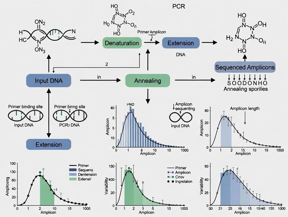
Polymerase Chain Reaction (PCR) is a fundamental step in preparing DNA samples for high-throughput amplicon sequencing. However, PCR is an imperfect process that introduces multiple forms of bias, skewing the representation of the original microbial community in sequencing results. These biases originate at multiple stages of the experimental workflow, from sample preservation to final sequencing, and can significantly impact downstream analyses and biological interpretations. Understanding these sources of bias is crucial for researchers aiming to generate robust, reproducible microbiota data.
The following diagram illustrates the complete amplicon sequencing workflow and identifies key sources of bias at each experimental stage:
Troubleshooting Guides: Identifying and Mitigating Bias at Each Stage
Sample Collection and Preservation Bias
Problem: Microbial community changes between sample collection and DNA extraction.
Question: How do different sample preservation methods affect the integrity of microbial community composition, and what is the optimal approach?
Answer: Sample preservation method significantly impacts microbial community representation. Immediate freezing at -80°C is considered the gold standard but presents logistical challenges for large-scale or remote studies [1].
Experimental Evidence:
- Comparison Study: A 2023 study compared immediate freezing with two stabilization buffers (OMNIgene·GUT and Zymo Research) stored at room temperature for 3-5 days [1].
- Findings: Stabilization buffers limited Enterobacteriaceae overgrowth compared to unpreserved samples but still showed differences from immediately frozen samples, with higher Bacteroidota and lower Actinobacteriota and Firmicutes abundance [1].
- Recommendation: For large-scale studies where cold chain maintenance is challenging, stabilization systems provide a acceptable compromise, though immediate freezing remains optimal [1].
DNA Extraction Bias
Problem: Differential cell lysis efficiency across microbial taxa.
Question: How does the DNA extraction method, particularly cell disruption technique, introduce bias in microbiome studies?
Answer: The method used for cell disruption is a major contributor to variation in microbiota composition, with mechanical methods generally providing more comprehensive lysis across diverse taxa [1].
Experimental Protocol:
- Mechanical Disruption: Use repeated bead-beating with pre-assembled tubes containing 0.5g zirconia/silica beads (0.1mm) and five glass beads (2.7mm) [1].
- Sample Processing: Add 0.25g fecal material and 700μL S.T.A.R. buffer to beads, or 1mL of stabilization buffer for preserved samples [1].
- Validation: Compare mechanical vs. enzymatic lysis (using lysis buffer with Proteinase K at 95°C for 5min followed by 56°C incubation) on the same sample to assess efficiency [1].
- Result: Mechanical disruption typically recovers a more diverse representation of microbial taxa, particularly those with robust cell walls [1].
PCR Amplification Bias
Problem: Differential amplification of community DNA templates during PCR.
Question: What are the primary sources of PCR amplification bias, and how can they be minimized?
Answer: PCR amplification bias arises from multiple sources including primer-template mismatches, GC content, secondary structures, and stochastic effects, which can skew relative abundances up to fivefold [2] [3].
Quantitative Data on PCR Bias Sources:
Table 1: Relative Impact of Different PCR Bias Sources
| Bias Source | Impact Level | Cycle Phase Most Affected | Key Findings |
|---|---|---|---|
| PCR Stochasticity | High | Early cycles | Major force skewing sequence representation in low-input samples [3] |
| Primer-Template Mismatches | High | First 3 cycles | Single nucleotide mismatches can lead to preferential amplification up to 10-fold [4] |
| GC Content | Variable | Mid-late cycles | Depletes loci with GC content >65% to ~1/100th of mid-GC references [5] |
| Secondary Structures | Moderate-High | All cycles | Significant association between amplification efficiencies and secondary structure energy [2] |
| Polymerase Errors | Low (but cumulative) | Late cycles | Common in later cycles but confined to small copy numbers [3] |
| Template Switching | Low | Late cycles | Rare and confined to low copy numbers [3] |
Experimental Protocol for Bias Assessment:
- Calibration Experiment: Pool aliquots of extracted DNA from each study sample into a single calibration sample [4].
- Cycle Gradient: Split the calibration sample into aliquots and amplify each for a predetermined number of PCR cycles (e.g., 22, 24, 26, 28, 30 cycles) [2] [4].
- Sequencing and Modeling: Sequence all aliquots and use log-ratio linear models to infer initial composition and amplification efficiencies [4].
- Application: Apply derived correction factors to experimental samples amplified with standard cycles [4].
Primer-Related Bias
Problem: Inefficient or biased amplification due to primer-template mismatches.
Question: How do degenerate primers contribute to amplification bias, and what are the alternatives?
Answer: While degenerate primers are designed to increase coverage of diverse templates, they can substantially reduce reaction performance and introduce bias through inefficient annealing and primer depletion [6].
Experimental Evidence:
- Performance Comparison: A 2025 study compared degenerate vs. non-degenerate primers using qPCR and computational modeling [6].
- Findings: Non-degenerate primers produced amplicons significantly better than their degenerate counterparts when amplifying either consensus or non-consensus targets [6].
- Alternative Approach: "Thermal-bias PCR" uses only two non-degenerate primers with a large difference in annealing temperatures to isolate targeting and amplification stages, allowing proportional amplification of mismatched targets [6].
Sequencing Platform Bias
Problem: Platform-specific errors and representation bias.
Question: How do different sequencing platforms contribute to errors in amplicon sequencing data?
Answer: Different sequencing platforms exhibit distinct error profiles, with Illumina platforms predominantly showing substitution errors rather than the homopolymer errors characteristic of 454 pyrosequencing [7].
Experimental Evidence:
- Error Profile Analysis: A 2015 study analyzed error patterns across multiple library preparation methods and sequencing conditions [7].
- Platform Comparison: Illumina systems show substitution errors correlated with specific sequence patterns (inverted repeats and GGC sequences) and are affected by phasing/pre-phasing issues [7].
- Error Correction: Quality trimming combined with error correction (BayesHammer) followed by read overlapping (PANDAseq) reduces substitution error rates by an average of 93% [7].
Quantitative Data and Optimization Strategies
PCR Cycle Optimization
Quantitative Impact of PCR Cycles:
- Cycle Number Effect: Increasing from 20 to 25 PCR cycles can inflate UMI counts by 10-15% due to PCR errors being misinterpreted as unique molecules [8].
- Community Richness: Community richness decreases by approximately four-fold between cycles 10 and 15 alone in environmental DNA studies [4].
- Optimal Range: For 16S rRNA gene amplification, ~25 cycles is typically optimal, with higher cycles increasing contaminant detection in negative controls [1].
Table 2: PCR Optimization Strategies and Their Effects
| Optimization Strategy | Protocol Adjustment | Impact on Bias |
|---|---|---|
| Limited Cycles | 25-30 cycles instead of 35-40 | Reduces late-cycle artifacts and polymerase errors [1] |
| Modified Denaturation | Extended denaturation (80s instead of 10s at 98°C) | Improves amplification of GC-rich templates [5] |
| Additives | 2M betaine | Reduces GC bias, stabilizes DNA denaturation [5] |
| Polymerase Selection | AccuPrime Taq HiFi instead of Phusion HF | Improves amplification evenness across GC spectrum [5] |
| Thermocycler Settings | Slower ramp speeds (2.2°C/s vs 6°C/s) | Allows more complete denaturation of GC-rich templates [5] |
| Input DNA | ~125pg input DNA | Reduces effect of contaminants while maintaining library complexity [1] |
Unique Molecular Identifiers (UMIs) for Error Correction
Problem: PCR errors and amplification bias affecting molecular quantification.
Question: How can UMIs mitigate PCR amplification bias, and what are the limitations of current approaches?
Answer: UMIs distinguish original molecules before amplification, theoretically removing PCR biases, but PCR errors within UMIs themselves can lead to inaccurate molecular counting [8].
Experimental Evidence:
- Error Assessment: Increasing PCR cycles from 20 to 25 led to a substantial increase in errors within common molecular identifiers (CMIs), causing transcript overcounting [8].
- Novel Solution: Homotrimeric nucleotide blocks for UMI synthesis provide error correction through a 'majority vote' method, significantly improving accuracy [8].
- Performance: Homotrimeric correction achieved 98.45%, 99.64%, and 99.03% correct CMI calls for Illumina, PacBio, and Oxford Nanopore Technologies platforms, respectively, outperforming monomer-based UMI-tools [8].
Research Reagent Solutions
Table 3: Essential Research Reagents for Bias Mitigation
| Reagent/Category | Specific Examples | Function in Bias Reduction |
|---|---|---|
| Stabilization Buffers | OMNIgene·GUT, Zymo Research DNA/RNA Shield | Preserves microbial community structure at room temperature [1] |
| Mechanical Beads | Zirconia/silica beads (0.1mm) with glass beads (2.7mm) | Ensures efficient cell disruption across diverse taxa [1] |
| High-Fidelity Polymerases | AccuPrime Taq HiFi, Q5, Kapa HiFi | Reduces polymerase errors and improves amplification evenness [7] [5] |
| PCR Additives | Betaine, DMSO | Reduces GC bias and stabilizes DNA denaturation [5] |
| Non-Degenerate Primers | Targeted V4 16S rRNA primers | Improves amplification efficiency and reduces spurious products [6] |
| UMI Systems | Homotrimeric UMI designs | Enables correction of PCR and sequencing errors [8] |
Frequently Asked Questions (FAQs)
Q1: What is the single most impactful step I can take to reduce PCR bias in my amplicon sequencing workflow? A: Limiting PCR cycles to the minimum necessary for library detection (typically 25-30 cycles) has one of the most significant impacts, as late-cycle amplification exponentially increases stochastic effects and favors already-dominant templates [4] [1].
Q2: How can I determine if my observed community differences are biological or technical in origin? A: Implement a calibration experiment using pooled samples across a PCR cycle gradient [4], include replicate extractions and amplifications, sequence mock communities, and use positive controls throughout your workflow to distinguish technical variation from biological signals [1].
Q3: Are there computational methods to correct for PCR biases after sequencing? A: Yes, multiple computational approaches exist, including:
- Log-ratio linear models that use cycle gradient data to estimate and correct for taxon-specific amplification efficiencies [4].
- UMI-based error correction tools (e.g., UMI-tools, homotrimeric correction) that identify and collapse PCR duplicates [8].
- Denoising algorithms that correct PCR errors and identify biological sequences [2].
Q4: How does GC content specifically affect amplification efficiency? A: GC content influences denaturation efficiency (high-GC templates require more complete denaturation), primer binding stability, and secondary structure formation. Templates with GC content >65% can be depleted to 1/100th of mid-GC templates under standard conditions, but this can be mitigated with longer denaturation times and additives like betaine [5].
Q5: What is the recommended approach for sample preservation in large-scale epidemiological studies where immediate freezing is logistically challenging? A: DNA stabilization buffers such as OMNIgene·GUT or Zymo Research DNA/RNA Shield provide a practical compromise, limiting major community shifts while allowing room temperature storage and transportation [1]. However, researchers should validate their chosen method against immediate freezing for their specific sample type.
How does mRNA enrichment introduce bias in my RNA-seq data?
mRNA enrichment is a critical first step in many RNA-seq workflows and is a significant source of bias. The most common method uses oligo-dT beads to capture polyadenylated RNA. However, this method inherently introduces 3'-end capture bias, where coverage is dramatically skewed toward the 3' end of transcripts [9]. This bias can mask important biological information located in the 5' regions, such as alternative transcription start sites or upstream open reading frames (uORFs) [10].
Furthermore, oligo-dT-based enrichment is unsuitable for prokaryotic samples or degraded RNA, such as that from Formalin-Fixed Paraffin-Embedded (FFPE) tissues, as it requires intact poly(A) tails [9]. In these cases, ribosomal RNA (rRNA) depletion is the preferred method. While rRNA removal mitigates the 3'-bias, its efficiency can vary across different RNA species, potentially leading to an underrepresentation of certain transcripts [9].
Table 1: mRNA Enrichment Methods and Associated Biases
| Enrichment Method | Principle | Primary Bias Introduced | Recommended Applications |
|---|---|---|---|
| Oligo-dT Selection | Hybridization to poly-A tail | Strong 3'-end bias; requires intact RNA | High-quality eukaryotic RNA; standard mRNA-seq |
| rRNA Depletion | Removal of ribosomal RNA | Variable efficiency across transcripts; less 3' bias | Prokaryotic RNA; degraded RNA (e.g., FFPE); whole-transcriptome analysis |
What are the consequences of RNA fragmentation bias?
Fragmentation is necessary to generate fragments of appropriate size for sequencing. The method of fragmentation can significantly impact the uniformity of sequence coverage. Early RNA-seq protocols often used RNase III for fragmentation, which is not completely random and can lead to reduced library complexity [9]. Biased fragmentation creates hotspots where fragments begin and end, which can be mistaken for biological signals and complicates the detection of splice variants and exact transcript boundaries [11].
To achieve more uniform coverage, it is recommended to use chemical treatment (e.g., zinc) for RNA fragmentation [9]. Alternatively, a more robust approach involves reverse transcribing intact RNA first and then fragmenting the resulting cDNA using mechanical or enzymatic methods [9]. This post-cDNA synthesis fragmentation helps generate more randomly distributed fragments.
How do priming strategies affect my sequencing results?
The choice of primers during reverse transcription and amplification is a major source of bias.
- Random Hexamer Priming: While designed to bind randomly across transcripts, random hexamers can anneal with varying efficiencies due to sequence context and secondary structure. This leads to uneven coverage along the transcript length and mispriming events [9] [10].
- Oligo-dT Priming: This method primes from the poly-A tail, resulting in strong 3' bias and poor coverage of the 5' ends of long transcripts [9] [10].
- Degenerate Primers: In amplicon sequencing, degenerate primer pools (containing mixed nucleotides) are used to amplify diverse templates. However, these pools can act as reaction inhibitors and are inefficient, paradoxically suppressing the amplification of both rare and consensus targets [6].
Experimental Solution: Thermal-Bias PCR A modern solution to priming bias is the "thermal-bias PCR" protocol, which uses only two non-degenerate primers in a single reaction. It exploits a large difference in annealing temperatures to separate the template targeting and library amplification stages, allowing proportional amplification of even mismatched targets [6].
Table 2: Priming Methods and Their Characteristics
| Priming Method | Common Use | Advantages | Disadvantages |
|---|---|---|---|
| Oligo-dT | Reverse Transcription | Specific for poly-A+ RNA; simple | Strong 3' bias; unsuitable for degraded RNA |
| Random Hexamers | Reverse Transcription / Whole Transcriptome Amplification | Covers non-poly-A RNA; less 3' bias | Uneven coverage; mispriming; sequence-dependent bias |
| Degenerate Primers | Amplicon Sequencing (e.g., 16S rRNA) | Theoretically broader taxonomic reach | Reduced overall efficiency; can inhibit amplification |
| Sequence-Specific | Targeted Amplicon Sequencing | High specificity | Limited to known target sequences |
What is the impact of PCR amplification on my differential expression analysis?
PCR amplification is a primary source of bias in sequencing library preparation, significantly impacting the accuracy of quantitative analyses like differential expression.
- Sequence-Dependent Bias: PCR does not amplify all sequences equally. Fragments with very high or very low GC content are often amplified less efficiently, leading to their underrepresentation in the final library [12] [13]. This can distort the true expression levels of these transcripts.
- Over-Amplification and Duplicates: Excessive PCR cycles lead to "overcycling," which increases artifacts, errors, and the rate of PCR duplicates [14] [15]. A critical point is that a large fraction of computationally identified duplicates are not PCR duplicates but natural duplicates caused by random sampling and fragmentation bias [11]. Therefore, the computational removal of all duplicates can actually worsen the accuracy of differential expression analysis by removing genuine biological information [11].
- Impact on Detection Power: Amplification bias adds technical noise, which reduces the statistical power to detect differentially expressed genes and can inflate the false discovery rate (FDR) [11].
Table 3: Quantitative Impact of PCR Amplification on RNA-seq Data
| Aspect | Impact of PCR Amplification | Consequence for Differential Expression |
|---|---|---|
| Accuracy | Under-representation of extreme GC content transcripts | Altered fold-change estimates for affected genes |
| Precision | Introduction of technical noise due to biased amplification | Reduced power to detect true differences |
| Duplicate Reads | Generation of PCR duplicates, but also loss of natural duplicates | Computational duplicate removal can worsen FDR |
What are the best practices to mitigate amplification bias?
Several strategies, both experimental and computational, can be employed to reduce the impact of amplification bias.
- Optimize PCR Components: The choice of DNA polymerase is critical. Studies have shown that enzymes like Kapa HiFi DNA Polymerase provide more uniform genomic coverage across a wide range of GC contents compared to other enzymes [12]. For extremely AT- or GC-rich templates, PCR additives like tetramethyleneammonium chloride (TMAC) or betaine can be used to improve amplification efficiency [9] [12].
- Minimize PCR Cycles: The most direct way to reduce PCR bias is to reduce the number of amplification cycles. Use the minimum number of cycles required to generate sufficient library yield [9] [14]. For high-input samples, consider PCR-free library preparation protocols [12].
- Utilize Unique Molecular Identifiers (UMIs): UMIs are random oligonucleotide sequences that are added to each molecule before any amplification steps. This allows for the bioinformatic identification and correction of PCR duplicates, generating accurate, absolute counts of the original RNA molecules [11]. Recent advances include using homotrimeric nucleotide blocks to create UMIs with built-in error-correcting capabilities, which further improve the accuracy of molecule counting by mitigating PCR-associated sequencing errors [8].
- Employ Alternative Amplification Methods: For low-input and single-cell RNA-seq, methods like Phi29 DNA polymerase-based amplification (multiple displacement amplification) can be used. This isothermal method has high processivity and can be less biased than PCR-based methods for certain applications [10]. Another approach is semirandom primed PCR (SMA), which uses oligonucleotides with random 3' sequences and a universal 5' sequence for uniform amplification, providing relatively uniform coverage of full-length transcripts [10].
Experimental Protocol: Thermal-Bias PCR for Reduced Priming Bias
This protocol, adapted from current research, uses non-degenerate primers and a two-stage temperature process to minimize bias [6].
Principle: A low-temperature annealing step allows the non-degenerate primer to bind to both matched and mismatched template targets. A subsequent high-temperature priming and extension step uses a second primer to selectively and efficiently amplify only the successfully targeted fragments.
Workflow Diagram:
Steps:
- Reaction Setup: Prepare a standard PCR mixture containing the mixed-template genomic DNA, two non-degenerate primers, a high-fidelity DNA polymerase, dNTPs, and buffer.
- Initial Denaturation: 98°C for 2 minutes.
- Thermal-Bias Cycling (15-25 cycles):
- Denaturation: 98°C for 10 seconds.
- Low-Temperature Annealing: 45-50°C for 30 seconds. This step allows the targeting primer to hybridize stably to both consensus and non-consensus templates.
- High-Temperature Priming & Extension: 72°C for 30 seconds. At this temperature, a second primer binds specifically to the newly synthesized strand for efficient and controlled amplification.
- Final Extension: 72°C for 5 minutes.
- Library Completion: The resulting amplicon can be purified and processed for sequencing. This method allows for the reproducible production of amplicon sequencing libraries that maintain the proportional representation of rare members in the community [6].
The Scientist's Toolkit: Key Reagent Solutions
Table 4: Essential Reagents for Mitigating Library Preparation Bias
| Reagent / Kit | Function | Role in Bias Mitigation | Key Feature |
|---|---|---|---|
| Kapa HiFi DNA Polymerase | PCR Amplification | Provides uniform coverage across varying GC content | High-fidelity enzyme optimized for NGS |
| mirVana miRNA Isolation Kit | RNA Extraction | Isolves high-quality RNA, including small RNAs | Provides high-yield and high-quality RNA from various sources [9] |
| UMI Adapters (e.g., Homotrimer Design) | Library Barcoding | Enables accurate counting and correction of PCR duplicates & errors | Random barcode sequence added pre-amplification; trimer design allows error correction [8] |
| SeqPlex Enhanced WTA / WGA Kits | Whole Transcriptome/Genome Amplification | Amplifies low-input/degraded samples with minimal sequence bias | Uses enhanced random primers for comprehensive coverage [16] |
| CircLigase | ssDNA Circligation | Circularizes cDNA for Phi29-based amplification | Allows amplification of short fragments in circularization-based methods [10] |
| Tetramethylammonium chloride (TMAC) | PCR Additive | Stabilizes AT-rich templates; reduces mispriming | Improves amplification efficiency of AT-rich regions [9] [12] |
Quantitative Data on PCR Bias
The following tables summarize key experimental data on how PCR cycle number and enzyme choice impact the accuracy and representation of sequencing results.
Table 1: Impact of PCR Cycle Number on Sequencing Outcomes in Low Biomass Samples
| Sample Type | PCR Cycles | Key Finding | Effect on Richness/Beta-Diversity |
|---|---|---|---|
| Bovine Milk [17] | 25, 30, 35, 40 | Increased sequencing coverage with higher cycles | No significant differences detected |
| Murine Pelage [17] | 25 vs 40 | Increased sequencing coverage with higher cycles | No significant differences detected |
| Murine Blood [17] | 25 vs 40 | Increased sequencing coverage with higher cycles | No significant differences detected |
Table 2: Effect of PCR Cycle Number and Protocol on Sequence Artifacts in 16S rRNA Gene Libraries
| Clone Library | No. of PCR Cycles | % Chimeric Sequences | % Unique 16S rRNA Sequences (100% similarity) | Library Coverage (%) (after artifact removal) |
|---|---|---|---|---|
| Standard [18] | 35 | 13% | 76% | 64% |
| Modified [18] | 15 + 3 reconditioning | 3% | 48% | 89.3% |
Table 3: Polymerase Enzyme Performance Across Genetic Marker Systems of Varying Complexity
| Enzyme | % Correct Reads (Test 1: Simple Locus) | % Correct Reads (Test 2: Single-Copy Nuclear) | % Correct Reads (Test 3: Multi-Gene Family) |
|---|---|---|---|
| Phusion [19] | 88-92% | 84% | 65-71% |
| Pwo [19] | 88-92% | - | - |
| KapHF [19] | 88-92% | - | - |
| FastStart [19] | - | - | 65-71% |
| Biotaq [19] | 50-53% | 2% | 17-20% |
Table 4: Impact of PCR Errors on Unique Molecular Identifier (UMI) Accuracy
| Sequencing Platform | % CMIs Correctly Called (Before Correction) | % CMIs Correctly Called (After Homotrimer Correction) |
|---|---|---|
| Illumina [8] | 73.36% | 98.45% |
| PacBio [8] | 68.08% | 99.64% |
| ONT (latest chemistry) [8] | 89.95% | 99.03% |
Experimental Protocols
Protocol: Investigating PCR Artifacts in Repetitive DNA Sequences
This protocol is adapted from research investigating the molecular mechanisms of PCR failure and artifact formation when amplifying repetitive DNA, such as TALE binding domains [20].
- Primary Objective: To analyze the formation of deletion artifacts and hybrid repeats during PCR amplification of highly repetitive DNA sequences.
- Sample Preparation:
- Template: Use pure plasmid DNA containing the repetitive sequence of interest (e.g., a TALE assembly in a vector like pTAL2).
- Primers: Design primers that flank the repetitive DNA region.
- PCR Amplification:
- Reaction Setup: Set up standard PCR reactions using a proofreading or non-proofreading DNA polymerase (e.g., Taq).
- Cycling Conditions: Use standard cycling conditions: initial denaturation at 98°C for 3 minutes, followed by 30-35 cycles of denaturation (98°C for 15 seconds), annealing (50-60°C for 30 seconds), and extension (72°C for 30 seconds per kb), with a final extension at 72°C for 7 minutes.
- Optimization Attempts: The protocol may include optimization steps such as the addition of DMSO, MgCl2 optimization, and testing different annealing temperatures, which typically fail to resolve artifacts in this specific context [20].
- Analysis:
- Gel Electrophoresis: Analyze PCR products on an agarose gel. Successful amplification of the repetitive region typically results in a "laddering" effect, with multiple bands appearing below and above the expected size, rather than a single clean band.
- Cloning and Sequencing: Isolate individual bands from the gel, clone them into a sequencing vector (e.g., pTOPO), and sequence multiple independent clones.
- Data Interpretation: Sequence analysis reveals that the artifact bands consist of hybrid repeats, where the polymerase has "skipped" over internal repeats, joining distant repeat units together. This is informative for generating models of artifact formation [20].
Protocol: Evaluating PCR Cycle Number for Low Microbial Biomass Samples
This protocol is designed for optimizing 16S rRNA gene amplicon sequencing from samples with low bacterial biomass and high host DNA content, such as milk, blood, or skin [17].
- Primary Objective: To determine the effect of increased PCR cycle number on sequencing coverage and community representation in low biomass samples.
- Sample Collection and DNA Extraction:
- Collect samples (e.g., aseptically collected milk, furred pelage, blood in EDTA tubes) and store at -20°C until processing.
- Extract DNA using a kit designed for complex samples (e.g., PowerFecal DNA Isolation Kit), incorporating a mechanical lysis step (e.g., TissueLyser II) to ensure efficient cell disruption.
- Quantify DNA via fluorometry (e.g., Qubit with dsDNA BR assay).
- Library Preparation with Variable Cycles:
- Target Region: Amplify the V4 region of the 16S rRNA gene using universal primers (e.g., U515F/806R) flanked by Illumina adapter sequences.
- PCR Reaction: Use a high-fidelity DNA polymerase (e.g., Phusion). Reactions should include 100 ng of metagenomic DNA, primers, dNTPs, and polymerase in the manufacturer's recommended buffer.
- Cycling Conditions: Use a touchdown or standard cycling protocol with a variable number of cycles. For matched sample DNA, create separate libraries amplified with different cycle numbers (e.g., 25, 30, 35, and 40 cycles) [17].
- Purification: Pool and purify amplicons using a magnetic bead-based clean-up system (e.g., Axygen Axyprep MagPCR clean-up beads).
- Sequencing and Data Analysis:
- Sequence the libraries on an Illumina MiSeq platform.
- Analysis: Compare coverage per sample, detected richness (alpha-diversity), and community structure (beta-diversity) between libraries generated with different cycle numbers.
Workflow: UMI-Based Error Correction for Accurate Molecular Counting
The following diagram illustrates an experimental workflow that uses error-correcting homotrimeric Unique Molecular Identifiers (UMIs) to account for PCR errors in sequencing data.
Troubleshooting Guides and FAQs
Frequently Asked Questions (FAQs)
Q1: Why does my PCR of repetitive DNA sequences (like TALEs) produce a ladder of bands instead of a single product? A: This laddering effect is a classic symptom of PCR amplification across highly repetitive sequences. The artifacts are caused by the DNA polymerase dissociating and misaligning with a different, homologous repeat unit on the template strand during synthesis. This leads to the generation of hybrid repeats and deletions, which manifest as multiple bands on a gel in increments roughly corresponding to the size of a single repeat unit (e.g., ~100 bp) [20]. Standard optimization (DMSO, Mg2+) often fails, and cloning/sequencing of individual bands is required to confirm the nature of these artifacts.
Q2: For low biomass samples like blood or milk, should I use a high number of PCR cycles to ensure I get enough product for sequencing? A: Yes, but with caution. While increasing PCR cycle number (e.g., to 35 or 40 cycles) is a valid and often necessary strategy to generate sufficient library coverage from low biomass samples, it does increase the risk of accumulating errors and artifacts [17]. The key finding from recent studies is that while higher cycles increase coverage, they may not significantly skew metrics of microbial richness or beta-diversity in these sample types. However, the increased signal must be balanced against the potential for higher noise, and rigorous negative controls are essential to distinguish true signal from contamination or artifacts [17].
Q3: How can I minimize PCR bias and errors in my amplicon sequencing library prep? A: A multi-pronged approach is most effective:
- Enzyme Choice: Select a high-fidelity polymerase (e.g., Q5, Phusion) that has been demonstrated to yield a high proportion of correct sequences in complex systems [21] [19].
- Cycle Number: Use the minimum number of PCR cycles required to generate sufficient library yield [18] [21].
- Modified Protocols: For community analysis, consider using a "reconditioning PCR" step (a few cycles with a fresh reaction mixture) to reduce heteroduplex molecules and chimeras [18].
- UMI Integration: For absolute molecular counting, incorporate error-correcting UMIs (e.g., homotrimeric UMIs) before amplification to digitally track and correct for PCR errors and biases in downstream bioinformatics analysis [8].
Q4: My PCR has multiple bands or a smear. What are the primary causes and solutions? A: Nonspecific amplification is a common issue. The main causes and solutions include [22] [21]:
- Annealing Temperature Too Low: Increase the annealing temperature in a step-wise manner or use a gradient cycler to find the optimal temperature.
- Poor Primer Design: Verify primer specificity and avoid self-complementarity or primers with complementary 3' ends.
- Excess Enzyme or Mg2+: Review and optimize the concentration of both the DNA polymerase and Mg2+ in the reaction.
- Template Quality/Quantity: Use high-quality, pure template DNA and ensure the concentration is not too high.
- Hot-Start Polymerase: Use a hot-start polymerase to prevent nonspecific amplification during reaction setup.
The Scientist's Toolkit: Essential Research Reagents
Table 5: Key Reagents for Managing PCR Bias
| Reagent / Solution | Function in Mitigating PCR Bias |
|---|---|
| High-Fidelity DNA Polymerases (e.g., Q5, Phusion) | Enzymes with proofreading (3'→5' exonuclease) activity that significantly reduce nucleotide misincorporation rates, leading to a higher proportion of correct sequences [21] [19]. |
| Hot-Start DNA Polymerases | Enzymes that are inactive until a high-temperature activation step, preventing nonspecific amplification and primer-dimer formation during reaction setup, thereby improving specificity and yield [22] [21]. |
| Unique Molecular Identifiers (UMIs) | Random oligonucleotide sequences used to uniquely tag individual RNA/DNA molecules before any amplification steps. This allows bioinformatic correction of PCR amplification biases and digital counting of original molecules [8]. |
| Error-Correcting UMIs (e.g., Homotrimer) | A specific UMI design where the random sequence is synthesized in blocks of three identical nucleotides (trimers). This allows for a "majority vote" correction method, dramatically improving the accuracy of UMI sequences after PCR and sequencing [8]. |
| PCR Additives (e.g., DMSO, GC Enhancers) | Co-solvents that help denature GC-rich templates and resolve secondary structures, promoting more uniform amplification of difficult sequences and improving overall coverage [22] [21]. |
| Pre-Plated, Breakaway PCR Panels | Pre-formulated, ready-to-use reaction panels that reduce manual assay preparation time, minimize pipetting errors and cross-contamination risk, and improve reproducibility across experiments [23]. |
In amplicon sequencing studies, the assumption that final sequencing data accurately represents the original template composition is often violated due to Polymerase Chain Reaction (PCR) bias. Sequence-intrinsic factors—specifically GC content, secondary structures, and primer-template mismatches—systematically distort amplification efficiency, leading to quantitative inaccuracies that compromise ecological and molecular interpretations [24] [25]. PCR bias manifests when certain DNA templates amplify more efficiently than others due to their inherent sequence properties, creating a distorted representation of the original template mixture in the final sequencing library [25] [5].
The impact of this bias extends beyond technical artifacts to affect biological conclusions. Recent research demonstrates that PCR bias significantly influences widely used ecological metrics, including Shannon diversity and Weighted-Unifrac, while perturbation-invariant measures remain more robust [24]. This review establishes a technical support framework within the broader thesis of mitigating PCR bias in amplicon sequencing, providing researchers with actionable troubleshooting guidelines, experimental protocols, and reagent solutions to recognize, quantify, and minimize these sequence-intrinsic distortions.
Technical FAQs: Addressing Common Experimental Challenges
How does GC content specifically influence PCR amplification efficiency?
GC-rich templates (typically defined as >60% GC content) present three major challenges during amplification. First, the triple hydrogen bonds in G-C base pairs confer higher thermostability, requiring more energy for denaturation and potentially leading to incomplete strand separation during cycling [26]. Second, these regions readily form stable secondary structures such as hairpins that physically block polymerase progression. Third, GC-rich sequences promote non-specific primer binding and primer-dimer formation [26].
Table 1: Quantitative Effects of GC Content on PCR Amplification
| GC Content Range | Amplification Efficiency Relative to Mid-GC Templates | Primary Challenge | Recommended Mitigation Strategy |
|---|---|---|---|
| <20% GC | Reduced to ~10% of reference level [5] | Low template stability, polymerase slippage | Increase primer specificity, add betaine [5] |
| 40-60% GC (balanced) | Optimal (reference level) [27] | Minimal bias | Standard protocols typically effective |
| 65-80% GC | Severely reduced to ~1% of reference level [5] | Incomplete denaturation, secondary structures | Extended denaturation times, specialized polymerases, additives [26] [5] |
| >80% GC | Nearly eliminated without optimization [5] | Extreme thermostability, complex structures | Combination of polymerase selection, additives, and thermal profile optimization [26] |
The suppression of amplification becomes dramatically more severe at GC contents exceeding 65%, with loci above 80% GC potentially depleted to one-hundredth of their pre-amplification abundance after just 10 PCR cycles when using standard protocols [5]. This bias follows a characteristic profile where mid-GC content templates (approximately 11-56% GC) typically amplify efficiently, creating a "plateau" of reliable amplification, while both extremely low-GC and high-GC fragments are systematically underrepresented [5].
What specific secondary structures most significantly inhibit amplification, and where must they be avoided?
Secondary structures that form in the template DNA, particularly near primer-binding sites, critically impact amplification efficiency by competitively inhibiting primer binding [28]. The most problematic structures include:
- Hairpins with long stems and small loops: When formed inside the amplicon, these structures cause particularly dramatic suppression of amplification efficiency. Research demonstrates that hairpins with 20-bp stems can completely prevent target amplification, yielding no detectable product [28].
- Stable structures near primer-binding sites: Secondary structures forming within approximately 60 bases both inside and outside the amplicon boundary can significantly interfere with primer annealing and extension [28].
Table 2: Effect of Hairpin Structures on qPCR Amplification Efficiency
| Hairpin Location | Stem Length | Loop Size | Amplification Efficiency | Mechanism of Interference |
|---|---|---|---|---|
| Inside amplicon | 10 bp | 5-10 nt | Moderate suppression | Polymerase stalling during elongation |
| Inside amplicon | 20 bp | 5-10 nt | No amplification [28] | Complete blocking of polymerase progression |
| Outside amplicon | 10 bp | 5-10 nt | Mild suppression | Competitive inhibition of primer binding [28] |
| Outside amplicon | 20 bp | 5-10 nt | Severe suppression | Steric hindrance of primer access to template |
| Near primer-binding site (<10 bp) | >8 bp | Any size | Severe suppression | Direct competition with primer annealing [28] |
The magnitude of amplification suppression increases with longer stem lengths and smaller loop sizes. Hairpins formed inside the amplicon cause more dramatic suppression than those outside, with 20-bp stem structures completely eliminating targeted amplification [28]. These effects are primarily attributed to competitive inhibition of primer binding to the template, as confirmed by melting temperature measurements [28].
How do primer-template mismatches impact amplification, and does location matter?
Mismatches between primer and template sequences introduce substantial amplification bias, particularly in complex template systems like microbial community profiling [25]. The impact of a mismatch is highly dependent on its position relative to the primer's 3' end:
- 3' end mismatches (-1 to -3 positions): Most detrimental, often reducing or preventing primer extension entirely due to impaired polymerase initiation [25].
- Middle region mismatches (~-8 position): Moderate impact, potentially reducing annealing efficiency but still permitting some amplification.
- 5' end mismatches (~-14 position): Least detrimental, often tolerated with minimal impact on amplification efficiency [25].
In standard PCR, perfect match primer-template interactions are strongly favored, especially when mismatches occur near the 3' end [25]. However, in complex natural samples with diverse templates, mismatch amplifications can paradoxically dominate when using heavily degenerate primer pools, leading to unexpected distortion of template representation [25].
Troubleshooting Guides
Problem: Poor Amplification of GC-Rich Templates
GC-rich regions (>60% GC) resist denaturation and form secondary structures that cause polymerases to stall, resulting in blank gels, smeared bands, or low yield [26].
Workflow for Troubleshooting GC-Rich Amplification
Step 1: Polymerase and Buffer Selection
- Choose polymerases specifically optimized for GC-rich templates (e.g., OneTaq DNA Polymerase with GC Buffer or Q5 High-Fidelity DNA Polymerase) [26].
- Utilize master mixes containing GC enhancers that help disrupt secondary structures.
- For standalone polymerases, add GC enhancer supplements (typically 5-20% of reaction volume).
Step 2: Thermal Profile Optimization
- Extend denaturation time: increase initial denaturation from 30 seconds to 3 minutes and cycle denaturation from 10 seconds to 80 seconds [5].
- Implement a thermal gradient to determine optimal annealing temperature.
- Consider using a "hot start" protocol with higher initial denaturation temperature.
Step 3: Additive Implementation
- Test betaine (1-1.3M final concentration) to reduce secondary structure formation [5].
- Evaluate DMSO (2-10%) to lower melting temperatures and disrupt stable structures [26].
- Avoid overusing additives, as they can inhibit polymerase activity at high concentrations.
Step 4: Magnesium Concentration Titration
- Perform MgCl₂ titration in 0.5 mM increments between 1.0-4.0 mM [26].
- Balance sufficient magnesium for polymerase activity (typically 1.5-2.0 mM) with the need to reduce non-specific binding.
Problem: Secondary Structure Interference
Stable secondary structures in templates competitively inhibit primer binding and block polymerase progression, particularly in regions with inverted repeats or hairpin-forming potential [28] [29].
Protocol: Systematic Evaluation of Secondary Structure Interference
Sequence Analysis Phase
- Scan approximately 60 bases on both sides of primer-binding sites using tools like Mfold or the UNAFold Tool [27].
- Identify potential hairpins with stem lengths >8 bp, particularly those near primer annealing sites.
- Check for homologous regions that might facilitate terminal hairpin formation and self-priming extension [29].
Experimental Verification
- Run PCR products on agarose gel to detect unusual banding patterns or smears indicating structural interference.
- Compare sequencing results from both directions; discrepancies may indicate structure-dependent elongation artifacts [29].
Remediation Strategies
- Redesign primers to avoid structured regions when possible.
- Incorporate additives like betaine or DMSO to destabilize secondary structures.
- Increase annealing temperature to favor specific primer binding over structure formation.
- Use polymerases with high processivity to overcome structural barriers.
Problem: Amplification Bias from Primer-Template Mismatches
In complex template mixtures, primer-template mismatches cause differential amplification efficiencies that distort the representation of original templates in final sequencing libraries [25] [30].
Table 3: Strategies for Minimizing Mismatch-Induced Bias
| Approach | Protocol | Advantages | Limitations |
|---|---|---|---|
| Degenerate Primer Pools | Include mixed nucleotides at variable positions in primer sequence [30] | Broad theoretical coverage of sequence variants | Can reduce overall reaction efficiency; may introduce new biases [30] |
| Reduced Cycling | Limit PCR to 20-25 cycles [25] | Minimizes late-cycle stochastic effects | May yield insufficient product for sequencing |
| Specialized PCR Methods | Implement Deconstructed PCR (DePCR) or Thermal-bias PCR [25] [30] | Empirically reduces bias; preserves template ratios | Additional processing steps; requires optimization |
| Touchdown PCR | Start with high annealing temperature, decrease incrementally | Improves specificity in early cycles | Does not address primer depletion issues |
| Polymerase Selection | Use high-fidelity, mismatch-tolerant enzymes | Some tolerance to minor mismatches | Limited effect on severe mismatches, especially at 3' end |
Protocol: Deconstructed PCR (DePCR) for Bias Reduction
DePCR separates linear copying of source templates from exponential amplification, preserving information about original primer-template interactions while reducing bias [25].
Linear Copying Phase
- Set up reaction with DNA template and forward primer only.
- Run 1-2 cycles with extended annealing/extension times.
- This creates complementary strands representing the original template mixture.
Exponential Amplification Phase
- Add reverse primer to the same reaction (or clean product and set up new reaction).
- Perform standard PCR cycling (20-30 cycles).
- The exponential amplification begins from the copied templates rather than the original genomic DNA.
Analysis
- Sequence final products and compare diversity metrics to standard PCR.
- DePCR demonstrates significantly lower distortion relative to standard PCR when mismatches are present [25].
Research Reagent Solutions
Table 4: Essential Reagents for Addressing Sequence-Intrinsic PCR Bias
| Reagent Category | Specific Examples | Mechanism of Action | Ideal Application Context |
|---|---|---|---|
| Specialized Polymerases | OneTaq DNA Polymerase with GC Buffer, Q5 High-Fidelity DNA Polymerase with GC Enhancer [26] | Improved processivity through structured regions; enhanced fidelity | GC-rich templates; complex secondary structures |
| PCR Additives | Betaine (1-1.3M), DMSO (2-10%), 7-deaza-2'-deoxyguanosine [26] [5] | Reduce secondary structure formation; lower template melting temperature | Hairpin-prone sequences; extremely GC-rich targets |
| Buffer Components | MgCl₂ (1.0-4.0 mM, optimized), specialized GC enhancers [26] | Cofactor for polymerase activity; destabilize G-C bonds | Fine-tuning reaction conditions for specific templates |
| High-Fidelity Master Mixes | Q5 High-Fidelity 2X Master Mix, OneTaq Hot Start 2X Master Mix with GC Buffer [26] | Convenience; optimized formulations for challenging templates | Standardized workflows; screening multiple targets |
| Modified Nucleotides | Phosphorothioate bonds at 3' primer ends [25] | Reduce nucleolytic degradation of primers | Long amplification cycles; complex template mixtures |
Advanced Methodologies
Thermal-Bias PCR Protocol for Complex Templates
Thermal-bias PCR represents a recent advancement that uses temperature manipulation rather than degenerate primers to amplify diverse templates while maintaining their relative abundances [30].
Experimental Workflow:
Primer Design
- Design non-degenerate primers based on consensus sequences.
- Avoid degeneracy while ensuring reasonable coverage of expected variants.
Reaction Setup
- Prepare PCR mix with non-degenerate primers, template DNA, and GC-enhanced polymerase formulation.
- Include appropriate additives based on template characteristics.
Thermal Cycling
- Initial denaturation: 98°C for 3 minutes.
- 5-10 cycles with high annealing temperature (e.g., 68-72°C) to favor specific priming.
- 20-25 cycles with lower annealing temperature (e.g., 55-60°C) to allow limited mismatch tolerance.
- Final extension: 72°C for 5 minutes.
Validation
- Quantify amplicon yield and distribution.
- Sequence and compare community structure to other methods.
- Thermal-bias PCR allows proportional amplification of targets containing substantial mismatches while using only two non-degenerate primers in a single reaction [30].
Quantitative Assessment of PCR Bias
Protocol: Using qPCR to Measure Amplification Bias Across GC Spectrum
Reference Template Preparation
qPCR Assay Design
- Develop short amplicon assays (50-69 bp) spanning the GC range.
- Ensure amplicons are sufficiently short to avoid internal secondary structure interference.
Amplification and Analysis
- Amplify reference templates under test conditions.
- Quantify abundance of each locus relative to a standard curve of input DNA.
- Normalize quantities relative to mid-GC reference amplicons (48-52% GC).
Bias Calculation
- Plot normalized quantity against GC content for each condition.
- Calculate bias magnitude as the deviation from ideal flat distribution.
- Compare different polymerases, additives, and cycling conditions to identify optimal parameters [5].
Addressing sequence-intrinsic factors in PCR amplification requires a multifaceted approach that begins with recognizing potential sources of bias and implements systematic troubleshooting strategies. The most reliable research outcomes emerge from methodologies that proactively address GC content challenges, secondary structure formation, and primer-template mismatches through appropriate reagent selection, protocol optimization, and validation techniques.
By integrating these troubleshooting guides, experimental protocols, and reagent solutions into amplicon sequencing workflows, researchers can significantly improve the quantitative accuracy of their studies and draw more reliable biological conclusions. The continued development of methods like Deconstructed PCR and Thermal-bias PCR highlights the importance of maintaining template representation while achieving specific amplification, ultimately supporting the broader thesis of reducing PCR bias in amplicon sequencing research.
Bias-Busting Strategies: Methodological Advances and Practical Applications
In amplicon sequencing studies, the polymerase chain reaction (PCR) is a critical step for amplifying target DNA regions from complex samples. However, standard PCR protocols can introduce significant amplification bias, distorting the true biological representation of different DNA templates in a sample [6] [31]. This bias manifests as the under-representation or complete dropout of specific sequences, such as those with extreme GC content or primer-binding site mismatches, ultimately compromising the accuracy of downstream sequencing data [31]. This guide details wet-lab optimization strategies—focusing on polymerase selection, chemical additives, and thermocycling protocols—to minimize these biases and generate more representative amplicon libraries for your research.
Frequently Asked Questions (FAQs) on PCR Bias
1. What is the biggest source of bias during library preparation for amplicon sequencing? Research has identified that the PCR amplification step itself during library preparation is the most discriminatory stage. One study traced genomic sequences with GC content ranging from 6% to 90% and found that as few as ten PCR cycles could deplete loci with a GC content >65% to about 1/100th of the mid-GC reference loci. Amplicons with very low GC content (<12%) were also significantly diminished [31].
2. Can using degenerate primers reduce bias? While degenerate primers (pools containing mixed nucleotide sequences) are often used to amplify targets with variations in their primer-binding sites, they can inadvertently reduce overall PCR efficiency and distort representation. Non-degenerate primers can sometimes produce better results, and novel methods like "thermal-bias" PCR are being developed to amplify mismatched targets without degenerate primers, leading to libraries that better maintain the proportional representation of rare sequences [6].
3. My thermocycler's manual mentions "ramp rate." Does this really affect my results? Yes, the temperature ramp rate of your thermocycler can be a critical hidden factor. Studies show that slower default ramp speeds can significantly improve the amplification of GC-rich templates. Simply switching from a fast-ramping to a slow-ramping instrument extended the GC-content plateau from 56% to 84% before seeing a drop in amplification efficiency [31]. This highlights the need to optimize and document your thermocycling equipment and protocols.
Troubleshooting Guide: Overcoming Common PCR Challenges
Use the following tables to diagnose and resolve common issues that contribute to PCR bias and amplification failure.
Table 1: Troubleshooting No or Low Amplification Product
| Possible Cause | Recommended Optimization Strategy |
|---|---|
| Incorrect Annealing Temperature | Recalculate primer Tm and test a gradient, starting 3–5°C below the lowest Tm [32] [33]. |
| Poor Primer Design | Verify specificity, avoid self-complementarity, and ensure primers have a GC content of 40–60% and a Tm within 5°C of each other [34] [33]. |
| Complex Template (e.g., High GC) | Use a polymerase designed for GC-rich targets. Add enhancers like 1–10% DMSO or 0.5 M–2.5 M betaine [22] [34] [35]. |
| Suboptimal Denaturation | Increase denaturation time and/or temperature. For GC-rich templates, extend the denaturation time during cycling [32] [31]. |
| PCR Inhibitors Present | Re-purify the template DNA or dilute the sample to reduce inhibitor concentration [22] [35]. |
Table 2: Troubleshooting Non-Specific Products and Smearing
| Possible Cause | Recommended Optimization Strategy |
|---|---|
| Low Annealing Temperature | Increase the annealing temperature in increments of 2°C to improve specificity [33] [35]. |
| Excessive Cycle Number | Reduce the number of PCR cycles (typically to 25–35), as overcycling increases non-specific product accumulation [32] [35]. |
| Too Much Template or Enzyme | Reduce the amount of input template or DNA polymerase as per manufacturer guidelines [22] [35]. |
| Primer Dimer Formation | Use a hot-start DNA polymerase to prevent activity at room temperature and set up reactions on ice [22] [35]. |
| Long Annealing Time | Shorten the annealing time (e.g., to 5–15 seconds) to minimize primer binding to non-specific sequences [35]. |
Optimized Experimental Protocols
Protocol 1: Thermal-Bias PCR for Reducing Primer-Bias
This protocol uses two non-degenerate primers with a large difference in annealing temperatures to stably amplify targets containing mismatches in their primer-binding sites, avoiding the inefficiencies of degenerate primers [6].
Workflow Overview:
Materials:
- DNA Template: 1–1000 ng of mixed-genome sample.
- Primers: Two non-degenerate primers designed for the target region, with a calculated Tm difference of >10°C.
- High-Fidelity DNA Polymerase: e.g., Q5 or Phusion.
- Appropriate 10X Reaction Buffer.
Method:
- Reaction Setup: Prepare a master mix containing buffer, dNTPs, DNA polymerase, and the low-Tm primer only. Add the DNA template.
- Initial Amplification Stage (5–10 cycles):
- Denaturation: 98°C for 10 seconds.
- Annealing: Use a temperature 3°C below the Tm of the low-Tm primer. This allows it to bind to both consensus and non-consensus targets.
- Extension: 72°C for 30 seconds/kb.
- Second Amplification Stage: Add the high-Tm primer to the reaction tube.
- Main Amplification Stage (20–25 cycles):
- Denaturation: 98°C for 10 seconds.
- Annealing/Extension: Use a single temperature suitable for the high-Tm primer (e.g., 72°C). The low-Tm primer will no longer bind, and only the correctly extended products from the first stage are amplified.
- Final Extension: 72°C for 5 minutes.
Protocol 2: Mitigating GC-Bias in Amplicon Libraries
This protocol optimizes denaturation and incorporates betaine to evenly amplify sequences across a wide GC spectrum [31].
Workflow Overview:
Materials:
- DNA Template: Composite sample with a range of GC contents.
- DNA Polymerase: A robust, high-fidelity enzyme such as Phusion or AccuPrime Taq HiFi.
- 5M Betaine Solution: Sterile-filtered.
Method:
- Reaction Setup: Prepare a standard master mix and add betaine to a final concentration of 0.5–2 M.
- Initial Denaturation: Perform at 98°C for 3 minutes (a significant increase from the typical 30 seconds) to ensure complete separation of GC-rich duplexes.
- Amplification Cycles (25–35 cycles):
- Denaturation: 98°C for 60–80 seconds per cycle (extended from the typical 10–30 seconds).
- Annealing: Temperature optimized for your primer set.
- Extension: 72°C for 30 seconds/kb.
- Final Extension: 72°C for 5–10 minutes.
The Scientist's Toolkit: Key Research Reagent Solutions
Table 3: Essential Reagents for PCR Bias Minimization
| Reagent / Material | Function in Bias Reduction | Example Use Case |
|---|---|---|
| High-Fidelity DNA Polymerase | Reduces misincorporation errors due to proofreading (3'→5' exonuclease) activity, leading to more accurate amplification [33]. | Cloning and sequencing applications where sequence accuracy is critical [33]. |
| Polymerase Blends (e.g., AccuPrime Taq HiFi) | Combines polymerases for improved efficiency and uniformity when amplifying complex mixed templates or difficult GC-rich targets [31]. | Generating even coverage across genomic loci with diverse base compositions [31]. |
| Hot-Start DNA Polymerase | Remains inactive until a high-temperature activation step, preventing non-specific priming and primer-dimer formation at lower temperatures [22]. | Improving specificity and yield in reactions prone to mispriming or when using complex templates [22] [35]. |
| Betaine | A chemical additive that equalizes the melting temperature of DNA, improving the amplification efficiency of GC-rich templates [31] [34]. | Added at 0.5–2 M to rescue amplification of high-GC targets that fail with standard protocols [31]. |
| DMSO | Disrupts secondary structures and reduces DNA melting temperature, helping to amplify templates with strong secondary structures or high GC content [32] [34]. | Used at 1–10% to assist in denaturing complex templates [34]. |
In amplicon sequencing studies, PCR bias is a significant challenge that can distort sequence representation and compromise the accuracy of quantitative results. Traditional methods often rely on degenerate primer pools—mixtures of primers with varying bases at specific positions—to target diverse sequences. However, this approach can introduce amplification biases, favoring certain templates over others. This technical support center provides troubleshooting guides and FAQs to help researchers address specific issues related to PCR bias and adopt advanced primer design strategies for more reliable and accurate amplicon sequencing.
Frequently Asked Questions (FAQs)
1. What are the main sources of PCR bias in amplicon sequencing? PCR bias in amplicon sequencing arises from several sources. The major forces skewing sequence representation are PCR stochasticity (the random sampling of molecules during early amplification cycles) and polymerase errors, which become very common in later PCR cycles but typically remain at low copy numbers [3]. Other significant factors include:
- GC Bias: Sequences with very high or very low GC content can amplify less efficiently. This has been identified as a principal source of bias during the library amplification step [31].
- Template Switching: A process where chimeric sequences are formed during amplification, though this is typically rare and confined to low copy numbers [3].
- Primer Mismatches: Variation in primer binding sites, especially when using universal or degenerate primers, leads to differential amplification efficiency [13].
2. How do degenerate primers contribute to amplification bias? While degenerate primers (pools of primers with nucleotide variations) are designed to broaden the range of amplifiable templates, they introduce several issues. They often ignore primer specificity, which can lead to false positives in applications like viral subtyping [36]. The different primers within a degenerate pool have varying melting temperatures (Tm) and binding efficiencies, which can cause uneven amplification of target sequences [13]. Furthermore, calculating the thermodynamic properties of a degenerate pool is complex, and heuristic methods based on mismatch counts can be misleading for predicting actual hybridization efficiency [36].
3. What are the key advantages of non-degenerate, targeted primer design? Targeted, non-degenerate approaches offer greater specificity and predictability. They allow for the design of primers with optimized and uniform thermodynamic properties, such as melting temperature, which leads to more balanced amplification [37]. These methods minimize off-target amplification and the formation of chimeras by ensuring primers are specific to their intended target [38]. By moving the design process away from consensus sequences and towards evaluating individual primers against diverse templates, these approaches better account for sequence variation and avoid biases introduced by degenerate bases [37].
4. Which modern tools can help design targeted, non-degenerate primers? Several advanced bioinformatics tools have been developed to address the limitations of degenerate primers:
- PMPrimer: A Python-based tool that automatically designs multiplex PCR primer pairs using a statistical filter to identify conserved regions based on Shannon's entropy, tolerates gaps, and evaluates primers based on template coverage and taxon specificity [37].
- varVAMP: A command-line tool for designing degenerate primers for viral genomes. It addresses the "maximum coverage degenerate primer design" problem by finding a trade-off between specificity and sensitivity, using a penalty system that incorporates primer parameters, 3’ mismatches, and degeneracy [38].
- Thermodynamic-Based Methods: New methods propose moving beyond simple mismatch counting. They use suffix arrays and local alignment to identify candidate regions, followed by rigorous thermodynamic analysis to evaluate the hybridization efficiency of primers against all potential targets, ensuring high specificity and sensitivity [36].
5. How can I minimize GC bias in my amplicon sequencing library preparation? GC bias can be significantly reduced by optimizing the PCR conditions during library preparation. Key steps include [31]:
- Using Betaine: Adding 2M betaine to the PCR reaction can help rescue amplification of extremely GC-rich fragments.
- Extending Denaturation Times: Simply extending the initial denaturation step and the denaturation step during each cycle can overcome the detrimental effects of fast temperature ramp rates on thermocyclers, improving the amplification of GC-rich templates.
- Optimizing Polymerase Blends: Substituting polymerases with specialized blends (e.g., AccuPrime Taq HiFi) can also contribute to more uniform amplification across a wide GC spectrum.
Troubleshooting Guides
Problem: Inaccurate Taxonomic Abundance Estimates from Metabarcoding Data
Potential Causes and Solutions:
- Cause: Primer-induced bias from variable binding sites.
- Solution: Shift to markers with more conserved priming sites or use tools like PMPrimer to design primers in regions with high sequence conservation, as determined by low Shannon's entropy [37] [13]. For highly diverse targets, consider a varVAMP-like approach that designs multiple discrete, non-degenerate primer sets to cover different variants, minimizing the need for high degeneracy [38].
- Cause: Amplification bias from PCR cycle number.
- Solution: Reduce the number of PCR cycles in the initial, locus-specific amplification round. Surprisingly, simply reducing cycles may not be sufficient on its own [13]. Combine cycle reduction with increased template concentration (e.g., 60 ng in a 10 µl reaction) to maximize the starting molecule number and reduce stochastic effects [13].
- Cause: Locus copy number variation (CNV).
- Solution: Be aware that CNV of the target locus between taxa will affect abundance estimates in both amplicon-based and PCR-free methods [13]. If a correlation between input DNA and read count can be established, apply taxon-specific correction factors to the read counts to improve abundance estimates [13].
Problem: Poor Amplification of Targets with Extreme GC Content
Potential Causes and Solutions:
- Cause: Incomplete denaturation of high-GC templates due to fast thermocycling.
- Solution: Optimize the thermocycling profile. Extend the denaturation time during each cycle (e.g., from 10 s to 80 s) and use a thermocycler with a slower ramp speed to ensure complete denaturation of GC-rich templates [31].
- Cause: Non-optimal polymerase or reaction chemistry.
- Solution: Use a PCR additive like 2M betaine. Furthermore, test alternative polymerase formulations, such as the AccuPrime Taq HiFi blend, which may perform better across a wide range of GC contents [31].
Problem: Designing Pan-Specific Primers for Highly Variable Viral Genomes
Potential Causes and Solutions:
- Cause: High genomic variability makes finding conserved regions difficult.
- Solution: Use a tool like varVAMP that is specifically designed for variable viral genomes. It uses a k-mer-based approach on consensus sequences derived from a multiple sequence alignment and employs Dijkstra's algorithm to find an optimal tiling path of amplicons with minimal primer penalties [38].
- Cause: Traditional degenerate primers lead to false positives or poor sensitivity.
- Solution: Employ a thermodynamics-driven design method. These methods use local alignment to find candidate primer binding sites across whole genomes and then perform a rigorous thermodynamic analysis to evaluate the true binding affinity, ensuring specificity and sensitivity beyond simple mismatch counting [36].
Experimental Protocols & Data
This protocol is designed to reduce the under-representation of sequences with extreme GC content during the library amplification step.
Reaction Setup:
- Use 15 ng of adapter-ligated DNA library.
- Set up a 10 µl PCR reaction using the AccuPrime Pfx SuperMix or a similar robust polymerase blend.
- Add forward and reverse primers to a final concentration of 0.5 µM each.
- Critical Addition: Include a final concentration of 2M betaine.
Thermocycling Conditions:
- Initial Denaturation: 3 minutes at 95°C.
- Cycling (10-18 cycles):
- Denaturation: 80 seconds at 95°C. Note: This extended denaturation time is crucial for GC-rich templates.
- Annealing: 30 seconds at 58°C.
- Extension: 30 seconds at 68°C.
- Final Extension: 5 minutes at 68°C.
Clean-up: Purify the PCR product using Agencourt RNAClean XP beads or a similar solid-phase reversible immobilization (SPRI) method before quantification and sequencing.
Table 1: Relative Impact of Different PCR-Induced Distortions on Sequence Representation [3]
| Source of Error | Relative Impact | Key Characteristics |
|---|---|---|
| PCR Stochasticity | Major | The primary force skewing sequence representation in low-input libraries; most significant for single-cell sequencing. |
| Polymerase Errors | Common but low impact | Very frequent in later PCR cycles, but erroneous sequences are confined to small copy numbers. |
| Template Switching | Minor | A rare event, typically confined to low copy numbers. |
| GC Bias | Variable | A significant source of bias during library PCR; effect can be minimized with protocol optimization [31]. |
Research Reagent Solutions
Table 2: Key Reagents for Mitigating PCR Bias in Amplicon Sequencing
| Reagent / Tool | Function / Application | Example / Note |
|---|---|---|
| Betaine | PCR additive that equalizes the amplification efficiency of templates with different GC contents by reducing the melting temperature disparity [31]. | Used at a final concentration of 2M. |
| AccuPrime Taq HiFi | A specialized blend of DNA polymerases noted for its performance in amplifying sequences with a broad range of GC content [31]. | An alternative to Phusion HF for GC-balanced amplification. |
| PMPrimer | Bioinformatics tool for automated design of multiplex PCR primers; uses Shannon's entropy to find conserved regions and evaluates template coverage [37]. | Python-based; useful for designing targeted primers for diverse templates like 16S rRNA or specific gene families. |
| varVAMP | Command-line tool for designing degenerate primers for tiled whole-genome sequencing of highly variable viruses; addresses the MC-DGD problem [38]. | Optimized for viral pathogen surveillance (e.g., SARS-CoV-2, HEV). |
Workflow Visualization
Primer Design Strategy Evolution
GC Bias Mitigation Strategies
Frequently Asked Questions (FAQs)
1. What are UMIs, and why are they crucial for amplicon sequencing? Unique Molecular Identifiers (UMIs) are short, random oligonucleotide sequences (typically 8-12 nucleotides long) that are ligated to individual DNA or RNA molecules before any PCR amplification steps [39] [40]. In amplicon sequencing, they are crucial for accurate molecular counting. After sequencing, reads sharing the same UMI are collapsed into a single read, which removes PCR duplicates and corrects for amplification biases, thereby improving the accuracy of quantitative applications like gene expression analysis or variant calling [8] [40] [41].
2. What are the primary sources of UMI errors? UMI errors originate from three major sources [39]:
- PCR Amplification Errors: Random nucleotide substitutions accumulate over multiple PCR cycles. With each cycle using previously synthesized products as templates, these errors can propagate, causing erroneous UMIs to be counted as distinct molecules [8] [39].
- Sequencing Errors: Incorrect base calls during sequencing lead to mismatches, insertions, or deletions in the UMI sequence. The error profile varies by platform: Illumina has low rates but mainly substitution errors, while long-read platforms like PacBio and Oxford Nanopore Technologies (ONT) are more susceptible to indels [39] [42].
- Oligonucleotide Synthesis Errors: These occur during the chemical manufacturing of the UMIs themselves, primarily involving truncations or unintended extensions due to the finite coupling efficiency of each synthesis step [39].
3. My UMI deduplication tool is running slowly and using a lot of memory. What could be the cause? Several factors can impact the performance of tools like UMI-tools [43]:
- Run Time: Shorter UMIs, higher sequencing error rates, and greater sequencing depth can all increase the "connectivity" between UMI sequences, leading to larger networks for the algorithm to resolve and longer processing times.
- Memory Usage: Processing chimeric read pairs or unmapped reads in paired-end sequencing modes can require keeping large buffers of data in memory, significantly increasing memory requirements.
4. How do homotrimeric UMIs correct errors, and when should I use them? Homotrimeric UMIs are an advanced design where each nucleotide in a conventional UMI is replaced by a triplet of identical bases (e.g., 'A' becomes 'AAA') [8] [39]. This creates internal redundancy. During analysis, a "majority vote" is applied to each triplet to correct single-base errors. For example, a sequenced 'ATA' triplet can be corrected to 'AAA' [8]. This design is particularly beneficial in scenarios prone to high error rates, such as single-cell RNA-seq with high PCR cycle numbers or long-read sequencing, as it significantly improves the accuracy of molecular counting [8].
5. What computational tools are available for UMI error correction, and how do I choose? The choice of tool depends on your UMI design and sequencing platform. The table below summarizes key tools:
Table 1: Comparison of UMI Deduplication Tools
| Tool Name | Key Features | Best For | Limitations |
|---|---|---|---|
| UMI-tools [43] [39] | Graph-based network, Hamming distance (substitutions) | Short-read data with monomeric UMIs and moderate error rates | Struggles with indel errors; can be slow with large datasets; single-threaded |
| UMI-nea [42] | Levenshtein distance (substitutions & indels), multithreading, adaptive filtering | Error-prone data (e.g., long-reads), ultra-deep sequencing, and structured UMIs | |
| Homotrimer Correction [8] | Majority voting and set cover optimization, built-in redundancy | Data generated with homotrimeric UMI designs, high PCR cycle conditions | Requires specific experimental design using homotrimer UMIs |
Troubleshooting Guides
Issue: Inflated Molecular Counts After UMI Deduplication
Potential Causes and Solutions:
High PCR Cycle Number:
- Cause: Excessive PCR cycles introduce and propagate errors within UMI sequences, causing a single original molecule to appear as multiple distinct molecules [8].
- Solution: Optimize your library preparation protocol to use the minimum number of PCR cycles necessary. Consider adopting error-resilient UMI designs like homotrimeric UMIs, which have been shown to maintain over 96% accuracy even at high PCR cycles (35 cycles), whereas monomeric UMI accuracy drops significantly [8].
Using an Inappropriate Computational Tool:
- Cause: Tools that only use Hamming distance (like UMI-tools) cannot correct for insertion and deletion (indel) errors, which are common in long-read sequencing [42].
- Solution: If you are using long-read sequencing or a UMI design prone to indels, switch to a tool that uses Levenshtein distance, such as UMI-nea, which can handle both substitutions and indels [42].
Issue: Poor Amplification of GC-Rich or GC-Poor Targets
Potential Causes and Solutions:
- Cause: This is a form of PCR amplification bias, where the standard PCR conditions and enzyme formulations do not efficiently denature or amplify templates with extreme GC content [5].
- Solutions:
- Optimize PCR Conditions: A study found that simply extending the denaturation time during thermocycling can significantly improve the representation of high-GC loci. Adding betaine (2M) and using polymerase blends like AccuPrime Taq HiFi can further create a more balanced amplification across a wide GC spectrum (e.g., 23% to 90% GC) [5].
- Avoid Degenerate Primers: While often used to capture diverse templates, degenerate primers can themselves be a source of bias and inhibit efficient amplification. Consider a "thermal-bias" PCR protocol that uses non-degenerate primers with a large difference in annealing temperatures to isolate targeting and amplification stages [6].
Experimental Protocols
Protocol 1: Validating UMI Error Correction Using a Common Molecular Identifier (CMI)
This protocol, adapted from a recent study, provides a robust method to quantify the accuracy of your UMI correction strategy [8].
1. Principle: A known, identical Common Molecular Identifier (CMI) is attached to every captured RNA molecule. In a perfect system, all transcripts should report this single CMI sequence. Any errors introduced during library prep or sequencing will create variant CMI sequences, allowing for precise measurement of the error rate and correction efficacy [8].
2. Reagents and Materials:
- Source Nucleic Acids: Equimolar mix of human and mouse cDNA.
- Common Molecular Identifier (CMI): A defined, unique oligonucleotide sequence.
- Library Prep Kit: Compatible with your sequencing platform (e.g., for Illumina, PacBio, or ONT).
- PCR Enzymes: Standard high-fidelity polymerase.
- Sequencing Platform: Access to Illumina, PacBio, or ONT sequencers.
3. Step-by-Step Procedure: a. Tagging: Attach the CMI to the 3' end of all RNA/cDNA molecules from the human/mouse mix. b. Amplification: Perform PCR amplification on the CMI-tagged library. c. Sequencing: Split the final library and sequence on multiple platforms (e.g., Illumina, PacBio, ONT). d. Data Analysis: i. Extract all CMI sequences from the sequencing data. ii. Calculate the percentage of CMIs that match the expected, correct sequence. iii. Apply your chosen UMI error-correction method (e.g., homotrimer majority vote) to the observed CMIs. iv. Re-calculate the percentage of correct CMIs post-correction.
4. Anticipated Results: The following table summarizes typical results from this experiment, demonstrating the high error-correction efficiency of the homotrimer method across platforms [8]:
Table 2: CMI Accuracy Before and After Homotrimer Error Correction
| Sequencing Platform | % Correct CMIs (Before Correction) | % Correct CMIs (After Homotrimer Correction) |
|---|---|---|
| Illumina | 73.36% | 98.45% |
| PacBio | 68.08% | 99.64% |
| ONT (Latest Chemistry) | 89.95% | 99.03% |
Protocol 2: Assessing the Impact of PCR Cycles on UMI Accuracy in Single-Cell RNA-seq
This protocol is designed to isolate and quantify the effect of PCR amplification on UMI error rates in a single-cell context [8].
1. Principle: Single-cell libraries are prepared, and an initial number of PCR cycles is performed. The product is then split and subjected to different numbers of additional PCR cycles. Comparing UMI counts and differential expression results between the low- and high-cycle libraries reveals the impact of PCR errors.
2. Reagents and Materials:
- Cells: A mix of human (e.g., JJN3) and mouse (e.g., 5TGM1) cell lines.
- Single-Cell Platform: 10X Chromium or Drop-seq system.
- Trimer Barcoded Beads: For incorporating error-correcting UMIs. a. Encapsulation and Reverse Transcription: Perform single-cell encapsulation and reverse transcription using a system like 10X Chromium or Drop-seq. b. Initial PCR: Carry out an initial set of 10 PCR cycles. c. Split and Amplify: Split the PCR product into aliquots. Perform additional PCR amplification on each aliquot to reach different final cycle totals (e.g., 20, 25, 30, 35 cycles). d. Sequencing and Analysis: Sequence the libraries. For each library, perform cell calling, UMI deduplication (using both standard and homotrimer methods), and differential expression analysis.
4. Anticipated Results:
- Libraries with higher PCR cycles will show inflated UMI counts when using standard (monomeric) UMI correction, falsely suggesting more transcripts [8].
- Differential expression analysis between libraries with different cycle counts (e.g., 20 vs. 25) will identify hundreds of significantly regulated transcripts with monomeric UMI correction, which are almost entirely eliminated when homotrimer UMI correction is applied, confirming they are artifacts of PCR errors [8].
Visualizations
Diagram 1: Workflow of Homotrimeric UMI Error Correction
Diagram 2: Experimental Workflow to Quantify PCR Errors with a CMI
The Scientist's Toolkit: Research Reagent Solutions
Table 3: Essential Reagents and Kits for UMI-Based Sequencing
| Item | Function/Description | Example Use Case |
|---|---|---|
| Homotrimeric UMI Oligos | Oligonucleotides designed with nucleotide triplets (e.g., AAA, CCC) to provide built-in error correction via majority voting. | Implementing advanced error correction in bulk or single-cell RNA-seq protocols to mitigate PCR errors [8]. |
| xGen cfDNA & FFPE Library Prep Kit | A library preparation kit designed for challenging samples, incorporating fixed UMI sequences for error correction. | Sequencing of circulating tumor DNA (ctDNA) or degraded DNA from FFPE samples, enabling sensitive variant detection [41]. |
| xGen NGS Amplicon Sequencing Panels | Pre-designed or custom panels of amplicons for targeted sequencing. | Efficiently targeting and sequencing specific genomic regions of interest for applications in cancer research and microbial ecology [44]. |
| AccuPrime Taq HiFi Polymerase | A blend of DNA polymerases noted for its high fidelity and performance in amplifying sequences with diverse GC content. | Generating balanced sequencing libraries with minimized GC bias [5]. |
| 10X Chromium / Drop-seq System | Single-cell RNA-seq platforms that use barcoded beads to label individual cells and their transcripts with UMIs. | Profiling gene expression at single-cell resolution from complex tissues or cell suspensions [8] [39]. |
Emerging Computational and Deep Learning Tools for Predicting and Correcting Sequence-Specific Amplification Efficiency
FAQs: Addressing PCR Amplification Bias
1. What is sequence-specific amplification bias and why is it a problem in amplicon sequencing? Sequence-specific amplification bias refers to the non-homogeneous amplification of different DNA templates during Polymerase Chain Reaction (PCR), which is a critical step in preparing libraries for amplicon sequencing. This results in skewed abundance data in sequencing results, compromising the accuracy and sensitivity of quantitative analyses. Even a template with an amplification efficiency just 5% below the average will be underrepresented by a factor of around two after only 12 PCR cycles [45]. This bias can lead to false negatives in variant calling, inaccurate quantification in transcriptomic studies, and misrepresentation of community structures in metabarcoding [46].
2. Beyond GC content, what sequence-specific factors contribute to poor amplification? While GC content has long been recognized as a major factor, recent deep learning models have identified that specific sequence motifs adjacent to adapter priming sites are closely associated with poor amplification efficiency. Research challenging long-standing PCR design assumptions has elucidated adapter-mediated self-priming as a major mechanism causing low amplification efficiency [45]. Furthermore, the use of degenerate primer pools, intended to increase target representation, can itself be a source of bias by reducing overall reaction efficiency and unpredictably biasing subsequent priming events [6].
3. How can deep learning models predict amplification efficiency from sequence data? Convolutional Neural Networks (CNNs) can be trained to predict sequence-specific amplification efficiencies based on sequence information alone. These models are trained on large, reliably annotated datasets derived from synthetic DNA pools. One such model achieved a high predictive performance with an AUROC (Area Under the Receiver Operating Characteristic curve) of 0.88 and an AUPRC (Area Under the Precision-Recall Curve) of 0.44. This allows for the in-silico screening and design of inherently homogeneous amplicon libraries before synthesis and wet-lab experimentation [45].
4. What are the wet-lab strategies to minimize PCR amplification bias? Several experimental strategies can mitigate bias:
- Thermal-bias PCR: A protocol that uses only two non-degenerate primers in a single reaction by exploiting a large difference in annealing temperatures to isolate the targeting and amplification stages. This allows for proportional amplification of targets containing mismatches in their primer binding sites [6].
- PCR-Free Workflows: Eliminating the amplification step altogether, though this requires higher amounts of input DNA [46].
- Unique Molecular Identifiers (UMIs): Random oligonucleotide sequences that tag individual molecules before amplification, allowing for bioinformatic correction of PCR duplicates [46]. Recent advances include using homotrimeric nucleotide blocks for UMIs, which provide an error-correcting solution that significantly improves the accuracy of counting sequenced molecules compared to traditional monomeric UMIs [8].
- Optimized PCR Chemistry and Cycling: Using polymerases engineered for difficult sequences, additives like betaine, and optimized thermocycling protocols with longer denaturation times can significantly reduce bias, especially for GC-rich templates [31].
5. How do computational tools correct for bias in sequenced data?
Computational tools can correct bias during data analysis. For data generated with UMIs, tools like UMI-tools and TRUmiCount use network-based algorithms to group reads originating from the same molecule. Homotrimeric UMI strategies implement a 'majority vote' method to correct PCR-induced errors within the UMI sequence itself, which has been shown to correct over 96% of errors and prevent inflated transcript counts in single-cell RNA sequencing [8]. Furthermore, bioinformatics normalization approaches can computationally correct for persistent coverage biases based on local sequence composition [46].
Troubleshooting Guide: Common Issues and Solutions
| Problem | Possible Cause | Solution |
|---|---|---|
| Low library complexity / high duplicate reads | Over-amplification by too many PCR cycles leading to dominance by the most efficient amplicons [14] [46]. | - Reduce the number of PCR cycles [31].- Use Unique Molecular Identifiers (UMIs) for accurate deduplication [8].- Switch to a PCR-free library preparation workflow if input DNA is sufficient [46]. |
| Under-representation of GC-rich or GC-poor regions | Incomplete denaturation of GC-rich templates or inefficient priming/extension for GC-poor templates [31]. | - Use a polymerase mixture formulated for high GC content [47].- Add enhancers like betaine (1-2 M) to the PCR reaction [31].- Optimize thermocycling conditions: extend denaturation time and slow the ramp rate [31]. |
| Skewed abundance in metabarcoding or multi-template PCR | Sequence-specific amplification efficiency differences and adapter-mediated self-priming [45]. | - Use deep learning models (e.g., 1D-CNN) to pre-screen and design balanced amplicon libraries [45].- Employ thermal-bias PCR protocols to improve amplification of mismatched targets [6].- Avoid overly degenerate primer pools; consider two-step amplification protocols [6]. |
| Inaccurate molecular counting in UMI-based assays | PCR errors within the UMI sequence itself, creating artificial molecular diversity [8]. | - Implement homotrimeric UMI designs for robust error correction [8].- Benchmark deduplication tools against a validated method. |
| No or low yield | PCR inhibitors, suboptimal primer design, or overly stringent cycling conditions [48] [47]. | - Re-purify the template DNA to remove inhibitors [14] [47].- Redesign primers and optimize annealing temperature [48] [47].- Use a hot-start polymerase to prevent non-specific amplification [48]. |
Experimental Protocol: Predicting Amplification Efficiency with a 1D-CNN
This protocol summarizes the methodology for training a deep learning model to predict sequence-specific amplification efficiency, as detailed in the referenced study [45].
Data Generation and Annotation
- Synthetic DNA Pool Design: Synthesize a pool of at least 12,000 random DNA sequences (e.g., 120-170 nt in length) flanked by constant adapter sequences (e.g., truncated Truseq adapters). A separate pool with constrained GC content (e.g., 50%) is recommended to control for this variable.
- Serial PCR Amplification: Subject the pool to multiple consecutive PCR reactions (e.g., 6 reactions of 15 cycles each). After each reaction, purify the product and take an aliquot for sequencing to track the change in each sequence's coverage over up to 90 cycles.
- Efficiency Calculation: For each sequence, fit its coverage trajectory over the PCR cycles to an exponential amplification model. The fit provides an estimated amplification efficiency (εi) for each sequence relative to the population mean.
Model Training
- Data Preparation: Format DNA sequences as one-hot encoded matrices (size: 4 x sequence length). Define a classification task, for example, by labeling the worst-performing 2% of sequences (low efficiency) as the positive class and the rest as negative.
- Model Architecture: Implement a 1D-Convolutional Neural Network (1D-CNN). The structure typically includes:
- Input Layer: Accepts one-hot encoded sequence.
- Convolutional Layers: Multiple layers with ReLU activation to detect sequence motifs.
- Pooling Layers: Max-pooling to reduce dimensionality.
- Fully Connected Layers: To combine features for the final prediction.
- Output Layer: Sigmoid activation for binary classification.
- Training: Train the model using the annotated dataset with a binary cross-entropy loss function and an optimizer like Adam. Use a separate validation set to monitor performance and prevent overfitting.
Model Interpretation with CluMo
- Framework: Use the CluMo (Motif Discovery via Attribution and Clustering) framework or a similar method to interpret the trained model.
- Process: Calculate attribution scores (e.g., using DeepLIFT or SHAP) to determine the contribution of each nucleotide position to the prediction of "poor amplification."
- Motif Discovery: Cluster the resulting important sequence segments to identify consensus motifs associated with low amplification efficiency. This can reveal biological mechanisms, such as adapter-mediated self-priming [45].
Workflow Diagram: From Sequence to Efficiency Prediction
Research Reagent Solutions
| Reagent / Tool | Function in Addressing PCR Bias |
|---|---|
| Synthetic DNA Pools | Provides large, well-defined datasets for training and validating deep learning models on amplification efficiency [45]. |
| High-Fidelity DNA Polymerase (e.g., Q5, Phusion) | Reduces error rates during amplification, crucial for maintaining UMI sequence integrity and minimizing misincorporation [48] [8]. |
| Homotrimeric UMI Oligonucleotides | Provides an error-correcting mechanism for accurate molecular counting by allowing a 'majority vote' correction of PCR errors within the UMI [8]. |
| Betaine | A chemical additive that equalizes the melting temperature of DNA, helping to improve the amplification efficiency of GC-rich templates [31]. |
| Non-Degenerate Primers | Used in thermal-bias PCR to avoid the inefficiencies and unpredictable biases introduced by highly degenerate primer pools [6]. |
| PCR-Free Library Prep Kits | Eliminates amplification bias entirely by bypassing the PCR step, though it requires higher input DNA [46]. |
From Problem to Solution: A Troubleshooting Guide for Optimal Amplicon Sequencing
FAQ: Addressing Common PCR Issues in Amplicon Sequencing
Why is there no amplification or a low yield of my PCR product?
Low yield or a complete lack of amplification can stem from several factors related to reaction components and cycling conditions.
- Cause: Suboptimal Reaction Components. Issues can include insufficient template DNA, poor template quality due to degradation or PCR inhibitors, suboptimal primer design or concentration, insufficient DNA polymerase, or incorrect Mg²⁺ concentration [22] [49].
- Solution: Verify template DNA quantity and quality using spectrophotometry or fluorometry [50]. For difficult templates like GC-rich sequences, use a polymerase specifically designed for such conditions and consider adding PCR enhancers like betaine [51] [5]. Ensure primers are well-designed and used at an appropriate concentration, typically between 0.1–1 µM [22].
- Cause: Suboptimal Thermal Cycling Conditions. An annealing temperature that is too high, an insufficient number of cycles, or insufficient denaturation can prevent amplification [52] [22].
- Solution: Optimize the annealing temperature in 1–2°C increments, using a gradient cycler if available [22] [49]. Increase the number of cycles, up to 40-45, for low-abundance templates [51] [52]. For GC-rich templates, increase the denaturation time and/or temperature to ensure complete strand separation [5].
Why do I have a high rate of PCR duplicates in my sequencing data?
PCR duplicates arise when multiple copies of the same original DNA molecule are sequenced, skewing quantitative representation.
- Cause: Limited Starting Material and Excessive PCR Cycling. The primary cause is having too few unique starting DNA molecules and over-amplifying them during library preparation to obtain sufficient material for sequencing [53]. With limited unique molecules, the probability that multiple copies of the same molecule will be sequenced increases significantly.
- Solution: The most effective strategy is to maximize the amount of unique input DNA at the start of library preparation. If input is limited, keep the number of PCR amplification cycles to an absolute minimum [53]. For example, one expert recommendation is to perform no more than 6 PCR cycles during library prep to maintain high library complexity and keep duplication rates low [53]. The table below illustrates how the number of unique starting molecules and PCR cycles influences the expected duplication rate.
Table: Impact of Input Material and PCR Cycles on Duplication Rates
| Unique Starting Molecules | PCR Cycles | Expected PCR Duplicate Rate | Explanation |
|---|---|---|---|
| High (e.g., 7e10) | Low (e.g., 6) | Very Low (~0.2%) | Vast pool of unique molecules minimizes chance of sampling duplicates [53] |
| Medium (e.g., 9e9) | Medium (e.g., 9) | Low (~1.7%) | Fewer unique molecules begin to increase duplication probability [53] |
| Low (e.g., 1e9) | High (e.g., 12) | High (~15%) | Limited diversity and over-amplification lead to frequent sampling of the same molecules [53] |
What are adapter dimers, and how do I remove them?
Adapter dimers are short, unwanted products formed by the ligation of sequencing adapters to themselves.
- Cause: Low Input or Inefficient Clean-up. Adapter dimers are common when using insufficient or degraded starting material, as there are not enough genuine DNA fragments for the adapters to ligate to [54]. An inefficient size selection clean-up step after adapter ligation can also leave them in the final library.
- Effects: Adapter dimers can cluster very efficiently on the flow cell and be sequenced, consuming a significant portion of your sequencing reads and potentially negatively impacting data quality and run performance [54]. It is recommended to keep them to 0.5% or lower of your library on patterned flow cells [54].
- Solution: Use an accurate fluorometric method to quantify input DNA and ensure you use the recommended amount [54]. To remove existing adapter dimers, perform an additional clean-up step using solid-phase reversible immobilization (SPRI) beads at a 0.8x to 1x ratio, which will selectively bind and remove the short dimer fragments [54].
How can I minimize amplification bias in my amplicon sequencing study?
PCR amplification bias skews the true representation of different sequences in your sample, which is a critical concern for quantitative applications like metabarcoding [3] [13].
- Strategy 1: Optimize PCR Enzymes and Conditions. PCR bias is a major force in skewing sequence representation [3] [5]. Using polymerases formulated for high fidelity and performance on difficult templates is crucial. Furthermore, simply extending the denaturation time during thermocycling can significantly improve the amplification of GC-rich fragments, which are often under-represented [5].
- Strategy 2: Use Degenerate Primers and Reduce Cycles. For metabarcoding, using primers with a high degree of degeneracy can help amplify across a broader taxonomic range by accounting for variation in priming sites [13]. While reducing PCR cycle numbers is a common suggestion to mitigate bias, one study on arthropod communities found that it did not have a strong effect and could actually make abundance predictions less predictable [13].
- Strategy 3: Apply Computational Correction. Since read abundance biases are often taxon-specific and predictable, bioinformatic tools can be used to calculate and apply correction factors to the data, thereby improving abundance estimates [13].
Table: Common PCR Inhibitors and Mitigation Strategies
| Inhibitor Type | Examples | Recommended Mitigation |
|---|---|---|
| Organic | Polysaccharides, humic acids, hemoglobin, heparin, polyphenols [51] | Dilute template DNA 100-fold; use polymerases with high inhibitor tolerance; purify template with specialized kits or ethanol precipitation [51] [22] |
| Inorganic | Calcium ions, EDTA [51] | Ensure Mg²⁺ concentration is optimized and exceeds the concentration of chelators like EDTA; re-purify template to remove salts [51] [22] |
Research Reagent Solutions
Table: Essential Reagents for Mitigating PCR Issues in Sequencing
| Reagent / Tool | Function / Application |
|---|---|
| High-Fidelity Hot-Start Polymerase | Increases specificity (reduces nonspecific bands and primer-dimers) and reduces error rates [22] [49]. |
| Polymerase for GC-Rich Templates | Specialized enzyme blends (e.g., AccuPrime Taq HiFi) and buffers with enhancers improve amplification of high-GC content regions [3] [5]. |
| PCR Additives (e.g., Betaine, BSA) | Betaine helps denature GC-rich templates [5]; BSA (Bovine Serum Albumin) can bind to and neutralize certain PCR inhibitors [50]. |
| SPRI Beads (e.g., AMPure XP) | Used for post-ligation clean-up to remove adapter dimers and for size selection [54] [13]. |
| Degenerate Primers | Contain mixed bases at variable positions to bind to conserved sites across diverse taxa, reducing amplification bias in metabarcoding [13]. |
Experimental Protocols for Key Experiments
Protocol 1: Minimizing GC Bias in Library Amplification
This protocol is adapted from a study that systematically optimized conditions to reduce base-composition bias during the PCR amplification step of Illumina library preparation [5].
- Reaction Setup: Set up the library amplification PCR using the AccuPrime Taq HiFi polymerase blend (or a similar robust, high-fidelity enzyme) [5].
- Thermocycling with Long Denaturation: Use the following modified thermocycling profile:
- Initial Denaturation: 3 minutes at 95°C.
- Cycling (10-12 cycles):
- Denaturation: 80 seconds at 95°C. Note: This extended denaturation is critical for complete melting of GC-rich fragments.
- Annealing: 30 seconds at 60°C.
- Extension: 60 seconds at 68°C.
- Final Extension: 5 minutes at 68°C.
- Use of Additives: Include 2M betaine in the PCR reaction to further destabilize secondary structures in GC-rich sequences [5].
- Validation: The efficiency of bias reduction can be validated by qPCR using a panel of amplicons with a wide range of GC contents (e.g., from 6% to 90% GC) [5].
Protocol 2: Evaluating Primer Performance and Bias in Metabarcoding
This protocol outlines a method to test how different primer sets affect amplification bias in a controlled mock community [13].
- Create a Mock Community: Prepare a DNA mock community by pooling genomic DNA from known taxa in defined proportions. This creates a ground truth for quantitative comparisons [13].
- Amplify with Multiple Primer Pairs: Amplify the same mock community sample using several different primer pairs targeting the same or different barcode loci. These can include primers with varying levels of degeneracy and those targeting regions with different levels of sequence conservation [13].
- Library Preparation and Sequencing: Prepare sequencing libraries from each amplification reaction and sequence them on a high-throughput platform [13].
- Bioinformatic Analysis: Process the sequencing data to determine the relative read abundance of each taxon for each primer set. Compare these results to the known proportions in the mock community to quantify the amplification bias and recovery efficiency of each primer pair [13].
Diagrams of Logical Relationships
Diagram 1: A troubleshooting map for common PCR failure modes, showing the logical flow from primary causes to specific solutions.
Diagram 2: The pathway leading to a high rate of PCR duplicates in next-generation sequencing data [53].
In amplicon sequencing studies, the accuracy of your results is profoundly influenced by the initial steps of library preparation. Biases introduced during polymerase chain reaction (PCR) amplification can skew the representation of different sequences in your final library, leading to inaccurate biological conclusions. This guide addresses three critical levers under your direct control—template concentration, PCR cycle number, and purification practices—to help you minimize amplification bias and generate more reliable, quantitative sequencing data.
FAQ: Template and Amplification
What is the optimal amount of DNA template to use in a PCR?
Using the correct amount of template DNA is a primary defense against PCR bias. Insufficient template leads to low yield and can necessitate excessive amplification cycles, while too much template can increase background and non-specific amplification [22]. The optimal quantity is not a single value but depends on the complexity and source of your DNA.
The following table summarizes recommended template amounts for various DNA sources to achieve approximately 10⁴ copies of your target, which is typically sufficient for detection in 25-30 cycles [55] [56] [57].
Table 1: Recommended DNA Template Input for PCR
| Template Type | Recommended Mass | Key Considerations |
|---|---|---|
| Plasmid or Viral DNA | 1 pg – 10 ng [55] | Lower complexity requires less input. |
| Genomic DNA | 1 ng – 1 µg [55] | Use 5–50 ng as a starting point for most applications; higher complexity requires more input [57]. |
| Human Genomic DNA | 10 – 100 ng [56] | For high-copy targets (e.g., housekeeping genes), 10 ng may be sufficient. |
| E. coli Genomic DNA | 100 pg – 1 ng [56] | Lower complexity than mammalian genomes. |
| PCR Product (re-amplification) | Diluted or purified product [57] | Unpurified products carry over reagents that can inhibit the new reaction; purification is best. |
How do I determine the correct number of PCR cycles?
The ideal number of PCR cycles balances the need for sufficient product yield with the risk of introducing bias and errors. Excessive cycling is a major source of PCR error and overcounting of molecules, especially in protocols using unique molecular identifiers (UMIs) [8].
Table 2: Guidelines for PCR Cycle Number
| Scenario | Recommended Cycles | Rationale |
|---|---|---|
| Routine Amplification | 25–35 cycles [22] | Provides a robust yield for standard applications. |
| Low Template Copy Number (<10 copies) | Up to 40 cycles [22] | Increased cycles are needed to generate a detectable amount of product. |
| Library Amplification for Sequencing | Use the minimum number that gives adequate yield [14] | Every additional cycle increases the duplication rate and the chance of errors [8]. PCR errors in UMIs can lead to inaccurate absolute molecule counts [8]. |
| Amplification with High-Fidelity Polymerases | Keep cycles to a minimum [22] | High numbers of cycles increase the cumulative chance of misincorporating nucleotides, even with high-fidelity enzymes. |
Optimization Tip: If your reaction requires more than 35 cycles to produce a visible product on a gel, investigate other potential issues like primer design, annealing temperature, or enzyme efficiency before proceeding [22].
What are the best practices for purifying PCR products for sequencing?
Effective purification is the final step in ensuring a high-quality sequencing library. Its main goals are to remove enzymes, salts, primers, primer-dimers, and non-specific products that can interfere with downstream sequencing and cause biased representation.
Key Purification Considerations:
- Remove Primer-Dimers and Small Fragments: These can compete for sequencing reagents and dominate the sequencing run, leading to poor data for your target amplicon. A sharp peak at ~70-90 bp on an electropherogram is a classic sign of adapter-dimer contamination [14].
- Minimize Sample Loss: Overly aggressive purification can selectively lose fragments of your desired size, changing the representation of different amplicons [14].
- Avoid Carryover of Inhibitors: Ensure complete removal of salts, ethanol, or other contaminants during the wash steps, as these can inhibit downstream enzymatic steps like sequencing [22] [14].
Methodology: Solid-State Reversible Immobilization (SPRI) Bead Cleanup This is a common and effective method for size selection and purification of sequencing libraries.
Reagents:
- SPRI Beads: Paramagnetic beads coated with a carboxylate polymer that binds DNA in the presence of a high concentration of PEG and salt.
- Fresh 80% Ethanol: Used for washing. Do not use old or diluted ethanol [14].
- Nuclease-Free Water or TE Buffer: For eluting the purified DNA.
Protocol:
- Bind: Combine the PCR reaction with a calculated volume of SPRI beads. The bead-to-sample ratio is critical for size selection. A common starting ratio is 0.8X to retain fragments above ~150-200 bp while removing primer-dimers.
- Incubate: Mix thoroughly and incubate at room temperature for 5-10 minutes to allow DNA binding.
- Separate: Place the tube on a magnetic stand until the solution clears. Carefully remove and discard the supernatant.
- Wash: With the tube still on the magnet, add fresh 80% ethanol to cover the beads. Incubate for 30 seconds, then remove and discard the ethanol. Repeat this wash a second time.
- Dry: Let the bead pellet air-dry for a few minutes until it appears matte (not shiny). Do not over-dry, as this will make resuspension difficult and reduce yield [14].
- Elute: Remove the tube from the magnet and resuspend the beads in your elution buffer. Incubate for 2 minutes, place back on the magnet, and transfer the purified DNA-containing supernatant to a new tube.
How can I experimentally track and minimize PCR bias in my workflow?
PCR amplification does not occur with uniform efficiency for all templates, a phenomenon known as PCR bias. This is especially problematic for amplicon sequencing, where the relative abundance of sequences must be preserved. Research has identified PCR as a principal source of bias, particularly for templates with extreme GC content [31].
Experimental Protocol: Using a Mock Community to Quantify Bias
A powerful strategy to diagnose bias in your wet-lab workflow is to use a standardized, known template mixture.
Key Reagent: Mock Microbial Community DNA. This is a controlled mixture of genomic DNA from known organisms (e.g., ATCC MSA-3001) [6]. The theoretical "true" abundance of each member is known, allowing you to compare your sequencing results to the expected profile.
Workflow:
- Amplify: Process the mock community DNA through your standard library preparation protocol.
- Sequence: Run the prepared library on your sequencer.
- Analyze: Bioinformatically determine the relative abundance of each organism in the mock community in your sequencing data.
- Compare: Calculate the deviation from the known, expected abundances. Significant over- or under-representation of specific members indicates technical bias in your protocol.
Mitigation Strategies Based on Analysis:
- If GC-rich templates are underrepresented: This is a common issue [31]. Consider:
- Switching Polymerases: Use a polymerase blend specifically designed for high GC content or long-range PCR [22] [56].
- Adding Enhancers: Include PCR additives like betaine (0.5-2.5 M) [31] [34], DMSO (1-10%) [56] [34], or formamide [34] to help denature stable secondary structures.
- Optimizing Thermocycling: Increase denaturation temperature (to 98°C) and/or duration [31] [56].
- If overall bias is high despite optimization: Evaluate the use of homotrimeric nucleotide blocks for synthesizing Unique Molecular Identifiers (UMIs). This novel approach provides an error-correcting solution that can significantly improve the accuracy of counting sequenced molecules by mitigating errors introduced during PCR amplification [8].
- If GC-rich templates are underrepresented: This is a common issue [31]. Consider:
The following diagram illustrates the logical workflow for diagnosing and correcting PCR amplification bias using a mock community.
The Scientist's Toolkit: Key Reagent Solutions
Selecting the right reagents is fundamental to successful PCR optimization. The following table details essential materials and their functions in the context of minimizing sequencing bias.
Table 3: Essential Research Reagents for PCR Optimization
| Reagent / Material | Function / Rationale | Optimization Notes |
|---|---|---|
| High-Fidelity DNA Polymerase | Reduces misincorporation of nucleotides, which is critical for sequence accuracy and minimizing erroneous UMI counts [22] [8]. | Often a blend of a polymerase with proofreading (3'→5' exonuclease) activity and a non-proofreading enzyme for robustness. |
| Hot-Start Polymerase | Remains inactive at room temperature, preventing non-specific priming and primer-dimer formation during reaction setup [22]. | Greatly improves specificity and yield, especially for complex templates. |
| PCR Additives (Betaine, DMSO) | Destabilize DNA secondary structure, promoting more uniform amplification of GC-rich regions and reducing GC-bias [31] [56] [34]. | Titrate concentration (e.g., DMSO 1-10%, Betaine 0.5-2.5 M); high concentrations can inhibit the polymerase [34]. |
| SPRI Beads | Enable efficient size selection and purification of amplicon libraries, removing primers, adapter-dimers, and other contaminants [14]. | The bead-to-sample ratio determines the size cutoff. Optimize this ratio for your target amplicon size. |
| Mock Community DNA | Provides a ground-truth standard for quantifying amplification bias and validating the entire amplicon sequencing workflow [6]. | Essential for quality control and protocol development. |
| Homotrimeric UMI Oligos | Provides an error-correcting solution for accurate molecule counting by allowing majority-rule correction of PCR-induced errors in the barcode sequence [8]. | Superior to traditional monomeric UMIs for correcting errors, especially with higher PCR cycle numbers. |
Frequently Asked Questions (FAQs)
FAQ 1: What is the fundamental difference between one-step and two-step PCR in amplicon sequencing library preparation?
In amplicon sequencing, the "one-step" and "two-step" refer to how sample indexing and amplification are handled.
- One-Step PCR: In this approach, the forward and reverse primers are long fusion primers that contain both the gene-targeting sequence (e.g., for the 16S rRNA gene) and the full set of sequencing adapters with sample indexes. The entire library is prepared in a single PCR reaction [58] [59].
- Two-Step PCR: This method involves an initial PCR with shorter, gene-specific primers to create the amplicon. The products are then used as template in a second, separate PCR reaction where primers add the full sequencing adapters and sample indexes [58] [59].
FAQ 2: Which protocol is better for assessing complex microbial communities, like those in soil?
Studies directly comparing the protocols have found that the one-step PCR approach performs better for assessing microbial diversity in complex samples like soil. Research shows that one-step PCR yields higher alpha-diversity indices and detects two to four times more unique taxa compared to the two-step method. It also provides better separation of communities in response to environmental changes, such as land use [58]. The two-step procedure can artificially simplify the perceived community by underestimating relatively minor, yet functionally important, taxa [58].
FAQ 3: What are the primary causes of PCR artifacts and bias in amplicon sequencing?
The major sources of artifacts and bias include:
- PCR Amplification Bias: Not all DNA templates are amplified with equal efficiency. This can lead to the inflation or deflation of the true proportions of different sequences in your final library [60] [5]. Factors influencing this include primer-template mismatches, GC content, and secondary structures [5].
- Over-Cycling: Using too many PCR cycles can lead to overamplification artifacts, a high duplicate rate, and increased errors due to polymerase misincorporation and unbalanced dNTP concentrations [61] [14] [22].
- Polymerase Fidelity: Lower-fidelity polymerases can introduce more sequence errors and contribute to chimera formation [59].
- Suboptimal Primers: Primers with degeneracy, while intended to cover a wider range of templates, can reduce overall reaction efficiency and act as inhibitors, thereby distorting representation [6].
- Cross-Talk: A low rate of index hopping (where reads are assigned to the wrong sample) can occur during sequencing, creating a background of artifacts [59].
FAQ 4: My amplicon sequencing library has a very high concentration of adapter dimers. What went wrong?
A prominent adapter dimer peak (typically seen at ~70-90 bp on an electropherogram) is often a result of inefficient ligation or an imbalanced adapter-to-insert molar ratio during library preparation. Excess adapters in the reaction promote adapter-dimer formation. This issue can also be exacerbated by overly aggressive purification that fails to remove these small fragments [14].
FAQ 5: How can I minimize the impact of PCR amplification bias in my experiments?
Several strategies can help minimize bias:
- Use High-Fidelity Polymerases: These enzymes reduce error rates and can limit chimera formation [59].
- Optimize PCR Cycles: Use the minimum number of PCR cycles necessary to generate sufficient library yield to prevent overamplification [14] [22].
- Consider Linear Amplification Methods: Novel protocols, like "thermal-bias PCR" or "sUMI-seq," use specialized primer designs to create self-annealing amplicons that undergo near-linear rather than exponential amplification, significantly reducing bias [6] [60].
- Employ Unique Molecular Identifiers (UMIs): When starting from DNA, methods like sUMI-seq incorporate barcodes before any amplification, allowing bioinformatic correction for both amplification bias and sequencing errors [60].
- Optimize Thermocycling Conditions: Extending denaturation times and using slower temperature ramp rates can improve the amplification of GC-rich templates that are often underrepresented [5].
Troubleshooting Guides
Table 1: Common PCR Artifacts and Solutions
| Artifact or Issue | Possible Causes | Recommended Solutions |
|---|---|---|
| Low Library Yield | Poor input DNA quality, contaminants (phenol, salts), inaccurate quantification, suboptimal adapter ligation [14] [22]. | Re-purify input DNA; use fluorometric quantification (Qubit) over UV absorbance; titrate adapter ratios; use polymerases with high tolerance to inhibitors [14] [22]. |
| High Adapter-Dimer Peak | Imbalanced adapter-to-insert ratio; inefficient ligation; inadequate cleanup to remove small fragments [14]. | Optimize adapter concentration; ensure fresh ligase and buffer; use bead-based cleanup with optimized ratios to exclude dimers [14]. |
| Nonspecific Amplification (Smearing/Bands) | Insufficiently stringent PCR conditions; primers binding nonspecifically; too much template or enzyme [61] [22]. | Increase annealing temperature; use hot-start polymerase; reduce number of cycles; optimize primer design and concentration; use touchdown PCR [61] [22]. |
| Underrepresentation of GC-Rich Templates | Overly fast thermocycling ramp rates; insufficient denaturation time; polymerase bias [5]. | Extend denaturation time; use slower ramp speeds; add PCR co-solvents like betaine; test alternative polymerase blends [5]. |
| Inaccurate Community Representation (Bias) | Over-cycling; use of degenerate primers; polymerase errors; primer mismatches [6] [60] [59]. | Minimize PCR cycles; use high-fidelity polymerase; consider non-degenerate primer protocols (e.g., thermal-bias PCR) or UMI-based methods (e.g., sUMI-seq) [6] [60] [59]. |
Table 2: Quantitative Comparison: One-Step vs. Two-Step PCR
This table summarizes key findings from a controlled study comparing one-step and two-step PCR protocols for 16S rRNA amplicon sequencing of soil microbial communities [58].
| Metric | One-Step PCR Performance | Two-Step PCR Performance |
|---|---|---|
| Alpha Diversity | Higher diversity indices | Lower diversity indices |
| Taxon Detection | Detected 2-4 times more unique taxa | Detected fewer unique taxa |
| Coverage Efficiency | Reached full coverage with ~104 sequences/sample | Required 105–109 sequences/sample for full coverage |
| Rank Abundance Coverage | Covered 100% of the distribution model | Covered only 38%-69% of the distribution model |
| Beta-Diversity Sensitivity | Better separation of communities by land use | Still showed a significant effect, but with less separation |
Experimental Protocols
Protocol 1: Standard One-Step Amplicon Library Preparation
This protocol is optimized for generating 16S rRNA amplicon libraries with fusion primers in a single reaction [58] [44].
- Primer Design: Design forward and reverse primers as long oligonucleotides containing, from 5' to 3':
- The P5 or P7 flow cell adapter sequence.
- A sample-specific index sequence (for multiplexing).
- The gene-specific sequencing primer binding site (e.g., the F1 or R1 sequence for Illumina).
- The gene-specific targeting sequence (e.g., the 16S V4 region).
- PCR Reaction Setup:
- Genomic DNA: 1-10 ng (or as optimized).
- Forward Primer (10 µM): 0.5 µL
- Reverse Primer (10 µM): 0.5 µL
- 2X High-Fidelity Master Mix: 12.5 µL
- Nuclease-free water to 25 µL.
- Thermocycling Conditions:
- Initial Denaturation: 98°C for 30 seconds.
- 25-35 Cycles of:
- Denaturation: 98°C for 10 seconds.
- Annealing: 50-60°C (primer-specific) for 15 seconds.
- Extension: 72°C for 30 seconds/kb.
- Final Extension: 72°C for 5 minutes.
- Hold: 4°C.
- Purification: Purify the final PCR product using a bead-based cleanup kit (e.g., AMPure XP) to remove primers, dimers, and salts. Elute in a low-EDTA TE buffer or nuclease-free water.
- Quantification and Pooling: Quantify the purified libraries using a fluorometric method. Pool equimolar amounts of each indexed library for sequencing.
Protocol 2: sUMI-seq for Bias-Corrected Amplicon Sequencing
This protocol outlines the sUMI-seq method, which uses unique molecular identifiers (UMIs) and linearized amplification to correct for amplification bias and sequencing errors when starting from DNA templates [60].
- Primer Design (sUMI-seq Primers): Design primers containing three key regions:
- Region 1: The target gene-specific sequence.
- Region 2: An 8 bp random UMI (barcode).
- Region 3: A common sequence that allows the PCR product to form self-annealing MALBAC-like loops.
- PCR1 (Linearized Amplification):
- Set up the first PCR reaction using the sUMI-seq primers.
- Cycle the reaction (5-20 cycles). The self-annealing property of the amplicons leads to preferential amplification of the original DNA template rather than the PCR products, resulting in near-linear amplification.
- Cleanup: Clean up the PCR1 product to remove unbound primers and primer dimers.
- PCR2 (Linearization and Sample Indexing):
- Use a second set of primers that bind to the common "Region 3" of the looped amplicons. These primers also contain the full Illumina P5/P7 adapters and sample indexes.
- This PCR linearizes the loops and generates the final sequencing-ready library.
- Bioinformatic Processing: Use a dedicated pipeline (e.g., from https://github.com/rbr1/sUMIprocessingpipeline) to:
- Identify reads sharing the same UMI.
- Correct sequencing errors by consensus building.
- Account for amplification frequency.
Workflow Diagrams
Diagram 1: Comparison of One-Step and Two-Step Amplicon Sequencing Workflows and Major Sources of Artifacts.
Diagram 2: sUMI-seq Workflow for Amplification Bias and Error Correction.
The Scientist's Toolkit: Research Reagent Solutions
Table 3: Essential Reagents for Minimizing PCR Artifacts
| Reagent / Solution | Function in Protocol | Key Consideration for Bias Reduction |
|---|---|---|
| High-Fidelity DNA Polymerase | Amplifies target regions with low error rates. | Reduces misincorporation and chimera formation. Essential for accurate sequence representation [59]. |
| Hot-Start Polymerase | Remains inactive until a high-temperature step, preventing non-specific amplification at lower temperatures. | Improves specificity and yield, reducing primer-dimer and spurious amplification [22]. |
| Ultra-Pure dNTPs | Provides balanced nucleotide concentrations for DNA synthesis. | Unbalanced dNTP concentrations increase PCR error rates. Use equimolar mixes [22]. |
| PCR Additives (e.g., Betaine, DMSO) | Co-solvents that destabilize DNA secondary structures. | Aids in denaturing GC-rich templates, improving their amplification and reducing GC-bias [5]. |
| Bead-Based Cleanup Kits (e.g., AMPure XP) | Selectively purifies DNA fragments by size. | Critical for removing adapter dimers and unincorporated primers. The bead-to-sample ratio must be optimized to prevent loss of desired fragments [14]. |
| UMI-Containing Primers | Provides a unique barcode to each original DNA molecule before amplification. | Enables bioinformatic correction for amplification bias and sequencing errors, as implemented in the sUMI-seq protocol [60]. |
| Non-Degenerate Primers | Primers with a single, specific sequence. | Can outperform degenerate primer pools in overall efficiency and reduce distortion in template representation [6]. |
FAQs on Amplicon Platform Performance and Troubleshooting
PCR amplification bias is a major challenge that can skew sequence representation. The primary sources and their solutions are summarized in the table below.
| Source of Bias | Impact on Data | Mitigation Strategy |
|---|---|---|
| PCR Stochasticity [3] | Major force skewing sequence representation after amplification of a pool of unique DNA amplicons, especially in low-input protocols. | Use high template concentrations and perform fewer PCR cycles to reduce random sampling effects [62] [3]. |
| GC Content [63] | Amplicons with >80% GC or >80% AT often exhibit low representation, leading to non-uniform coverage. | Use a polymerase and buffer system formulated for high-GC templates. For AT-rich targets, ensure proper primer design and denaturation protocols [64] [63]. |
| Primer Binding Efficiency [62] | Different primer binding energies can cause overamplification of specific templates, distorting true ratios in a community. | Use degenerate primers with balanced AT-GC content, optimize annealing temperature, and employ a multiplexed primer pool design to balance amplification [65] [62] [66]. |
| Template Switching [3] | Creates novel chimeric sequences, misrepresenting the original template population. | While found to have a minor impact in some studies, chimeras can be identified and removed bioinformatically with specialized tools [3]. |
How do I choose between short-read (Illumina) and long-read (Nanopore) platforms for my amplicon study?
The choice depends on the specific research goals, as each platform offers distinct advantages [66].
| Platform | Key Strengths | Key Limitations | Ideal Use Cases |
|---|---|---|---|
| Illumina | Short-Read [66]: Exceptionally high base-level accuracy (Q30+); ideal for detecting low-frequency single-nucleotide variants. | Inability to resolve long repetitive regions or complex structural variations [66]. | Detecting rare mutations, high-resolution microbiome profiling (e.g., 16S rRNA sequencing), and any application requiring the highest single-base confidence [66]. |
| Oxford Nanopore | Long-Read [67] [66] [68]: Reads thousands of bases; excellent for large structural variants, phasing mutations, and covering complex/repetitive regions. | Higher per-base error rate compared to Illumina, with errors more common in homopolymer regions and specific motifs like Dcm methylation sites [67] [68]. | Whole-genome sequencing of viruses or small genomes in single amplicons, resolving complex structural variations, and haplotype phasing [68]. |
For example, a recent HPV16 study used Nanopore to generate complete viral genomes from long amplicons (up to 7.7 kb), enabling comprehensive variant analysis and phylogenetic classification [68].
My amplicon sequencing shows poor uniformity, with some targets being lost. What is the cause?
Non-uniform coverage, such as the loss of short, long, GC-rich, or AT-rich amplicons, is a common issue. The table below outlines specific causes and corrective actions [63].
| Observation | Possible Cause | Recommended Action |
|---|---|---|
| Loss of short amplicons | Poor purification during library cleanup; over-denaturation. | Increase the bead-to-sample ratio (e.g., from 1.5X to 1.7X) during magnetic bead cleanups to retain small fragments. Avoid excessive digestion steps [63]. |
| Loss of long amplicons | Inefficient PCR amplification; insufficient sequencing flows. | Use a calibrated thermal cycler and ensure adequate primer annealing/extension times (e.g., an 8-minute combined step). Use an assay design optimized for long targets [63]. |
| Loss of AT-rich amplicons | Denaturation of the amplicon during library prep. | Optimize incubation temperatures during enzymatic steps. Note that amplicons with >80% AT are inherently challenging [63]. |
| Loss of GC-rich amplicons | Inadequate denaturation during PCR; inefficient amplification. | Use a high-fidelity polymerase formulated for GC-rich templates. Ensure your thermal cycler is calibrated for precise temperature control [64] [63]. |
I am getting low library yield. How can I fix this?
Low yield can stem from multiple points in the workflow. A systematic diagnostic approach is essential [14].
- Verify Input Quality and Quantity: Degraded DNA or contaminants (phenol, salts, EDTA) inhibit enzymes. Re-purify your sample and always use fluorometric quantification (e.g., Qubit) instead of photometric methods (e.g., Nanodrop), as the latter can overestimate concentration [67] [14].
- Optimize Ligation and Amplification: Suboptimal adapter-to-insert molar ratios can lead to adapter-dimer formation instead of productive ligation. Titrate your adapter concentration. Avoid over-amplification in the library PCR, as this leads to artifacts and high duplicate rates; if yield is low, it is better to go back and repeat the amplification from the ligation product than to over-cycle [14].
- Troubleshoot Purification: Using the wrong bead-to-sample ratio during magnetic bead cleanups is a common point of failure. An incorrect ratio can either exclude desired fragments or fail to remove unwanted adapter dimers. Follow kit instructions precisely and ensure beads are thoroughly resuspended before use [14].
Experimental Protocols for Improved Uniformity
Protocol 1: Designing a High-Resolution, Species-Specific Amplicon Assay
This protocol is adapted from a study on Staphylococcus aureus strain typing, which demonstrates how to design a custom, multiplexed amplicon assay for high-resolution genotyping directly from samples [65].
- Reference Genome Selection and Alignment: Download a diverse set of reference genomes for your target species from a database like RefSeq. Align these genomes to a single reference genome using a tool like NUCmer [65].
- Target Loci Identification:
- Mask Non-Specific Regions: Identify and mask genomic regions with high similarity to non-target species (e.g., other common commensals) to ensure primer specificity [65].
- Select Informative Targets: Use a greedy optimization tool like VaST to iteratively select a minimal set of target loci (e.g., 100 bp windows) that maximizes discriminatory power between the reference genomes. The goal is to find conserved regions that contain informative polymorphisms [65].
- Primer Design and Validation:
- Design primers to amplify the selected targets from highly conserved flanking sequences.
- Optimize all primers to work together in a single multiplex PCR by ensuring similar melting temperatures and minimizing potential primer-primer interactions.
- Validate the primer pool in silico and empirically to confirm specificity and uniform amplification [65].
Protocol 2: Long-Amplicon Whole-Genome Sequencing on the Nanopore Platform
This protocol, derived from a scalable HPV16 whole-genome sequencing workflow, leverages long-read technology for comprehensive genomic coverage [68].
- Primer Strategy for Genome Coverage:
- Near Full-Length Primer Set: Design primers to generate the largest possible amplicon, ideally close to the full length of the target genome (e.g., 7.7 kb for an 8 kb viral genome).
- Tiling Primer Set: Design a set of 2-4 primer pairs that yield overlapping amplicons (e.g., 2.1 kb, 3.9 kb, 2.6 kb) to ensure the entire genome is covered.
- Junction Primer Pair: If the target can integrate into a host genome, design a primer pair that spans the potential junction site to capture both episomal and integrated forms [68].
- Multiplexed Long-Range PCR:
- Perform separate PCR reactions for the full-length and each tiling amplicon using a high-fidelity, long-range DNA polymerase.
- Test sensitivity using a control sample with a known copy number to determine the minimum input requirement [68].
- Library Preparation and Sequencing:
- Pool the purified PCR products in equimolar ratios.
- Proceed with a standard Nanopore library preparation kit (e.g., Ligation Sequencing Kit).
- Sequence on a MinION or PromethION flow cell. The circular consensus sequencing capability of Nanopore can help improve accuracy [68].
- Variant Calling and Benchmarking:
- For a high-confidence variant set, benchmark variant callers. The HPV16 study found that Clair3 excelled in SNP calling (96.7% precision, 100% recall) while PEPPER showed a more balanced performance for indels, though indel calling remains challenging due to errors in homopolymer regions [68].
Workflow Diagrams
Amplicon Sequencing and Bias Mitigation Workflow
Technology Selection for Amplicon Sequencing
Research Reagent Solutions
Essential materials and reagents for implementing robust and scalable amplicon sequencing workflows.
| Item | Function & Application |
|---|---|
| High-Fidelity DNA Polymerase (e.g., Q5, Phusion) | Provides superior accuracy through proofreading activity (3'→5' exonuclease), essential for reducing polymerase errors in the final sequence data [64] [66]. |
| GC-Rich Polymerase/Buffer Systems | Specialized enzyme and buffer formulations that improve denaturation efficiency and amplification yield of difficult GC-rich templates, mitigating a major source of coverage bias [64]. |
| Magnetic Bead Purification Kits (e.g., AMPure XP) | Used for size selection and clean-up post-amplification and post-ligation. Critical for removing primer dimers, excess adapters, and for selecting the desired insert size, directly impacting library quality [14] [66]. |
| Fluorometric Quantitation Kits (e.g., Qubit dsDNA HS/BR Assay) | Provides highly accurate quantification of double-stranded DNA concentration. This is crucial for avoiding over- or under-loading in library prep, a common cause of failure when using less accurate UV absorbance methods [67] [14]. |
| Unique Dual Index (UDI) Adapter Kits | Allows multiplexing of many samples in a single sequencing run while minimizing index hopping artifacts. Each sample receives a unique combination of two indices, ensuring sample integrity and accurate demultiplexing [66]. |
Proof of Performance: Validating Methods and Comparing Platforms for Accurate Profiling
In amplicon sequencing studies, the polymerase chain reaction (PCR) is a critical yet substantial source of bias that can distort the observed composition of microbial communities. These amplification biases affect quantitative accuracy, potentially leading to erroneous biological conclusions. Mock communities—defined mixtures of microorganisms with known composition—serve as essential controls, providing a "ground truth" to benchmark performance, identify technical artifacts, and optimize protocols. This technical support center provides troubleshooting guides and FAQs to help researchers address specific issues related to PCR bias using mock communities.
Troubleshooting Guides & FAQs
PCR amplification can significantly distort the representation of species in a microbial community. The main sources of bias include:
- Primer-Template Mismatches: Variations in the primer binding sites of different taxa lead to differential amplification efficiencies. The use of highly degenerate primer pools to counter this can, paradoxically, reduce overall reaction efficiency and introduce new biases [6].
- GC Content: Templates with low mol% guanine-cytosine (GC) content are often preferentially amplified over those with high GC content [69].
- Gene Copy Number Variation: Species with multiple copies of the 16S rRNA gene can be overrepresented in the final sequencing data compared to their true biological abundance [69].
- Polymerase Fidelity: Errors introduced during PCR amplification can affect downstream quantification, particularly when using unique molecular identifiers (UMIs) [8].
How can I use mock communities to diagnose PCR bias in my workflow?
Mock communities allow you to pinpoint the step in your workflow where bias is introduced. Systematically compare the expected composition of your mock community to the observed sequencing results at different preparation stages [69]:
- Mixed PCR Products: Amplify 16S rRNA genes from single cultures and mix the PCR products before sequencing. This controls for bias introduced during sequencing itself.
- Mixed Extracted DNA: Extract and mix genomic DNA from individual members before PCR. This reveals bias introduced during the PCR amplification step.
- Mixed Whole Cells: Mix bacterial cells before any processing. This controls for bias from both DNA extraction and PCR amplification.
A significant deviation from the expected composition in the "mixed whole cells" and "mixed extracted DNA" samples, but not in the "mixed PCR products," indicates that PCR amplification is a major source of bias in your protocol [69].
What are the best practices for constructing and using mock communities?
To effectively benchmark your study, follow these guidelines for mock communities:
- Habitat Relevance: Construct mock communities using representatives from your habitat of interest to evaluate methodology-specific biases relevant to your samples [69] [70].
- Inclusion of Challenging Scenarios: Design communities that include taxonomically closely related species, species with varying genomic characteristics (GC content, genome size), and species with low sequence identity to known type strains [69].
- Proper Controls: Include both negative controls (e.g., reagent blanks) to monitor contamination and positive controls (mock communities) to assess technical bias and accuracy across every sequencing run [70] [71].
- Multiple Input Formats: Using different input formats (whole cells, genomic DNA, PCR products) helps isolate the source of technical bias [69] [72].
How can I correct for PCR amplification errors?
Beyond understanding bias, specific experimental and computational methods can correct for PCR errors:
- Homotrimeric Unique Molecular Identifiers (UMIs): Using UMIs synthesized with homotrimeric nucleotide blocks (sets of three identical nucleotides) allows for a "majority vote" error-correction method. This approach significantly improves the accuracy of counting sequenced molecules by correcting for PCR-induced errors in the UMI sequences themselves [8].
- Thermal-Bias PCR Protocol: This novel method uses only two non-degenerate primers in a single reaction by employing a large difference in annealing temperatures to separate the template-targeting and library-amplification stages. This protocol allows for more proportional amplification of targets, even those with substantial mismatches in their primer-binding sites [6].
- Bioinformatic Corrections: Choose bioinformatics pipelines that have been validated with mock communities. Some pipelines demonstrate high sensitivity and accuracy in taxonomic profiling [73]. Additionally, consider implementing gene copy number normalization during bioinformatic analysis to correct for overrepresentation of species with multiple 16S rRNA gene copies [69].
Experimental Protocols & Data
Detailed Methodology for Benchmarking PCR Bias
This protocol systematically evaluates bias introduced at different stages of amplicon sequencing [69].
1. Mock Community Preparation:
- Select bacterial isolates representative of your study habitat.
- Grow pure cultures overnight and standardize cell density (e.g., OD600 = 0.1).
- Create three types of mock community inputs:
- Mixed Whole Cells: Combine equal volumes of adjusted cell suspensions.
- Mixed Extracted DNA: Extract DNA from each pure culture individually using a standardized kit (e.g., Qiagen DNeasy Blood & Tissue Kit), then mix equal amounts of DNA.
- Mixed PCR Products: Perform PCR amplification of the full-length 16S rRNA gene from each individual DNA extract, then mix the purified PCR products.
2. DNA Extraction:
- For mixed whole cells, use a lysis buffer containing lysozyme (e.g., 25 mg/mL) incubated at 37°C for 30 minutes, followed by processing with a commercial DNA extraction kit [69].
- Include bead-beating steps for habitats with tough-to-lyse cells (e.g., soil, feces) [70].
3. PCR Amplification and Sequencing:
- Amplify the full-length 16S rRNA gene (or target region) using recommended primers.
- Use PacBio circular consensus sequencing (ccs) to generate high-fidelity (HiFi) reads or Illumina MiSeq for shorter reads.
- Include negative controls (no-template) to detect contamination.
4. Bioinformatic Analysis:
- Process sequences using a standardized pipeline (e.g., QIIME 2).
- Perform taxonomic assignment against a curated reference database (e.g., RefSeq) and compare the observed composition to the expected composition of the mock community.
Quantitative Data from Benchmarking Studies
The following tables summarize key quantitative findings from published mock community studies, highlighting the impact of various factors on sequencing accuracy.
Table 1: Impact of DNA Template Type on NGS Output Accuracy [72]
| DNA Template Type | Slope of Correlation (Input vs. Output) | R² Value | Interpretation |
|---|---|---|---|
| Recombinant Plasmid | 1.0082 | 0.9975 | Near-perfect correlation; most accurate |
| Genomic DNA (gDNA) | 0.8884 | 0.9894 | Good correlation but shows bias |
| PCR Product | 0.8585 | 0.9825 | Weakest correlation; least accurate |
Table 2: Factors Significantly Associated with NGS Output Bias [72]
| Factor | Type of Influence | Notes |
|---|---|---|
| GC Content of Target Region | Molecular | Low GC content often leads to preferential amplification [69]. |
| 16S rRNA Gene Copy Number | Genomic | Higher copy numbers cause overestimation of species abundance [69]. |
| gDNA Size | Physical | Larger genomes may introduce extraction and amplification biases. |
| Cell Wall Structure (Gram-type) | Physical | Gram-positive bacteria may require more rigorous lysis, leading to under-representation [74]. |
Table 3: Performance of Shotgun Metagenomic Classification Pipelines [73]
| Pipeline | Key Methodology | Reported Performance |
|---|---|---|
| bioBakery (MetaPhlAn4) | Marker gene & metagenome-assembled genomes (MAGs) | Best overall performance in accuracy metrics [73] |
| JAMS | Assembly-based, uses Kraken2 classifier | High sensitivity |
| WGSA2 | Optional assembly, uses Kraken2 classifier | High sensitivity |
| Woltka | Operational Genomic Unit (OGU) approach, phylogeny-based | Newer method with a different classification approach |
The Scientist's Toolkit: Key Research Reagent Solutions
Table 4: Essential Materials for Mock Community Experiments
| Item | Function | Example Products / Strains |
|---|---|---|
| Commercial Mock Communities | Pre-formulated ground truth for benchmarking | ATCC MSA-3001; ZymoBIOMICS Microbial Community Standards; NBRC Mock Communities [74] |
| DNA Extraction Kits | Standardized cell lysis and DNA purification | Qiagen DNeasy Blood & Tissue Kit; NEB Monarch HMW DNA Extraction Kit [69] |
| High-Fidelity Polymerase | Reduces PCR-introduced errors | NEBNext Ultra II Q5 Master Mix [6] |
| Validated Primer Sets | Amplification of target genes with minimal bias | Primers for full-length 16S rRNA gene (PacBio) or V3-V4 region (Illumina) [69] [6] |
| Bioinformatics Pipelines | Taxonomic profiling and bias assessment | QIIME 2; bioBakery; JAMS; WGSA2 [69] [73] |
Workflow Diagrams
The following diagram illustrates the core concepts of using mock communities to diagnose and correct PCR bias.
Figure 1: A workflow for diagnosing and correcting PCR bias using mock communities.
Figure 2: The two-stage Thermal-Bias PCR protocol for reducing amplification bias.
In amplicon sequencing studies, the choice of sequencing platform is a critical determinant of data quality and biological interpretation. A central challenge across all major platforms—Illumina, PacBio, and Oxford Nanopore Technologies (ONT)—is the management of PCR amplification bias, which can significantly distort the true representation of biological samples. This technical support center provides targeted guidance to help researchers navigate platform-specific limitations, implement effective bias mitigation strategies, and optimize their experimental outcomes for more reliable and reproducible results.
Platform Comparison at a Glance
The table below summarizes the key technical specifications and performance characteristics of the three major sequencing platforms for amplicon sequencing applications.
| Feature | Illumina | PacBio HiFi | Oxford Nanopore (ONT) |
|---|---|---|---|
| Read Type | Short reads | Long, high-fidelity reads | Long reads |
| Typical Amplicon Target | Single hypervariable regions (e.g., V3-V4) | Full-length 16S rRNA gene | Full-length 16S rRNA gene |
| Average Read Length | ~442 bp [75] | ~1,453 bp [75] | ~1,412 bp [75] |
| Key Advantage | High raw accuracy and output volume | High accuracy with long read length | Ultra-long reads, real-time analysis |
| Species-Level Resolution | 48% [75] | 63% [75] | 76% [75] |
| Common Bias/Error Profile | GC-bias, PCR stochasticity [3] [5] | Polymerase errors in late PCR cycles [76] | Higher raw error rate, PCR errors [75] [76] |
Troubleshooting Guides & FAQs
FAQ: Addressing Common Platform-Specific Challenges
Q1: Our Illumina 16S rRNA sequencing data shows inconsistent coverage and low diversity estimates. What could be the cause?
A: This is a classic symptom of PCR amplification bias, primarily caused by two factors:
- GC-Bias: Fragments with very high or very low GC content are often underrepresented. This can be mitigated by optimizing PCR conditions, such as using polymerases formulated for high-GC templates, adding betaine, or extending denaturation times [5].
- PCR Stochasticity: During the early cycles of PCR, the random sampling of which molecules get amplified can significantly skew the final representation of sequences, especially when starting with low input DNA [3]. Using a sufficient amount of high-quality input DNA is crucial to minimize this effect.
Q2: We are using PacBio HiFi for full-length 16S sequencing to get better species resolution, but many sequences are classified as "uncultured_bacterium." Is this a platform issue?
A: This is likely not a platform-specific error but a limitation of the reference database. While PacBio HiFi and ONT, with their long reads, improve species-level resolution compared to Illumina (63% and 76% vs. 48%, respectively) [75], their performance is ultimately constrained by the completeness and quality of annotations in databases like SILVA. A significant portion of environmental microbes remains uncharacterized, leading to ambiguous "uncultured" annotations [75].
Q3: Our nanopore sequencing data has a higher error rate. How can we improve basecalling accuracy for amplicon analysis?
A: ONT technology has seen rapid improvements. To enhance accuracy:
- Use the latest chemistry and flow cells (e.g., R10.4.1), which have significantly improved raw read accuracy to over 99% [77].
- Employ specialized bioinformatic pipelines designed for ONT 16S data, such as Emu, which are optimized to handle its error profile and generate fewer false positives [77].
- Note that while ONT's per-read accuracy is lower, its impact on the interpretation of well-represented taxa in community analyses may be minimal [77].
Troubleshooting Guide: Diagnosing PCR Amplification Bias
The following flowchart outlines a systematic approach to diagnose and address PCR amplification bias in your sequencing data.
Experimental Protocols for Bias Mitigation
Protocol 1: Implementing an Ultrasensitive Amplicon Barcoding (sUMI-seq) Approach
This protocol uses a secondary structure-assisted UMI incorporation method to minimize amplification bias and correct sequencing errors when starting from DNA templates [60].
Principle: Specialized primers generate self-annealing amplicons during an initial PCR, leading to near-linear rather than exponential amplification of the original DNA template. This dramatically reduces the preferential amplification of certain sequences.
Workflow:
PCR1 with sUMI-seq Primers:
- Use primers containing:
- A target-specific region (e.g., 16S V3-V4 or full-length).
- A Unique Molecular Identifier (UMI) barcode (e.g., 8 bp).
- A "MALBAC" region that enables loop formation.
- Perform limited cycles (5-20). The amplicons form looped structures that are less available for further amplification, favoring the original template.
- Clean up the product to remove excess primers and dimers.
- Use primers containing:
PCR2 - Linearization and Library Preparation:
- Use primers binding to the common MALBAC region to linearize the looped amplicons.
- This step also adds platform-specific adapters and sample indices for multiplexing.
Sequencing & Bioinformatic Processing:
- Sequence on your platform of choice (Illumina, PacBio, or ONT).
- Use a dedicated pipeline (e.g., from [60]) to group reads by their UMI, generate consensus sequences, and correct for amplification frequency and sequencing errors.
Protocol 2: Correcting PCR Errors with Homotrimeric UMIs
For sensitive quantification, especially in single-cell RNA sequencing or absolute counting of molecules, PCR errors can create artificial diversity. This protocol uses a novel UMI design for enhanced error correction [76].
Principle: UMIs are synthesized using homotrimeric nucleotide blocks (e.g., 'AAA', 'CCC', 'GGG', 'TTT'). Errors can be corrected via a "majority vote" system within each trimer block, which also provides tolerance to indel errors.
Workflow:
The Scientist's Toolkit: Essential Reagents & Materials
This table lists key solutions for preparing robust and bias-controlled amplicon sequencing libraries.
| Research Reagent Solution | Function | Considerations for Bias Mitigation |
|---|---|---|
| High-Fidelity DNA Polymerase | PCR amplification with low error rates. | Reduces polymerase errors that accumulate in late PCR cycles and inflate diversity [76]. |
| DNeasy PowerSoil Kit (QIAGEN) | DNA extraction from complex samples (feces, soil). | Effective removal of PCR inhibitors (e.g., humic acids) that cause biased amplification [75] [77]. |
| sUMI-seq Primers | Ultrasensitive amplicon barcoding from DNA. | Enables linear amplification and error correction, minimizing inflation/deflation of variant proportions [60]. |
| Homotrimeric UMI Adapters | Unique Molecular Identifiers for absolute counting. | Trimer-block design allows superior error correction of PCR and sequencing errors compared to standard UMIs [76]. |
| SILVA SSU Database | Reference database for 16S rRNA taxonomic assignment. | Essential for classification; its annotation quality limits species-level resolution regardless of platform [75]. |
| Agencourt RNAClean XP Beads | Solid-phase reversible immobilization (SPRI) bead-based cleanup. | Used for precise size selection and purification to remove adapter dimers and non-ligated products [3]. |
Troubleshooting Guides
Guide 1: Addressing PCR Amplification Bias in Amplicon Sequencing
Problem: My amplicon sequencing data shows dramatic shifts in taxa relative abundances, up to fivefold changes, compared to expected profiles. The community structure appears non-linearly distorted [2].
Explanation: In multi-template PCR, amplification efficiency is not uniform. This heterogeneity arises from several template-specific factors:
- Secondary Structure: The energy of secondary structures in the DNA template can significantly impede polymerase progression, leading to inefficient amplification [2].
- Primer-Template Binding: Differences in the binding energy between primers and target sequences, influenced by sequence mismatches or GC content, cause variable amplification efficiencies [2] [13].
- Compositional Nature: Amplicon data is compositional, meaning measurements are relative. An increase in one taxon's abundance will cause the relative proportions of all others to decrease, even if their absolute counts remain unchanged. This compositionality can lead to non-linear changes in relative abundances during PCR [2] [78].
Solution: Follow a systematic protocol to diagnose and mitigate this bias.
Step 1: Diagnose the Source of Bias
- Review Primer Design: Check for degeneracy and conservation of priming sites. Primers with high degeneracy or those targeting conserved regions can reduce bias [13].
- Analyze Template Characteristics: If possible, inspect templates for high GC content or sequences prone to forming stable secondary structures [2].
- Use Mock Communities: Sequence a known mock community alongside your samples. Discrepancies between observed and expected read counts will directly reveal your protocol's specific bias profile [13].
Step 2: Apply Wet-Lab Mitigation Techniques
- Optimize PCR Conditions:
- Reduce Cycle Number: Minimize the number of amplification cycles to the lowest possible number that still yields sufficient product for library construction. This reduces the exponential accumulation of bias [13] [18].
- Use High-Fidelity Polymerases: Employ polymerases with high processivity and proofreading activity to reduce error rates [2] [7].
- Modify Protocol: Incorporate a "reconditioning PCR" step—a few final cycles in a fresh reaction mixture—to minimize heteroduplex molecules [18].
- Consider Alternative Primers: If bias persists, switch to primer pairs with higher degeneracy or those targeting different, more conserved regions, though this may trade off some taxonomic resolution [13].
Step 3: Apply Computational Correction
- Use Bias-Correction Algorithms: Apply specialized methods like ANCOM-BC (for microbiome data), which models the sampling fraction and corrects for bias within a linear regression framework [78]. Alternatively, DEBIAS-M can infer taxon- and batch-specific multiplicative bias factors to correct data across studies [79].
Prevention for Future Experiments: Standardize your PCR protocol meticulously, including polymerase, cycle numbers, and reagent batches. However, be aware that even with standardized protocols, bias can still occur if the initial community composition varies, as the effect of bias is composition-dependent [2] [79].
Guide 2: Managing False Discoveries in Single-Cell Differential Expression Analysis
Problem: My single-cell RNA-seq differential expression (DE) analysis identifies hundreds of differentially expressed genes, but validation experiments reveal a high false discovery rate, particularly among highly abundant genes [80].
Explanation: This is a classic symptom of analyses that fail to account for biological variation between replicates.
- Pseudobulk Methods: Methods that aggregate counts from all cells within a biological replicate to form a "pseudobulk" sample, and then apply established bulk RNA-seq tools (like
edgeRorDESeq2) to these replicates, correctly account for between-replicate variation. These methods outperform those that compare individual cells across conditions [80]. - Single-Cell Method Pitfalls: Methods that treat individual cells as independent observations, rather than grouping them by the biological replicate they came from, mistakenly attribute natural variation between replicates to the experimental condition. This leads to a systematic bias where highly expressed genes are often falsely identified as differentially expressed [80].
Solution: Adopt an analysis workflow that properly incorporates the structure of biological replication.
Step 1: Implement a Pseudobulk Workflow
- Aggregate by Replicate: For each biological replicate (e.g., each individual mouse or each independently cultured batch), sum the gene expression counts from all cells of the same type to create a single pseudobulk profile.
- Apply Bulk RNA-seq Tools: Perform differential expression analysis on the matrix of pseudobulk profiles using robust tools like
edgeR,DESeq2, orlimma[80] [81]. - Ensure Proper Normalization: Use appropriate normalization methods for the chosen tool, such as the Trimmed Mean of M-values (TMM) for
edgeRor the geometric mean method forDESeq2, to account for differences in library size and composition [81].
Step 2: Validate Findings
- Cross-Check with Ground Truth: If available, compare your DE results with matched bulk RNA-seq data from the same biological samples [80].
- Be Skeptical of Highly Expressed Genes: Scrutinize DE genes that are already highly expressed, as these are common false positives in flawed analyses.
Diagram: Pseudobulk vs. Single-Cell DEA Workflow
Guide 3: Correcting for UMI Errors in Quantitative Sequencing
Problem: Despite using Unique Molecular Identifiers (UMIs) to count RNA molecules accurately, my absolute molecule counts seem inflated, and I observe spurious differential expression, especially after higher numbers of PCR cycles [8].
Explanation: UMIs are designed to correct for PCR amplification biases, but the UMIs themselves are susceptible to errors during PCR.
- PCR Errors in UMIs: Each cycle of PCR can introduce substitution errors (and less frequently, indels) into the UMI sequence. An erroneous UMI is counted as a unique molecule, leading to overcounting and inaccurate quantification [8].
- Impact on Downstream Analysis: This UMI error rate increases with the number of PCR cycles and can cause false positive calls in differential expression analysis, as transcript counts are artificially inflated in a cycle-dependent manner [8].
Solution: Implement an error-resilient UMI design and correction strategy.
Step 1: Use Error-Correcting UMIs
- Homotrimeric UMI Design: Instead of standard monomeric UMIs (where each base is random), synthesize UMIs using homotrimeric nucleotide blocks (e.g.,
AAA,CCC,GGG,TTT). This design allows for a "majority vote" error correction mechanism, where the consensus of the three nucleotides in a block is taken, dramatically improving error correction [8].
Step 2: Apply Computational Correction
- Leverage Specialized Tools: Process your sequencing data with tools that can utilize the homotrimeric structure for correction. This approach has been shown to outperform standard UMI correction tools like
UMI-toolsandTRUmiCount, especially in the presence of indel errors [8].
Step 3: Minimize PCR Cycles
- As with general amplification bias, keep the total number of PCR cycles as low as possible during library preparation to minimize the introduction of UMI errors in the first place [8].
Frequently Asked Questions (FAQs)
FAQ 1: What is the single most significant source of skew in sequence representation after PCR amplification? While GC bias is often discussed, in low-input sequencing libraries, PCR stochasticity is the dominant force skewing sequence representation. Polymerase errors are common in later cycles but typically confined to small copy numbers, while template switching and GC bias have minor effects in comparison [3]. PCR stochasticity refers to the random fluctuation in the number of offspring molecules for each sequence in every amplification cycle, which has a profound effect when molecule numbers are small.
FAQ 2: My microbiome data is compositional. Why does this matter for differential abundance testing? Microbiome sequencing data (e.g., 16S rRNA amplicon data) is compositional because you obtain relative abundances that sum to a constant (e.g., 1 or 100%). This means an increase in one taxon's proportion will cause the relative proportions of all others to decrease, even if their absolute abundances remain the same. Standard statistical methods (e.g., t-tests, ANOVA) assume data are independent and can produce inflated false discovery rates when applied directly to relative abundances [78]. Methods like ANCOM-BC are specifically designed to account for compositionality [78].
FAQ 3: Can I use batch-correction methods designed for transcriptomics on my microbiome data? While it is technically possible, it is often not advisable. Many batch-correction methods from transcriptomics make strong parametric assumptions that do not align well with the sparse, zero-inflated, and compositional nature of microbiome data [79]. Using them can introduce non-interpretable artifacts. It is better to use methods specifically designed for microbiomes, such as DEBIAS-M or ANCOM-BC, which model the taxon-specific multiplicative biases inherent in microbiome profiling protocols [78] [79].
FAQ 4: How does reducing PCR cycles help mitigate bias, and is there a downside? Reducing the number of PCR cycles limits the exponential amplification of initially small differences in amplification efficiency between templates. This prevents efficient templates from completely dominating the final product mixture, yielding a profile closer to the original template composition [13] [18]. The potential downside is that fewer cycles yield less product, which could jeopardize successful library preparation. This can be countered by increasing the initial template concentration [13].
Experimental Protocols
Protocol 1: Estimating PCR Amplification Bias Using a Cycle Series
This protocol is adapted from a study investigating the dynamics of microbial communities during PCR [2].
Objective: To quantify and model the impact of PCR amplification bias on the taxonomic profile of a complex microbial sample.
Key Reagents and Materials:
- Template DNA: Extracted genomic DNA from a microbial community (e.g., human stool sample).
- Primers: High-fidelity primers targeting a hypervariable region (e.g., 16S V4 rRNA primers F515/R806).
- PCR Master Mix: A high-fidelity polymerase kit (e.g., Encyclo polymerase) to minimize polymerase errors.
- qPCR Machine: A thermocycler capable of precise temperature control and housing many samples (e.g., Bio Rad T100).
- Sequencing Platform: Access to an Illumina MiSeq or similar HTS platform.
Methodology:
- Preliminary qPCR: Perform a qPCR assay with your template DNA and primers to determine the range of cycles that fall within the log-linear amplification phase. This identifies cycles where product is accumulating exponentially but is not yet saturated.
- Cycle Series PCR Setup:
- Prepare a single, large master mix containing all PCR components to minimize tube-to-tube variation.
- Aliquot the master mix into multiple PCR tubes (e.g., 12 replicates per cycle point).
- Run the PCR for a maximum number of cycles (e.g., 26 cycles).
- At predetermined cycle points (e.g., cycles 22, 23, 24, 25, and 26), carefully remove a set of replicate tubes from the thermocycler. To minimize thermal disturbance, limit the number of extraction events.
- The placement of tubes within the thermocycler should be randomized to control for spatial temperature variations.
- Library Preparation and Sequencing: Purify the products from each tube, prepare sequencing libraries, and sequence all samples on a single Illumina MiSeq run to avoid batch effects.
- Data Analysis:
- Process raw sequences using a pipeline like DADA2 to infer amplicon sequence variants (ASVs).
- Analyze the changes in relative abundance of each taxon across the cycle series.
- Fit a mathematical model (e.g., a Bayesian model with heterogeneous amplification efficiencies) to the observed dynamics to estimate taxon-specific amplification efficiencies and quantify bias [2].
Protocol 2: A Cross-Study Validation Benchmark for Microbiome Batch Correction
This protocol outlines a benchmark to evaluate the performance of batch-correction methods like DEBIAS-M [79].
Objective: To assess the ability of a bias-correction method to facilitate generalizable cross-study prediction.
Key Reagents and Materials:
- Public Microbiome Datasets: Multiple independent case-control studies with publicly available data for a specific condition (e.g., HIV, Colorectal Cancer). These should include raw count tables and associated metadata.
- Computing Environment: A Python/R environment with the necessary packages (e.g., DEBIAS-M, ComBat, ConQuR, voom-SNM) and machine learning libraries (e.g.,
scikit-learn).
Methodology:
- Data Curation: Compile publicly available datasets for a specific prediction task (e.g., HIV diagnosis from gut microbiome). Ensure uniform preprocessing of all raw data (rarefaction, taxonomy assignment).
- Define Batches: Treat each independent study as a separate "batch."
- Leave-One-Study-Out Cross-Validation:
- Iteratively designate one study as the hidden test set.
- Apply the batch-correction method (e.g., DEBIAS-M) to the remaining training studies to learn batch-specific correction factors.
- Apply the learned correction to the hidden test study.
- Train a predictive model (e.g., a linear classifier) on the corrected training data.
- Evaluate the trained model's performance (e.g., using Area Under the Receiver Operating Characteristic curve, auROC) on the corrected test study.
- Analysis and Comparison:
- Compare the median auROC and interquartile range (IQR) achieved by different correction methods against using raw, uncorrected data.
- The method that produces the highest and most robust cross-study prediction accuracy is considered the most effective at correcting for study-specific biases and enabling generalizable insights.
Data Presentation
Table 1: Comparison of Common Differential Abundance (DA) and Batch-Correction Methods
| Method Name | Field | Primary Function | Key Principle | Controls FDR | Provides Confidence Intervals |
|---|---|---|---|---|---|
| ANCOM-BC [78] | Microbiome | Differential Abundance | Models sampling fraction & corrects bias in a linear regression framework. | Yes | Yes |
| DEBIAS-M [79] | Microbiome | Batch Correction / Domain Adaptation | Infers taxon- and batch-specific multiplicative bias factors to minimize domain shift. | N/A | N/A |
| DESeq2 [81] | Transcriptomics | Differential Expression | Uses a negative binomial model and shrinkage estimators for dispersion and fold change. | Yes | Yes |
| edgeR [81] | Transcriptomics | Differential Expression | Uses a negative binomial model and empirical Bayes methods to estimate tagwise dispersion. | Yes | Yes |
| Pseudobulk + edgeR/DESeq2 [80] | Single-Cell Transcriptomics | Differential Expression | Aggregates single-cell data by biological replicate before applying bulk RNA-seq tools. | Yes | Yes |
| Homotrimer UMI Correction [8] | Quantitative Sequencing (Bulk & Single-Cell) | UMI Error Correction | Uses UMIs synthesized from homotrimer nucleotide blocks for majority-rule error correction. | N/A | N/A |
Table 2: Quantitative Impact of PCR Cycle Number on Sequencing Artifacts
This table summarizes data from a study that constructed 16S rRNA gene libraries using different PCR cycles [18].
| PCR Protocol | Total Cycles | % Chimeric Sequences | % Unique 16S rRNA Sequences (before correction) | Estimated Total Sequences (Chao-1) | Library Coverage (%) |
|---|---|---|---|---|---|
| Standard | 35 | 13% | 76% | 3,881 | 24% |
| Modified (with reconditioning step) | 18 (15+3) | 3% | 48% | 1,633 | 64% |
The Scientist's Toolkit: Research Reagent Solutions
| Reagent / Tool | Function in Bias Correction | Brief Explanation |
|---|---|---|
| High-Fidelity DNA Polymerase | Reduces PCR errors and UMI mutations. | Enzymes with proofreading activity (e.g., Q5, Kapa HiFi) exhibit lower error rates than standard Taq polymerase, minimizing sequence artifacts and errors in UMIs [8] [7]. |
| Degenerate Primers | Mitigates primer-binding bias. | Primers containing degenerate bases (e.g., W, R, N) at variable positions can bind to a wider range of template sequences, improving amplification uniformity across diverse taxa [13]. |
| Unique Molecular Identifiers (UMIs) | Corrects for PCR amplification bias and sampling noise. | Random oligonucleotide sequences added to each molecule before PCR allow bioinformatic identification and counting of original molecules, correcting for amplification disparities [8]. |
| Homotrimeric UMIs | Corrects PCR-induced errors within UMIs. | UMIs synthesized from blocks of three identical nucleotides (AAA, CCC, etc.) enable a "majority vote" correction, drastically improving accuracy over standard UMIs [8]. |
| Mock Communities | Gold standard for bias quantification. | Genomic DNA mixes of known composition allow researchers to measure the bias profile of their specific wet-lab and computational pipeline by comparing expected vs. observed abundances [13] [7]. |
| ANCOM-BC Software | Performs differential abundance analysis for microbiome data. | An R package that corrects for differences in sampling fractions and accounts for the compositional nature of data to identify differentially abundant taxa with valid statistical tests [78]. |
| DEBIAS-M Software | Corrects cross-study processing bias in microbiome data. | A Python method that learns interpretable, taxon-specific bias factors for each batch/study, enabling better integration and more generalizable predictive models [79]. |
Visualization of Concepts
Comparative Analysis of Bioinformatics Pipelines for UMI Error Correction and Bias Mitigation
Within amplicon sequencing studies, PCR amplification bias presents a significant challenge for accurate molecular quantification. Unique Molecular Identifiers (UMIs) are random oligonucleotide barcodes used to distinguish true biological molecules from PCR duplicates. However, errors introduced during PCR amplification and sequencing can create artifactual UMIs, leading to inflated molecular counts and compromised data integrity. This technical support center provides a comprehensive framework for troubleshooting UMI errors, comparing bioinformatics pipelines, and implementing robust experimental protocols to mitigate amplification bias in your research.
Understanding UMI Errors and Their Impact on Data Quality
What are the main sources of UMI errors?
- PCR Amplification Errors: Random nucleotide substitutions accumulate over multiple PCR cycles, with error rates increasing significantly with each cycle. These errors propagate through amplification and can become fixed in downstream sequencing data [8] [39].
- Sequencing Errors: Platform-dependent errors include substitutions (common in Illumina) and insertions/deletions (more prevalent in PacBio and Oxford Nanopore technologies) [39].
- Oligonucleotide Synthesis Errors: Chemical synthesis imperfections during UMI manufacturing lead to truncations and unintended extensions, with coupling efficiency of approximately 98-99% per step [39].
Why is UMI error correction particularly challenging?
UMI sequences are synthesized randomly without a predefined whitelist, making it inherently difficult to trace errors to their origin. This randomness complicates mathematical modeling and computational correction, as there is no reference for distinguishing true from erroneous UMIs [39].
Bioinformatics Pipelines for UMI Error Correction
Comparative Analysis of Computational Methods
Table 1: Bioinformatics Tools for UMI Error Correction
| Tool/Method | Algorithm Approach | Error Types Addressed | Key Features | Limitations |
|---|---|---|---|---|
| UMI-tools [82] | Network-based clustering using edit distances | Primarily substitution errors | Directional method accounts for UMI counts; Identifies central nodes in UMI networks | Less effective with indel errors and complex UMI settings |
| Homotrimer UMIs [8] | Majority voting within nucleotide triplets | Substitutions, some indel tolerance | Triple modular redundancy; Corrects single-base errors in each triplet | Increases oligonucleotide length |
| UMIc [83] | Consensus sequencing with quality and frequency weighting | Substitutions, sequencing errors | Alignment-free preprocessing; Considers base frequency and Phred quality | Requires R implementation; Limited to specific UMI configurations |
| TRUmiCount [8] | Hamming distance-based clustering | Substitution errors | Designed for specific UMI configurations | Cannot correct indel errors effectively |
| mclUMI [39] | Markov cluster algorithm | Substitution errors | Does not rely on fixed Hamming distance thresholds | Requires parameter tuning (expansion, inflation) |
Quantitative Performance Comparison
Table 2: Correction Performance Across Sequencing Platforms
| Sequencing Platform | Baseline CMI Accuracy (%) | After Homotrimer Correction (%) | Key Error Characteristics |
|---|---|---|---|
| Illumina [8] | 73.36 | 98.45 | Polymerase-dependent errors from bridge amplification |
| PacBio [8] | 68.08 | 99.64 | Errors from circular consensus sequencing |
| ONT (latest chemistry) [8] | 89.95 | 99.03 | Lower contribution from sequencing errors vs. PCR |
| Increased PCR Cycles [8] | Substantial decrease | 96-100% recovery | Error rate increases with PCR cycle number |
Experimental Protocols for UMI Implementation
Principle: Replace each nucleotide in conventional UMIs with triplets of identical bases (e.g., A becomes AAA, G becomes GGG) to create internal redundancy enabling majority voting for error correction.
Workflow:
- Library Preparation: Label RNA with homotrimeric UMIs at both ends for enhanced error detection
- PCR Amplification: Conduct amplification with appropriate cycle optimization
- Processing: Assess trimer nucleotide similarity and correct errors by adopting the most frequent nucleotide in each triplet
- Validation: Use Common Molecular Identifiers (CMIs) for accuracy assessment
Diagram 1: Homotrimer UMI Error Correction Workflow
Core Algorithm:
- Network Construction: Create graphs where nodes represent UMIs and edges connect UMIs separated by a single edit distance
- Adjacency Method: Resolve complex networks using node counts - remove most abundant node and all connected neighbors iteratively
- Directional Method: Apply formula na ≥ 2nb − 1 to connect nodes, assuming counts from sequencing errors should be lower
Three-Stage Process:
- Initial Read Correction: Generate consensus sequences for reads sharing identical UMIs using base frequency and quality metrics
- UMI Merging: Group similar UMIs using Hamming distances while considering sequence similarity
- Final Read Correction: Apply consensus generation to sequences from merged UMI groups
Troubleshooting Guides and FAQs
Frequently Asked Questions
Q: How many PCR cycles are safe to use without significantly impacting UMI accuracy?
A: The impact of PCR cycles on UMI errors is substantial and cumulative. Experiments show that increasing from 20 to 25 PCR cycles significantly increases UMI errors and inflates transcript counts [8]. The homotrimer approach maintains 96-100% CMI accuracy even up to 35 cycles, while monomer UMIs show progressive degradation. We recommend (1) using the minimum number of PCR cycles possible for your application, (2) implementing homotrimer UMIs for high-cycle applications, and (3) always reporting PCR cycle numbers in your methods section.
Q: What is the most effective approach for handling indel errors in UMIs?
A: Traditional monomer UMIs using Hamming distance (UMI-tools, TRUmiCount) cannot effectively correct indel errors due to single indels inflating Hamming distance beyond correctability [8]. The homotrimer approach provides better indel tolerance through its block-based structure. For datasets with significant indel errors, consider (1) homotrimer UMI designs, (2) platform-specific error profiles (PacBio and ONT have higher indel rates), and (3) tools specifically designed for indel-prone data.
Q: How do I choose between alignment-based and alignment-free UMI correction tools?
A: Consider your data type and computational resources:
- Alignment-based tools (UMI-tools, Picard): Better for integrated analysis with transcriptome alignment, useful when genomic context informs correction
- Alignment-free tools (UMIc, Calib): Faster processing for large datasets, suitable for preprocessing before alignment
For single-cell RNA-seq with large cell numbers (>10,000 cells), alignment-free tools may offer significant speed advantages [83].
Q: What wet-lab strategies can reduce UMI errors before computational correction?
A:
- Enzyme Selection: Use high-fidelity polymerases with proofreading capability
- PCR Optimization: Implement modified thermocycling protocols with extended denaturation times to improve GC-rich amplification [5]
- UMI Design: Consider structured UMIs (homotrimers) with built-in error correction
- Cycle Management: Minimize PCR cycles through adequate input material
Research Reagent Solutions
Table 3: Essential Materials for UMI Experiments
| Reagent/Tool | Function | Application Context |
|---|---|---|
| Homotrimer UMI Synthesis | Provides error-correcting barcode structure | All sequencing platforms (Illumina, PacBio, ONT) |
| Common Molecular Identifiers (CMI) | Validation control for accuracy assessment | Protocol optimization and troubleshooting |
| High-Fidelity Polymerase | Reduces PCR-induced nucleotide substitutions | All UMI applications, especially high-cycle protocols |
| xGen cfDNA & FFPE Library Prep Kit [41] | Fixed UMI sequences for error correction | Circulating tumor DNA, formalin-fixed samples |
| Betaine Additive [5] | Improves amplification of GC-rich regions | Mitigating base-composition bias |
| AccuPrime Taq HiFi Blend [5] | Alternative enzyme with better bias profile | Replacement for Phusion in GC-rich contexts |
Advanced Technical Considerations
Impact on Differential Expression Analysis
Inaccurate UMI correction directly impacts biological conclusions. Studies show 7.8% discordance in differentially expressed genes and 11% discordance in transcripts between monomer UMI correction and homotrimer approaches [8]. Homotrimer correction increases fold enrichment of biologically relevant gene ontology terms related to DNA replication and splicing, demonstrating improved accuracy in identifying meaningful biological signals.
Single-Cell Sequencing Considerations
Single-cell RNA-seq presents particular challenges due to limited input material requiring extensive amplification. Experiments show libraries subjected to 25 PCR cycles had greater numbers of UMIs compared to 20 cycles, demonstrating how PCR errors inflate transcript counts [8]. Homotrimer correction eliminated approximately 300 differentially regulated transcripts identified by monomer UMI correction, highlighting its superior accuracy in single-cell applications.
Effective UMI error correction requires both experimental and computational optimization. Based on current evidence:
- Implement structured UMIs (homotrimers) for new experimental designs, particularly for long-read sequencing or high-PCR cycle applications
- Combine multiple correction strategies - both wet-lab (enzyme selection, cycle optimization) and computational (network-based tools)
- Validate with CMIs when establishing new protocols to quantify baseline error rates
- Match correction algorithms to your sequencing platform - consider platform-specific error profiles
- Report detailed methods including UMI structure, PCR cycles, and correction software parameters to enable reproducibility
As sequencing technologies evolve toward higher throughput and single-cell applications scale, robust UMI error correction remains essential for accurate molecular quantification in amplicon sequencing studies.
Conclusion
PCR bias in amplicon sequencing is no longer an insurmountable obstacle but a manageable variable. A multi-pronged strategy that integrates careful experimental design—including optimized library preparation, judicious PCR cycling, and robust primer selection—with advanced technological solutions like error-correcting UMIs and bias-aware bioinformatics pipelines is essential for generating quantitative data. The future of accurate molecular counting lies in the continued development of PCR-free methods, the refinement of enzyme formulations and buffer systems, and the deeper integration of AI-driven predictive models into experimental workflows. For biomedical research and clinical diagnostics, embracing these comprehensive mitigation strategies is paramount to ensuring that amplicon sequencing fulfills its promise as a precise, reliable, and quantitatively accurate tool for discovery and application.
References
- 1. Reducing bias in microbiome research: Comparing ... [https://www.frontiersin.org/journals/microbiology/articles/10.3389/fmicb.2023.1094800/full]
- 2. Be aware of the allele-specific bias and compositional ... [https://pmc.ncbi.nlm.nih.gov/articles/PMC9438772/]
- 3. Sources of PCR-induced distortions in high-throughput ... [https://pmc.ncbi.nlm.nih.gov/articles/PMC4666380/]
- 4. Measuring and mitigating PCR bias in microbiota datasets [https://pmc.ncbi.nlm.nih.gov/articles/PMC8284789/]
- 5. Analyzing and minimizing PCR amplification bias in Illumina ... [https://genomebiology.biomedcentral.com/articles/10.1186/gb-2011-12-2-r18]
- 6. Thermal-bias PCR: generation of amplicon libraries without ... [https://pmc.ncbi.nlm.nih.gov/articles/PMC12558157/]
- 7. Insight into biases and sequencing errors for amplicon ... [https://pmc.ncbi.nlm.nih.gov/articles/PMC4381044/]
- 8. Correcting PCR amplification errors in unique molecular ... [https://www.nature.com/articles/s41592-024-02168-y]
- 9. Bias in RNA-seq Library Preparation: Current Challenges and Solutions - PMC [https://pmc.ncbi.nlm.nih.gov/articles/PMC8079181/]
- 10. Two methods for full-length RNA sequencing for low ... [https://pmc.ncbi.nlm.nih.gov/articles/PMC3545756/]
- 11. The impact of amplification on differential expression ... [https://www.nature.com/articles/srep25533]
- 12. Library Preparation for Next-Generation Sequencing: Dealing with PCR Bias [https://sequencing.roche.com/global/en/article-listing/library-preparation-for-next-generation-sequencing-dealing-with-pcr-bias.html]
- 13. Estimating and mitigating amplification bias in qualitative ... [https://www.nature.com/articles/s41598-017-17333-x]
- 14. How to Troubleshoot Sequencing Preparation Errors (NGS ... [https://www.cd-genomics.com/resource-sequencing-preparation-troubleshooting.html]
- 15. Troubleshooting your PCR [https://www.takarabio.com/learning-centers/pcr/faq/troubleshooting?srsltid=AfmBOoqgDcqX2e_icPdyJrRUgI021TRUkskj0ihVcyq8iaikwp4vQY9H]
- 16. NGS Library Preparation [https://www.sigmaaldrich.com/US/en/products/molecular-biology-and-functional-genomics/ngs-library-prep]
- 17. Influence of PCR cycle number on 16S rRNA gene ... [https://pmc.ncbi.nlm.nih.gov/articles/PMC7454008/]
- 18. PCR-Induced Sequence Artifacts and Bias [https://pmc.ncbi.nlm.nih.gov/articles/PMC1317340/]
- 19. Effect of the enzyme and PCR conditions on the quality ... [https://www.nature.com/articles/srep08056]
- 20. PCR amplification of repetitive DNA: a limitation to genome ... [https://pmc.ncbi.nlm.nih.gov/articles/PMC4031481/]
- 21. PCR Troubleshooting Guide [https://www.neb.com/en-us/tools-and-resources/troubleshooting-guides/pcr-troubleshooting-guide?srsltid=AfmBOopaDCUppJnQmEGl7sRI9QfJqO5PR999Hg9Bz4gAX3t4aHjRp-e8]
- 22. PCR Troubleshooting Guide | Thermo Fisher Scientific - US [https://www.thermofisher.com/us/en/home/life-science/cloning/cloning-learning-center/invitrogen-school-of-molecular-biology/pcr-education/pcr-reagents-enzymes/pcr-troubleshooting.html]
- 23. Identifying Common Bottlenecks in PCR Workflows [https://www.moleculardesigns.com/blog/identifying-common-bottlenecks-in-pcr-workflows]
- 24. PCR Bias Impacts Microbiome Ecological Analyses - PubMed [https://pubmed.ncbi.nlm.nih.gov/40766442/]
- 25. Quantitating primer-template interactions using ... [https://pmc.ncbi.nlm.nih.gov/articles/PMC11317036/]
- 26. Four tips for PCR amplification of GC-rich sequences [https://www.neb.com/en-us/nebinspired-blog/four-tips-for-pcr-amplification-of-gc-rich-sequences?srsltid=AfmBOoru2Pe7Kwn56pk2mVGaTork9l9wa-QTpYDSb6JK4N55nZx3TOJO]
- 27. Rules and Tips for PCR & qPCR Primer Design | IDT [https://www.idtdna.com/page/support-and-education/decoded-plus/how-to-design-primers-and-probes-for-pcr-and-qpcr/]
- 28. Effects of secondary structures of DNA templates on the ... [https://pubmed.ncbi.nlm.nih.gov/30101656/]
- 29. Amplicon Secondary Structure Formation and Elongation ... [https://www.longdom.org/open-access/amplicon-secondary-structure-formation-and-elongation-during-the-process-of-sequencing-32744.html]
- 30. Thermal-bias PCR: generation of amplicon libraries without ... [https://peerj.com/articles/20241/]
- 31. Analyzing and minimizing PCR amplification bias in Illumina ... [https://pmc.ncbi.nlm.nih.gov/articles/PMC3188800/]
- 32. PCR Cycling Parameters—Six Key Considerations for ... [https://www.thermofisher.com/us/en/home/life-science/cloning/cloning-learning-center/invitrogen-school-of-molecular-biology/pcr-education/pcr-reagents-enzymes/pcr-cycling-considerations.html]
- 33. PCR Troubleshooting Guide [https://www.neb.com/en-us/tools-and-resources/troubleshooting-guides/pcr-troubleshooting-guide?srsltid=AfmBOopeCGxrxIBKqdbVYc2qC5cvC8chtUXdIL2SuIggcKMZygu0TOUW]
- 34. Polymerase Chain Reaction: Basic Protocol Plus ... [https://pmc.ncbi.nlm.nih.gov/articles/PMC4846334/]
- 35. Troubleshooting your PCR [https://www.takarabio.com/learning-centers/pcr/faq/troubleshooting?srsltid=AfmBOopDiEdHT5-L5uErYOXZ3CKpDMkOsNcHJ4LwUZXmLMEoN21ZHacM]
- 36. Effective primer design for genotype and subtype detection of ... [https://bmcbioinformatics.biomedcentral.com/articles/10.1186/s12859-025-06251-9]
- 37. A tool to automatically design multiplex PCR primer pairs ... [https://www.nature.com/articles/s41598-023-43825-0]
- 38. varVAMP: degenerate primer design for tiled full genome ... [https://www.nature.com/articles/s41467-025-60175-9]
- 39. Rethinking UMI Errors in the Era of High-Resolution ... [https://sctrends.org/p/rethinking-umi-errors-in-the-era]
- 40. Principles of digital sequencing using unique molecular ... [https://www.sciencedirect.com/science/article/pii/S0098299724000128]
- 41. How do I use UMIs for error correction? | IDT [https://www.idtdna.com/pages/support/faqs/how-do-i-use-umis-for-error-correction]
- 42. UMI-nea: a fast, robust tool for reference-free ... [https://pmc.ncbi.nlm.nih.gov/articles/PMC12453673/]
- 43. FAQ — UMI-tools documentation - Read the Docs [https://umi-tools.readthedocs.io/en/latest/faq.html]
- 44. What you need to know about amplicons and ... [https://www.idtdna.com/page/support-and-education/decoded-plus/here-s-what-you-need-to-know-about-amplicons-and-amplicon-sequencing/]
- 45. Predicting sequence-specific amplification efficiency in ... [https://www.nature.com/articles/s41467-025-64221-4]
- 46. Reducing GC & PCR Bias in Whole-Genome Sequencing [https://www.revvity.com/blog/understanding-impact-gc-and-pcr-biases-whole-genome-sequencing]
- 47. Troubleshooting your PCR [https://www.takarabio.com/learning-centers/pcr/faq/troubleshooting?srsltid=AfmBOor_OvlBLyUVtBDN1PRbrXdxsr1Tz29K4upQwknAqzqxu7tKBycc]
- 48. PCR Troubleshooting Guide [https://www.neb.com/en-us/tools-and-resources/troubleshooting-guides/pcr-troubleshooting-guide?srsltid=AfmBOoqgPs5N-Ji5vOnPiYSEgqWFvyZY8dkwHgOSOiX3gAUQ8Dg9Dylo]
- 49. PCR Troubleshooting Guide [https://www.neb.com/en-us/tools-and-resources/troubleshooting-guides/pcr-troubleshooting-guide?srsltid=AfmBOorZ3w_pzLT_G_PiwS4U7gWPOgZaRiTahSdUPPgNimDVff_1gZ9c]
- 50. PCR Troubleshooting: Common Problems and Solutions [https://www.mybiosource.com/learn/pcr-troubleshooting-common-problems-and-solutions/]
- 51. Troubleshooting your PCR [https://www.takarabio.com/learning-centers/pcr/faq/troubleshooting?srsltid=AfmBOootujIcEwhmSlSMMkHSpvYoUa8TNsqg2Ht8fAbY-l66hoSuEJIt]
- 52. FAQ: Why do I have a low yield of PCR product? [https://www.neb.com/en-us/faqs/2012/01/02/why-do-i-have-a-low-yield-of-pcr-product1?srsltid=AfmBOopw4com9H1TEEdJELKD5OzeYuyCnNUTp8IZPrL47ORdSXTfgdBp]
- 53. How PCR duplicates arise in next-generation sequencing [https://www.cureffi.org/2012/12/11/how-pcr-duplicates-arise-in-next-generation-sequencing/]
- 54. Adapter dimers causes, effects, and how to remove them [https://knowledge.illumina.com/library-preparation/general/library-preparation-general-troubleshooting-list/000001911]
- 55. Guidelines for PCR Optimization with Taq DNA Polymerase [https://www.neb.com/en-us/tools-and-resources/usage-guidelines/guidelines-for-pcr-optimization-with-taq-dna-polymerase?srsltid=AfmBOoqYGLmviPOE_jqtf8qPH8qtGAKTfj4DSI-5JDWdmG8aAHNeX6Z8]
- 56. Optimizing your PCR [https://www.takarabio.com/learning-centers/pcr/faq/optimization?srsltid=AfmBOoqSNN6yh0cCpiAOrswj-dsNG3at3lhDfSDymAikWcZCie1Jfpvw]
- 57. PCR Setup—Six Critical Components to Consider [https://www.thermofisher.com/us/en/home/life-science/cloning/cloning-learning-center/invitrogen-school-of-molecular-biology/pcr-education/pcr-reagents-enzymes/pcr-component-considerations.html]
- 58. One-step PCR amplicon sequencing libraries perform ... [https://pubmed.ncbi.nlm.nih.gov/35998308/]
- 59. Multi-factorial examination of amplicon sequencing workflows ... [https://bmcmicrobiol.biomedcentral.com/articles/10.1186/s12866-023-02851-8]
- 60. Ultrasensitive amplicon barcoding for next-generation ... [https://www.nature.com/articles/s41598-020-67290-1]
- 61. Troubleshooting your PCR [https://www.takarabio.com/learning-centers/pcr/faq/troubleshooting?srsltid=AfmBOoqZxp8G6KGhSUpRJn9rhNvJJF7N1tWwbw8Yo9m-SzJsjWmiW48m]
- 62. Bias in Template-to-Product Ratios in Multitemplate PCR [https://pmc.ncbi.nlm.nih.gov/articles/PMC106531/]
- 63. Troubleshooting Guides [https://www.ampliseq.com/help/troubleshooting.action]
- 64. Troubleshooting your PCR [https://www.takarabio.com/learning-centers/pcr/faq/troubleshooting?srsltid=AfmBOorfAZgHwG0T02ujvAlml_VV9bOE3Xq29H1hoHgWhD7EPLKN5yDU]
- 65. High-throughput targeted amplicon screening tool for ... [https://pmc.ncbi.nlm.nih.gov/articles/PMC12198441/]
- 66. How Amplicon Sequencing Works: From Primer Design ... [https://www.cd-genomics.com/resource-how-amplicon-sequencing-works.html]
- 67. Troubleshooting & result interpretation [https://eurofinsgenomics.eu/en/next-generation-sequencing/applications/clonal-amplicon-sequencing/linear-amplicon-data-interpretation/]
- 68. Scalable long-read nanopore HPV16 amplicon-based ... [https://www.nature.com/articles/s41598-025-18664-w]
- 69. Impact of DNA extraction, PCR amplification, sequencing, and ... [https://bmcmicrobiol.biomedcentral.com/articles/10.1186/s12866-024-03677-8]
- 70. A blueprint for contemporary studies of microbiomes - PMC [https://pmc.ncbi.nlm.nih.gov/articles/PMC11977902/]
- 71. Mock community assessment and quality control? [https://forum.qiime2.org/t/mock-community-assessment-and-quality-control/18776]
- 72. Mock communities to assess biases in next-generation ... [https://www.acm.or.kr/2801-03/]
- 73. Mock community taxonomic classification performance of ... [https://www.nature.com/articles/s41597-023-02877-7]
- 74. Characterization and Demonstration of Mock Communities as ... [https://pmc.ncbi.nlm.nih.gov/articles/PMC8941912/]
- 75. Comparative analysis of Illumina, PacBio, and nanopore ... [https://www.frontiersin.org/journals/microbiomes/articles/10.3389/frmbi.2025.1587712/full]
- 76. Correcting PCR amplification errors in unique molecular ... [https://pmc.ncbi.nlm.nih.gov/articles/PMC10927542/]
- 77. Comparative evaluation of sequencing platforms [https://pmc.ncbi.nlm.nih.gov/articles/PMC12365774/]
- 78. Analysis of compositions of microbiomes with bias correction [https://www.nature.com/articles/s41467-020-17041-7]
- 79. Processing-bias correction with DEBIAS-M improves cross ... [https://pmc.ncbi.nlm.nih.gov/articles/PMC10888995/]
- 80. Confronting false discoveries in single-cell differential ... [https://www.nature.com/articles/s41467-021-25960-2]
- 81. Differential gene expression analysis pipelines and ... [https://pmc.ncbi.nlm.nih.gov/articles/PMC10951429/]
- 82. UMI-tools: modeling sequencing errors in Unique Molecular ... [https://pmc.ncbi.nlm.nih.gov/articles/PMC5340976/]
- 83. UMIc: A Preprocessing Method for UMI Deduplication and ... [https://www.frontiersin.org/journals/genetics/articles/10.3389/fgene.2021.660366/full]