Illuminating Ocean Carbon Cycling: How FRET Glycan Probes Track Microbial Sugar Degradation in Marine Environments
This article provides a comprehensive guide for researchers on the application of Förster Resonance Energy Transfer (FRET)-based glycan probes to study microbial polysaccharide degradation in marine ecosystems.
Illuminating Ocean Carbon Cycling: How FRET Glycan Probes Track Microbial Sugar Degradation in Marine Environments
Abstract
This article provides a comprehensive guide for researchers on the application of Förster Resonance Energy Transfer (FRET)-based glycan probes to study microbial polysaccharide degradation in marine ecosystems. It covers the fundamental principles of marine glycan diversity and microbial catabolism, details the design, synthesis, and in-situ application protocols for FRET probes, addresses common experimental challenges and optimization strategies, and evaluates the technique's validation against established methods like mass spectrometry and bioassays. The synthesis offers actionable insights for scientists and drug discovery professionals aiming to explore marine microbial metabolism, enzyme discovery, and the implications for biogeochemical cycling and biomedicine.
The Hidden Sugar Feast: Understanding Marine Glycans and Microbial Metabolism
Introduction to Marine Dissolved Organic Matter (DOM) and the Central Role of Glycans
Marine dissolved organic matter (DOM) is one of the largest active carbon reservoirs on Earth, comparable in size to atmospheric CO₂. Within this complex mixture, carbohydrates, particularly glycans, constitute a significant fraction of the labile and semi-labile carbon. Microbial degradation of these glycans is a critical pathway in the ocean's biological pump. Research utilizing Förster Resonance Energy Transfer (FRET)-based glycan probes provides a powerful method to track this degradation in real-time, offering insights into microbial metabolism and carbon turnover. These applications are central to advancing our understanding of ocean biogeochemistry and informing marine biodiscovery efforts for novel enzymes.
Table 1: Global Pools and Fluxes of Marine Carbon, Highlighting DOM and Glycans
| Parameter | Estimated Magnitude | Significance |
|---|---|---|
| Total Ocean Dissolved Organic Carbon (DOC) | ~662 Pg C | Largest active organic carbon pool on Earth. |
| Labile/Semi-labile DOC (turnover <100y) | ~10-20% of total DOC | Key reservoir for microbial metabolism and carbon cycling. |
| Carbohydrates in Surface Ocean DOM | 10-30% of high-molecular-weight DOM | Major identifiable bioavailable component. |
| Typical Concentrations of Total Dissolved Carbohydrates | 10-200 µg C L⁻¹ (Surface) | Varies with productivity and depth. |
| Primary Microbial Uptake Mechanism | TonB-dependent transporters (TBDTs) for large glycans | Initial step in degradation by heterotrophic bacteria. |
Table 2: Characteristics of FRET Glycan Probes for Microbial Degradation Tracking
| Probe Property | Typical Design/Value | Functional Role |
|---|---|---|
| Fluorophore Pair | Cy3/Cy5, Alexa Fluor 488/555, or similar | Donor and acceptor for FRET signal. |
| Linker/Spacer | PEG or alkyl chain (e.g., 6-12 atoms) | Separates fluorophores to set initial FRET efficiency. |
| Glycan Substrate | Laminarin, xylan, arabinogalactan, etc. | Specific microbial enzyme target; defines probe specificity. |
| Initial FRET Efficiency | 70-95% | High initial signal indicates intact probe. |
| Signal Change upon Cleavage | Loss of FRET, increase in donor emission | Direct readout of enzymatic hydrolysis. |
| Detection Limit (Enzyme Activity) | Low pM to nM range | Enables tracking of low-abundance microbial processes. |
Experimental Protocols
Protocol 1: Synthesis of FRET-Quenched Glycan Probes Objective: To conjugate donor and acceptor fluorophores to a defined glycan substrate for FRET-based activity sensing.
- Glycan Derivatization: Dissolve 5 mg of purified polysaccharide (e.g., laminarin from Laminaria digitata) in 1 mL of anhydrous DMSO. Add 10 molar equivalents of a diamine linker (e.g., ethylenediamine or hexamethylenediamine) and 5 equivalents of cyanoborohydride. React at 60°C for 48h under argon.
- Purification: Precipitate the aminated glycan in ice-cold ethanol (10 mL). Centrifuge at 10,000 x g for 15 min. Wash pellet twice with 80% ethanol and dry under vacuum.
- Fluorophore Conjugation: Redissolve the aminated glycan in 0.5 mL of 0.1 M sodium bicarbonate buffer (pH 8.5). Add a 3:1 molar ratio of NHS-ester donor fluorophore (e.g., Cy3) to glycan amine groups. React for 2h at room temperature, protected from light.
- Acceptor Conjugation & Purification: Add a 5:1 molar ratio of NHS-ester acceptor fluorophore (e.g., Cy5) to the reaction. Incubate overnight. Separate the dual-labeled probe from free dye using size-exclusion chromatography (Sephadex G-25) in ultrapure water or ammonium acetate buffer.
- Validation: Verify labeling ratio (donor:acceptor ~1:1) by UV-Vis spectroscopy using fluorophore extinction coefficients. Confirm substrate integrity via NMR or monosaccharide analysis.
Protocol 2: Real-Time Tracking of Microbial Glycan Degradation in Seawater Assays Objective: To measure in situ glycan hydrolase activity in environmental samples using FRET probes.
- Sample Collection & Preparation: Collect seawater using Niskin bottles. Pre-filter through 3 μm pore-size polycarbonate filters to remove large particles and grazers. Process samples immediately or store at 4°C for <24h.
- Assay Setup: In a black, flat-bottom 96-well plate, add 200 μL of filtered seawater per well. Include negative controls (autoclaved seawater) and substrate-free blanks (seawater only). Prepare probe stock solutions in ultra-pure water.
- Kinetic Measurement: Add FRET-glycan probe to a final concentration of 100 nM directly to sample wells using a multi-channel pipette. Mix gently.
- Fluorescence Detection: Immediately place plate in a pre-heated (e.g., in situ temperature) microplate reader with fluorescence capability. Monitor donor emission (e.g., Cy3 at ~570 nm) with excitation at the donor's excitation peak (e.g., 550 nm) every 2-5 minutes for 6-24 hours. The increase in donor fluorescence signal is proportional to glycan cleavage.
- Data Analysis: Calculate hydrolysis rates from the linear portion of the fluorescence increase over time, using a standard curve of free donor fluorophore for quantification. Normalize rates to sample volume and time (e.g., nmol L⁻¹ h⁻¹).
Visualization: Pathways and Workflows
The Scientist's Toolkit: Key Research Reagents & Materials
Table 3: Essential Reagents for FRET-Glycan Probe Research
| Item | Function & Application | Example/Notes |
|---|---|---|
| Defined Polysaccharides | Serve as the enzymatic target substrate for probe construction. | Laminarin (β-1,3-glucan), Xylan, Arabinogalactan, Pectin. |
| Amino-Reactive Fluorophores | Conjugate to glycans to create the FRET pair. | Cy3/Cy5 NHS esters, Alexa Fluor 488/555 NHS esters. |
| Size-Exclusion Chromatography Media | Purify conjugated probes from unreacted dyes and reagents. | Sephadex G-25, Bio-Gel P-6, PD-10 Desalting Columns. |
| Black Multi-Well Assay Plates | Enable sensitive fluorescence detection with minimal crosstalk. | 96-well or 384-well, flat-bottom, polypropylene or polystyrene. |
| Fluorescence Microplate Reader | Measures kinetic fluorescence changes in high-throughput format. | Requires appropriate filters/optics for donor/acceptor pair. |
| Membrane Filters (0.2 μm, 3 μm) | Fractionate seawater to isolate free-living microbes. | Polycarbonate or PES filters, sterile. |
| Anhydrous Solvents & Linkers | Facilitate chemical derivatization of glycans. | Anhydrous DMSO, diamino linkers, cyanoborohydride. |
This application note details key microbial taxa and enzymatic pathways responsible for marine polysaccharide degradation, framed within a research thesis utilizing Förster Resonance Energy Transfer (FRET) glycan probes. These probes enable real-time tracking of enzymatic cleavage events in situ, offering unprecedented insight into carbon cycling dynamics in oceanic environments.
Marine polysaccharide degradation is dominated by specific bacterial clades within the Bacteroidota (particularly Flavobacteriaceae and Cytophagaceae), Gammaproteobacteria (e.g., Alteromonadaceae, Vibrionaceae), and Alphaproteobacteria (e.g., Rhodobacteraceae). Their substrate specialization is crucial for niche partitioning.
Table 1: Primary Marine Polysaccharide-Degrading Bacterial Clades and Substrates
| Bacterial Clade | Key Polysaccharide Substrates | Primary Hydrolytic Loci | Ecological Niche |
|---|---|---|---|
| Flavobacteriaceae | Alginate, laminarin, xylan, pectin, sulfated polysaccharides | Polysaccharide Utilization Loci (PULs) | Particle-associated, algal blooms |
| Cytophagaceae | Cellulose, chitin, mixed-linkage glucans | PULs and Sus-like systems | Detrital particles, sediments |
| Alteromonadaceae | Alginate, laminarin, agar, chitin | Genomic islands, CAZyme clusters | Free-living, particle responders |
| Vibrionaceae | Chitin, N-acetylglucosamine polymers | Chitin utilization regulons | Zooplankton associations, chitin particles |
| Rhodobacteraceae | Ulvan, laminarin, arabinogalactans | Transporters and peripheral CAZymes | Ubiquitous free-living, diverse substrates |
Core Enzymatic Machinery: CAZymes
Carbohydrate-Active enZymes (CAZymes) are categorized in the CAZy database. Key classes for marine polysaccharide degradation include Glycoside Hydrolases (GHs), Polysaccharide Lyases (PLs), Carbohydrate Esterases (CEs), and Auxiliary Activities (AAs). These are often co-localized in PULs for coordinated expression.
Table 2: Major CAZyme Families Involved in Degradation of Common Marine Glycans
| Polysaccharide | Source | Key CAZyme Families | Bond Cleavage Type |
|---|---|---|---|
| Laminarin | Diatoms, Brown Algae | GH16, GH17, GH158 | β-1,3- and β-1,6-glycosidic |
| Alginate | Brown Algae | PL6, PL7, PL17, PL18 | β-elimination of 1,4-linkages |
| Chitin | Arthropods, Fungi | GH18, GH19, GH20, CE4 | Hydrolysis of β-1,4-N-acetylglucosamine |
| Agar/Carrageenan | Red Algae | GH16, GH50, GH86, GH117, PL22 | Hydrolysis and β-elimination |
| Ulvan | Green Algae | PL24, PL25, GH78, GH105 | β-elimination and hydrolysis |
| Xylan | Seagrasses, Algae | GH10, GH11, GH30, CE1, CE2 | Hydrolysis of β-1,4-xylose |
Protocol: Tracking Degradation with FRET Glycan Probes
This protocol describes the use of custom-synthesized, double-labeled FRET glycan probes to measure enzymatic hydrolysis rates in environmental samples or pure enzyme assays.
A. Probe Preparation
- Substrate: Synthesize or procure glycan probes (e.g., laminarin- or alginate-oligosaccharides) labeled with a FRET pair (e.g., Donor: 5-Carboxyfluorescein (5-FAM); Acceptor: Black Hole Quencher 1 (BHQ1)) at opposing ends.
- Storage: Reconstitute lyophilized probes in nuclease-free water to a 1 mM stock. Aliquot and store at -20°C protected from light.
B. Experimental Setup for Kinetic Assays
- Sample Source: Environmental seawater (0.22 µm filtered to capture dissolved enzymes) or bacterial culture supernatant.
- Reaction Mix (200 µL total in black 96-well plate):
- 190 µL of sample or assay buffer (e.g., 50 mM Tris-HCl, pH 7.5, 100 mM NaCl, 2 mM CaCl₂ for alginate lyases).
- 10 µL of FRET glycan probe stock (final concentration 50 µM).
- Controls:
- Negative: Boiled/inactivated sample.
- Background: Probe in buffer only.
- Measurement: Use a fluorescence plate reader with appropriate filters (excitation: 485-495 nm, emission: 515-525 nm for FAM). Monitor fluorescence increase every 30-60 seconds for 1-2 hours at controlled temperature (e.g., in situ ocean temp or 20°C).
- Data Analysis: Subtract background and negative control signals. Calculate initial velocities (RFU/sec) from the linear phase. Normalize to protein content or cell count if applicable.
C. Protocol for In Situ Profiling
- Deployment: Utilize submersible incubation devices equipped with microplate capability or syringe-based injection systems to mix FRET probes with seawater at depth.
- Fixation: At timed intervals, fix subsamples with 2% final concentration paraformaldehyde for 15 min, then flash-freeze in liquid nitrogen for later flow cytometry or single-cell activity sorting to link degradation to specific microbial populations.
The Scientist's Toolkit: Key Research Reagents
Table 3: Essential Reagents for FRET-Based Marine Glycan Degradation Studies
| Item | Function/Description | Example/Catalog |
|---|---|---|
| FRET Glycan Probes | Oligosaccharide substrates labeled with donor/quencher pair for real-time hydrolysis measurement. | Custom synthesis (e.g., MetaBio, Dextra); Laminarin-FRET (FAM/BHQ1). |
| CAZyme Reference Standards | Purified recombinant enzymes for assay validation and positive controls. | Recombinant Zobellia galactanivorans β-agarase (GH16). |
| Marine Broth Media | For cultivation of model marine polysaccharide degraders (e.g., Flavobacterium, Vibrio spp.). | Difco Marine Broth 2216. |
| Polysaccharide Substrates (Native) | Unlabeled high molecular weight polymers for enrichment cultures and enzyme induction. | Laminarin from Laminaria digitata (Sigma L9634), Sodium Alginate. |
| Fluorogenic Methylumbelliferyl (MUF) Substrates | Simpler, commercially available substrates for screening glycosidase activities (e.g., MUF-β-glucoside). | Sigma-Aldrich MUF-glycoside library. |
| Trace Metal & Vitamin Mix | Essential supplement for preparing artificial seawater media for oligotrophic marine isolates. | ATCC Vitamin & Trace Element Supplements. |
| Fluorescence Plate Reader | Instrument for kinetic measurement of FRET probe cleavage in high-throughput format. | BioTek Synergy H1 or equivalent with temperature control. |
Visualizing Key Concepts and Workflows
Diagram 1: Microbial Polysaccharide Degradation Pathway and FRET Probe Detection
Diagram 2: FRET Probe Experimental Workflow
Diagram 3: FRET Probe Quenching and Activation Mechanism
Within the broader thesis on developing FRET glycan probes for tracking microbial polysaccharide degradation in ocean microbiomes, understanding the structural complexity of the primary substrates is paramount. Marine polysaccharides like alginate, laminarin, and xylan represent a vast reservoir of fixed carbon. Their diverse and often heterogeneous structures dictate the specificity of microbial enzymatic machinery, influencing carbon cycling rates. This application note details the structural features of these key glycans and provides protocols for their preparation and analysis, which are foundational for subsequent probe synthesis and degradation assays.
Structural Data & Quantitative Comparison
Table 1: Structural Characteristics of Key Marine Polysaccharides
| Polysaccharide | Primary Source | Monomeric Composition & Linkage | Key Structural Features | Average Molecular Weight Range | Solubility in Aqueous Systems |
|---|---|---|---|---|---|
| Alginate | Brown algae (Phaeophyceae) | β-D-mannuronate (M) and α-L-guluronate (G); 1→4 linkages. | Heteropolymer; Block structures (M-, G-, and MG-blocks); G-blocks bind Ca²⁺, forming gels. | 50 - 200 kDa (viscous) | Soluble in neutral/alkaline water; insoluble at low pH. |
| Laminarin | Brown algae (e.g., Laminaria sp.) | β-D-glucose; primarily 1→3 linkages with some 1→6 branch points. | Linear β-1,3-glucan with occasional β-1,6 branches; may be terminated with mannitol (M-series) or glucose (G-series). | 3 - 5 kDa (low viscosity) | Cold water soluble; forms colloidal solutions. |
| Xylan (Marine) | Red/Green algae, Seagrasses | β-D-xylose; 1→4 backbone; substitutions with glucuronic acid, arabinose, methyl groups. | Highly substituted; backbone of β-1,4-xylose; degree of substitution varies by source, affecting solubility. | 10 - 50 kDa | Solubility varies; often requires alkaline conditions for full solubilization. |
Application Notes & Protocols
Protocol 1: Preparation and Purification of Marine Polysaccharides from Biomass
Objective: To extract high-purity alginate, laminarin, and xylan from marine biomass for use as standards or substrate pools.
Materials (Research Reagent Solutions):
- Algal Biomass: Dried, milled Laminaria sp. (alginate/laminarin), Palmaria palmata (xylan source).
- Extraction Buffer A (Alginate): 0.1M HCl, pH ~2. Precipitates alginates as alginic acid.
- Extraction Buffer B (Laminarin): Hot water (70°C). Selectively solubilizes low-MW laminarin.
- Extraction Buffer C (Xylan): 1M KOH with 1% (w/v) NaBH₄. Dissolves hemicelluloses under reducing conditions.
- Calcium Chloride (CaCl₂) Solution: 2% (w/v). For alginate gelation and purification.
- Ethanol (EtOH): 96% and 70% (v/v). For precipitation and washing.
- Dialysis Tubing: MWCO 3.5 kDa and 12-14 kDa.
- Size-Exclusion Chromatography (SEC) Columns: Sephacryl S-300 HR (for alginate), S-100 HR (for laminarin/xylan).
Procedure:
- Biomass Pretreatment: Defat dried, milled algae (10g) using 2:1 chloroform:methanol (3x, 1hr each). Air-dry.
- Sequential Extraction:
- Laminarin: Resuspend biomass in 200 mL of hot (70°C) Extraction Buffer B for 2h with stirring. Centrifuge (10,000 x g, 20 min). Retain supernatant (A).
- Alginate: Treat residual pellet with 200 mL of Extraction Buffer A for 2h at RT. Centrifuge. The insoluble pellet contains alginic acid. Neutralize with Na₂CO₃ to solubilize as sodium alginate. Retain supernatant (B).
- Xylan: Treat final residual pellet with 200 mL of Extraction Buffer C for 4h under N₂ atmosphere. Centrifuge. Neutralize supernatant (C) with glacial acetic acid.
- Purification:
- Precipitate laminarin from supernatant A using 3 volumes of 96% EtOH at 4°C overnight. Pellet, wash with 70% EtOH.
- For alginate (supernatant B), add CaCl₂ to 2% to form a gel. Homogenize gel, wash, then solubilize in 0.1M EDTA. Dialyze (MWCO 12-14 kDa) against Milli-Q water.
- Precipitate xylan from neutralized supernatant C with 3 volumes of 96% EtOH. Pellet, wash.
- Final Processing: Dissolve all pellets in appropriate buffers, dialyze exhaustively (MWCO 3.5 kDa for laminarin/xylan), and lyophilize. Optional: Further fractionate by SEC for molecular weight homogeneity.
Protocol 2: Enzymatic Hydrolysis Profiling for Substrate Characterization
Objective: To characterize polysaccharide structure by profiling the oligosaccharide products generated by specific glycoside hydrolases.
Materials:
- Purified Polysaccharide Substrates: From Protocol 1.
- Glycoside Hydrolases: Alginate lyase (EC 4.2.2.3), β-1,3-glucanase (EC 3.2.1.39), endo-β-1,4-xylanase (EC 3.2.1.8).
- Reaction Buffers: 50 mM HEPES, pH 7.5 (alginate); 50 mM NaOAc, pH 5.5 (laminarin/xylan).
- High-Performance Anion-Exchange Chromatography with Pulsed Amperometric Detection (HPAEC-PAD) System.
Procedure:
- Reaction Setup: Prepare 1 mL reactions containing 0.5% (w/v) substrate in appropriate buffer. Initiate by adding 0.1 U of enzyme.
- Incubation: Incubate at 30°C (marine relevant) with shaking. Withdraw 100 µL aliquots at t=0, 5, 15, 30, 60, 120 min.
- Reaction Quenching: Immediately boil aliquots for 5 min to inactivate the enzyme.
- Analysis: Centrifuge quenched samples. Analyze supernatant via HPAEC-PAD (e.g., CarboPac PA200 column) using a gradient of NaOAc in NaOH. Identify oligosaccharide peaks against available standards.
- Data Interpretation: The time-course release of specific oligomers (e.g., unsaturated dimers from alginate lyase) provides information on substrate block structure, branching frequency, and enzyme mode of action.
Visualizations
Polysaccharide Extraction & Analysis Workflow
FRET Probe Design in Thesis Context
The Scientist's Toolkit: Research Reagent Solutions
Table 2: Essential Materials for Marine Glycan Analysis & Probe Development
| Item | Function/Benefit | Key Consideration for Marine Glycans |
|---|---|---|
| Glycoside Hydrolase Kits (e.g., CAZyme panels) | High-activity, recombinant enzymes for controlled substrate hydrolysis and oligosaccharide generation. | Select enzymes from marine microbes (e.g., Saccharophagus degradans, Formosa agariphila) for ecological relevance. |
| Defined Oligosaccharide Standards (Alginate DP2-DP6, Laminari-oligosaccharides, Xylo-oligosaccharides) | Essential for calibrating analytical systems (HPAEC-PAD, MS) and identifying hydrolysis products. | Source standards that reflect marine structures (e.g., M/G blocks for alginate, mannitol-terminated laminarin). |
| FRET Quencher/Acceptor Pairs (e.g., DABCYL/EDANS, Cy3/Cy5) | Covalently attached to synthetic glycan chains to create degradation-sensitive probes. | Linker chemistry must not interfere with enzyme recognition; requires pure, characterized oligosaccharide cores. |
| HPAEC-PAD System | Gold-standard for separating and detecting non-derivatized oligosaccharides with high sensitivity. | Optimize NaOH/NaOAc gradients for each glycan class; marine samples may contain interfering salts. |
| Size-Exclusion Chromatography (SEC) Media (e.g., Sephacryl, Superdex series) | Fractionates polysaccharides by hydrodynamic volume, critical for obtaining defined MW ranges. | Use high-salt buffers (e.g., 0.1-0.3M NaCl) to prevent aggregation of charged glycans like alginate. |
| Marine-Specific Lysis Buffers | Extract intracellular enzymes or glycans from marine microbial cultures without denaturation. | Often contain compatible solutes (e.g., betaine) and are isotonic with seawater to maintain activity. |
1.0 Introduction: The Need for In Situ Tracking in Microbial Glycan Cycling
Within marine microbial ecology, understanding the spatiotemporal dynamics of polysaccharide degradation is critical for modeling global carbon cycling. Traditional bulk measurements, while foundational, average population-level processes, obscuring critical heterogeneity and localized activity. This application note, framed within thesis research on Förster Resonance Energy Transfer (FRET)-based glycan probes, argues for the necessity of in situ tracking to overcome the limitations of bulk techniques. These limitations hinder our ability to link specific microbial actors to substrate turnover in complex consortia, a linkage essential for both fundamental oceanography and the discovery of novel microbial enzymes for biotech and drug development.
2.0 Limitations of Traditional Bulk Measurement Techniques: A Quantitative Summary
Bulk methods provide essential data but lack resolution at the scale of individual cells or microenvironments.
Table 1: Key Limitations of Bulk Measurement Techniques for Microbial Glycan Degradation
| Technique | Primary Measurement | Key Limitations for Glycan Degradation Studies | Impact on Thesis Research Context |
|---|---|---|---|
| Total Organic Carbon (TOC) / Substrate Depletion | Loss of substrate from media. | Cannot attribute degradation to specific taxa in a consortium; insensitive to initial degradation steps (e.g., hydrolysis vs. uptake). | Fails to identify which microbial species are actively hydrolyzing FRET-glycan probes in a mixed sample. |
| Enzyme Assays (Spectrophotometric) | Activity of extracted enzymes or crude lysates. | Removes spatial context (extracellular vs. periplasmic); may miss activity dependent on intact cell machinery or membrane transporters. | Does not report on in vivo localization of glycan hydrolase activity or real-time kinetics in live cells. |
| PCR/qPCR of Gene Markers | Abundance of glycoside hydrolase (GH) genes. | Measures genetic potential, not actual enzyme expression or activity. | Cannot confirm if GH genes are functionally expressed and actively degrading target glycans in real-time. |
| Metatranscriptomics | Community-wide gene expression (mRNA). | Resource-intensive; correlates expression with potential, not direct activity; post-transcriptional regulation is missed. | Does not provide a direct, quantitative readout of glycan hydrolysis rates by active cells. |
| Bulk Fluorescence (e.g., LIBER) | Fluorescent signal from entire sample. | Averages signal across active cells, inactive cells, and debris; cannot resolve single-cell activity distributions. | Obscures heterogeneity in degradation capability within a microbial population exposed to FRET probes. |
3.0 Application Notes: The FRET-Glycan Probe Approach for In Situ Tracking
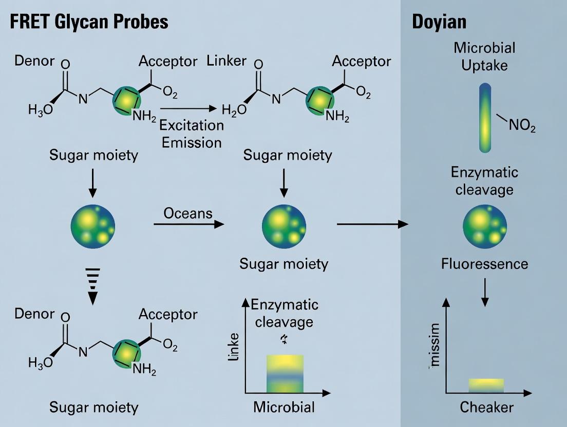
FRET-glycan probes consist of a specific polysaccharide (e.g., laminarin, xylan) labeled with a donor (e.g., Cy3) and an acceptor (e.g., Cy5) fluorophore in close proximity. Upon enzymatic hydrolysis, the fluorophores separate, leading to a loss of FRET and an increase in donor emission. This provides a direct, in situ optical readout of degradation activity.
3.1 Key Advantages Over Bulk Methods:
- Spatial Resolution: Activity can be localized to individual cells or particles via microscopy (e.g., FISH-FRET).
- Temporal Resolution: Real-time kinetics of degradation can be monitored in live cells.
- Functional Specificity: Signal is generated only upon cleavage of the specific glycosidic bond within the probe.
- Single-Cell Heterogeneity: Activity distributions across populations can be quantified.
4.0 Experimental Protocols
4.1 Protocol: Synthesis of Double-Labeled FRET-Glycan Probes (e.g., Laminarin)
- Objective: To create a polysaccharide substrate with donor and acceptor fluorophores for FRET-based degradation assays.
- Reagents: Pure polysaccharide (e.g., laminarin from Eisenia bicyclis), Cy3 NHS Ester, Cy5 NHS Ester, anhydrous DMSO, 0.1M Sodium Bicarbonate Buffer (pH 8.3), Sephadex G-25 column, Size-Exclusion Chromatography (SEC) columns.
- Procedure:
- Activation: Dissolve 10 mg of laminarin in 1 mL of 0.1M sodium bicarbonate buffer (pH 8.3).
- Donor Labeling: Add a 5-fold molar excess of Cy3-NHS ester (from DMSO stock) to the polysaccharide solution. React for 2 hours at 25°C in the dark with gentle mixing.
- Purification (Step 1): Purify the Cy3-laminarin conjugate using a Sephadex G-25 column equilibrated with Milli-Q water to remove free dye. Lyophilize the product.
- Acceptor Labeling: Re-dissolve the Cy3-laminarin in fresh bicarbonate buffer. Add a 10-fold molar excess of Cy5-NHS ester. React for 2 hours at 25°C in the dark.
- Purification (Step 2): Purify the dual-labeled product via SEC (e.g., Superdex 30 Increase) to separate double-labeled polymers from single-labeled and unreacted species. Confirm labeling ratio and FRET efficiency via absorbance and fluorescence spectrometry.
4.2 Protocol: In Situ Tracking of Microbial Glycan Degradation via Flow Cytometry
- Objective: To quantify single-cell degradation activity within a marine microbial community.
- Reagents: Marine microbial sample (enrichment culture or natural assemblage), FRET-glycan probe (from Protocol 4.1), sterile artificial seawater (ASW) medium, nucleic acid stain (e.g., SYBR Green I for total cells), flow cytometer with 488nm and 640nm lasers.
- Procedure:
- Sample Incubation: Dilute microbial sample in ASW to ~10^6 cells/mL. Add FRET-glycan probe to a final concentration of 50-100 nM. Include a no-probe control and a heat-killed cell control.
- Time-Course: Incubate at in situ temperature in the dark. Collect aliquots at T=0, 30, 60, 120, and 240 minutes.
- Staining: Fix aliquots with 0.5% formaldehyde (final conc.) for 15 min. Stain with SYBR Green I (1:10,000 dilution) for 15 min to identify all cells.
- Flow Cytometry Acquisition: Analyze samples using a flow cytometer. Use the 488nm laser to excite SYBR Green (detect at 530/30 nm) and Cy3 (detect at 580/30 nm). Use the 640nm laser to excite Cy5 (detect at 670/30 nm).
- Gating & Analysis: Gate on SYBR-positive events (total cells). Plot Cy3 vs. Cy5 fluorescence. Active degraders show high Cy3 (donor) and low Cy5 (acceptor) fluorescence due to FRET loss. Calculate the donor/acceptor ratio for each cell as a metric of degradation activity.
5.0 The Scientist's Toolkit: Key Research Reagent Solutions
Table 2: Essential Materials for FRET-Glycan Degradation Studies
| Item | Function & Relevance |
|---|---|
| Custom FRET-Glycan Probes | Core substrate for in situ activity detection. Available with various glycan backbones (alginate, chitin, laminarin) to target specific enzyme classes. |
| Marine Microbial Community DNA/RNA Stabilization Kits | Preserve community structure and gene expression at the point of sampling for correlative 'omics analyses. |
| Flow Cytometer with 405, 488, 640 nm Lasers | Enables multi-parameter detection of cell scatter, nucleic acid stains, and FRET probe signals for high-throughput single-cell analysis. |
| Fluorescence-Activated Cell Sorting (FACS) Capability | Allows physical sorting of active (high donor/low acceptor) vs. inactive cell populations for downstream cultivation or genomic analysis. |
| Coupled FISH-FRET Probe Sets | Combines phylogenetic identification (FISH probes) with activity detection (FRET probe) to link function to identity in situ via microscopy. |
| Microfluidic Single-Cell Cultivation Devices | Enables isolation and growth of cells sorted based on in situ degradation activity, overcoming cultivation bias. |
6.0 Visualizations
Title: Limitations of Bulk Techniques Lead to Obscured Heterogeneity
Title: FRET Probe Activation via Glycan Hydrolysis
Title: Single-Cell Activity Workflow via Flow Cytometry
Förster Resonance Energy Transfer (FRET) is a non-radiative process where energy from an excited donor fluorophore is transferred to a nearby acceptor fluorophore. This efficiency of this transfer is exquisitely sensitive to the inverse sixth power of the distance separating the two fluorophores, making it a powerful "molecular ruler" for measuring distances in the 1-10 nm range. Within the context of tracking microbial sugar degradation in oceans, FRET-based glycan probes allow researchers to visualize and quantify the enzymatic cleavage of complex polysaccharides in real-time. As marine microbes secrete hydrolytic enzymes to break down glycans, the disruption of a FRET pair integrated into the glycan structure generates a quantifiable fluorescent signal, providing insights into carbon cycling dynamics.
Core Physics & Quantitative Relationships
FRET efficiency (E) is governed by the Förster equation:
[ E = \frac{1}{1 + (r/R_0)^6} ]
Where r is the donor-acceptor distance and R₀ is the Förster radius (the distance at which efficiency is 50%). R₀ depends on the spectral properties of the fluorophores:
[ R0^6 = \frac{9QD(\ln 10)\kappa^2 J}{128\pi^5N_A n^4} ]
Table 1: Key Parameters & Their Impact on FRET Efficiency
| Parameter | Symbol | Typical Range/Value | Influence on FRET |
|---|---|---|---|
| Donor-Acceptor Distance | r | 1 – 10 nm | Primary determinant. E drops drastically as r increases beyond R₀. |
| Förster Radius | R₀ | 3 – 6 nm (commonly) | Defines the measurement scale. Larger R₀ increases usable distance range. |
| Spectral Overlap Integral | J | Varies (M⁻¹cm⁻¹nm⁴) | Larger overlap => larger R₀ => higher potential efficiency. |
| Donor Quantum Yield | Q_D | 0 – 1 | Higher yield => larger R₀. |
| Orientation Factor | κ² | 0 – 4 | Assumed 2/3 for freely rotating dipoles. Can introduce error if restricted. |
| Refractive Index | n | ~1.33 – 1.4 (aqueous) | Environmental factor; lower n => larger R₀. |
Table 2: Common FRET Pairs for Biochemical Probes
| Donor | Acceptor | R₀ (nm) | Application Notes |
|---|---|---|---|
| Cy3 | Cy5 | ~5.4 | Bright, photostable; common for oligonucleotide/ glycan labeling. |
| GFP (CFP variant) | YFP | ~4.9 – 5.2 | Genetically encodable; used in live-cell biosensors. |
| Alexa Fluor 488 | Alexa Fluor 594 | ~5.5 | High brightness and photostability; good for in vitro assays. |
| mTurquoise2 | Venus | ~6.1 | Improved genetically encoded pair with higher quantum yield. |
| FRET-Quencher Pair | Example: Cy3 / BHQ-2 | Variable | Acceptor is a non-fluorescent quencher; signal is donor emission loss. |
Diagram 1: FRET Process & Distance Dependence
Protocol: FRET-Based Assay for Monitoring Glycan Degradation by Marine Extracts
This protocol details the use of dual-labeled glycan probes to detect hydrolytic activity in environmental samples, such as seawater or microbial culture supernatants.
A. Materials & Reagent Preparation
The Scientist's Toolkit: Essential Reagents for FRET Glycan Assays
| Item | Function & Specification |
|---|---|
| FRET Glycan Probe | Synthetic oligosaccharide (e.g., laminarin, xylan) labeled with donor (Cy3) and acceptor (Cy5) at specific positions. Cleavage separates the pair, reducing FRET. |
| Marine Sample | Filtered (0.22 µm) seawater or concentrated extracellular enzyme fraction from microbial cultures. |
| Control Enzymes | Purified endo-1,3-β-glucanase (for laminarin probe) or xylanase (for xylan probe) for positive control. |
| Assay Buffer (pH ~8) | Mimics seawater conditions: 50 mM HEPES, 400 mM NaCl, 10 mM MgCl₂, 0.01% BSA, pH 8.0. |
| Microplate Reader | Capable of temperature control and sequential excitation/emission readings (e.g., donor excitation/acceptor emission for FRET channel). |
| Black 96- or 384-well Plates | Low fluorescence background plates for optimal signal-to-noise. |
| Data Analysis Software | For fitting kinetic curves and calculating degradation rates (e.g., Prism, custom Python/R scripts). |
B. Step-by-Step Experimental Procedure
Probe Reconstitution & Dilution:
- Centrifuge the lyophilized FRET-glycan probe vial briefly.
- Reconstitute in ultrapure water or DMSO (as specified by manufacturer) to create a 100 µM stock solution. Aliquot and store at -80°C.
- On the day of the experiment, dilute the stock in assay buffer to a 200 nM working solution. Keep on ice in the dark.
Sample & Reaction Plate Setup (in triplicate):
- Prepare a master mix containing the assay buffer and the FRET-glycan probe working solution.
- Pipette 90 µL of the master mix into each well of a black microplate.
- Add 10 µL of the following to designated wells:
- Test Samples: Filtered marine extract.
- Positive Control: 10 µL of purified enzyme solution (known activity).
- Negative Control: 10 µL of heat-inactivated (95°C, 10 min) marine extract or buffer only.
- Gently mix by pipetting or plate shaking. Seal the plate to prevent evaporation.
Kinetic Measurement:
- Place the plate in a pre-warmed (e.g., in-situ ocean temperature: 15°C or 25°C) microplate reader.
- Program the reader to take cyclic measurements every 1-5 minutes for 1-3 hours.
- FRET Signal: Excite the donor (e.g., Cy3 at 535 nm) and measure emission from the acceptor (e.g., Cy5 at 670 nm).
- Donor Control Signal (Optional): In a separate cycle, excite the donor and measure donor emission (e.g., Cy3 at 570 nm) to monitor direct donor quenching.
Termination & Analysis (Optional Endpoint):
- After the kinetic run, the reaction can be stopped by adding a quenching solution (e.g., 100 µL of 1 M Na2CO3) or by freezing.
C. Data Analysis & Interpretation
- FRET Ratio Calculation: For each time point, calculate the signal ratio: Acceptor Emission (FRET channel) / Donor Emission (Donor channel). This ratio normalizes for well-to-well variations in probe concentration and quenching.
- Kinetic Curve Fitting: Plot the FRET ratio (or raw FRET channel fluorescence) versus time. Fit the initial linear phase to determine the rate of signal change (RFU/min).
- Quantifying Activity: Compare the slope of the test sample to a standard curve generated with known amounts of purified enzyme to express activity as enzyme equivalents per liter of seawater.
Diagram 2: FRET Glycan Degradation Assay Workflow
Application Note: Designing Probes for Marine Polysaccharides
The effectiveness of the assay hinges on probe design. Key considerations include:
- Linkage Specificity: The fluorophores should bracket the specific glycosidic bond targeted by the enzyme of interest (e.g., β-1,4 for cellulases, β-1,3 for laminarinases).
- Labeling Chemistry: Amine-reactive NHS esters of dyes can label aminoglycan derivatives. Click chemistry is an alternative for more specific conjugation.
- Quenching Mechanism: Using a dark quencher (e.g., BHQ-2) as the acceptor simplifies detection to monitoring only donor fluorescence recovery upon cleavage.
Table 3: Example FRET-Glycan Probes for Marine Research
| Target Polysaccharide | Donor-Acceptor Pair | Labeling Positions | Detected Enzyme Class |
|---|---|---|---|
| Laminarin (β-1,3-glucan) | Cy3 / Cy5 | On reducing and non-reducing ends, or internal via modified glucose | Endo-1,3-β-glucanase |
| Xylan (β-1,4-xylose) | Alexa Fluor 488 / Alexa Fluor 594 | On xylose residues flanking cleavage site | Endo-1,4-β-xylanase |
| Alginate (Poly G/M blocks) | FITC / TRITC | On uronic acid residues | Alginate lyase |
| Porphyran (agarose substitute) | mTurquoise2 / Venus (genetically encoded) | Expressed as fusion within designer substrate | Porphyranase |
Application Notes
Within the thesis research on tracking microbial polysaccharide degradation in marine ecosystems, the design of a Förster Resonance Energy Transfer (FRET)-based synthetic glycan probe is critical. This enables real-time, sensitive quantification of hydrolytic enzyme activities in complex environmental samples, providing insights into carbon cycling dynamics. The core concept involves synthesizing a polysaccharide-mimetic oligosaccharide flanked by a donor-acceptor fluorophore pair. Upon intact substrate mimic cleavage by a target microbial enzyme (e.g., laminarinase, xylanase), the FRET pair separates, leading to a measurable change in fluorescence emission ratio.
Quantitative Design & Performance Parameters
Table 1: Key Spectral & Biochemical Parameters for a Model Laminarin FRET Probe
| Parameter | Donor Fluorophore (e.g., Cy3) | Acceptor Fluorophore (e.g., Cy5) | Synthetic Glycan Substrate |
|---|---|---|---|
| Excitation Max (nm) | 550 | 649 | N/A |
| Emission Max (nm) | 570 | 670 | N/A |
| FRET Efficiency (R0 in Å) | ~60 Å (for Cy3-Cy5 pair) | ||
| Linker/Spacer | Aminohexanoic acid & PEG | Aminohexanoic acid & PEG | β-1,3-glucan oligosaccharide (DP~8-12) |
| Cleavage Site | N/A | N/A | Specific glycosidic bond (e.g., β-1,3) |
| Kinetic Readout | Increase in donor emission (I~570~) & decrease in acceptor emission (I~670~) upon cleavage. | ||
| Assay Sensitivity (Enzyme) | Detectable activity in picomolar range for purified enzymes. | ||
| Environmental Application | Can be spiked into seawater samples to measure community-level enzymatic rates (nmol/L/hr). |
Table 2: Advantages Over Natural Substrate Assays
| Aspect | Natural Glycan (e.g., FITC-Laminarin) | Synthetic FRET-Glycan Mimic |
|---|---|---|
| Signal Mechanism | Fluorescence de-quenching or release of small fluorophore. | Rationetric FRET change (internal calibration). |
| Specificity | Measures end-product release; can be less specific. | Can be designed for specific bond cleavage (endo- vs exo-acting). |
| Signal-to-Noise Ratio | Moderate, susceptible to environmental quenching. | High, due to dual-wavelength rationetric measurement. |
| Real-Time Kinetics | Yes, but may require secondary detection. | Excellent, direct continuous measurement. |
| Probe Stability | Variable, susceptible to non-specific degradation. | High, with designed synthetic backbone. |
Detailed Protocols
Protocol 1: Synthesis of a β-1,3-Glucan FRET Probe (Laminarin Mimic)
Objective: Chemoenzymatic synthesis of a defined-length β-1,3-linked gluco-oligosaccharide labeled at the reducing end with Cy3 and at the non-reducing end with Cy5.
Research Reagent Solutions:
| Item | Function |
|---|---|
| Peracetylated Glucose β-Glycosyl Fluoride | Building block for iterative glycosylation. |
| Glycosynthase Mutant (e.g., of Humicola insolens Ce17B) | Engineered glycosidase that catalyzes synthesis of β-1,3 linkages without hydrolysis. |
| Cy3B-NH~2~ & Cy5-NH~2~ | Bright, stable amine-reactive fluorophores with optimal spectral overlap for FRET. |
| Amino-PEG~3~-Alkyne & Azido-PEG~3~-Amino | Heterobifunctional linkers for "click chemistry" conjugation and spacer introduction. |
| Cu(I) TBTA Catalyst | Catalyzes the azide-alkyne cycloaddition (CuAAC) "click" reaction. |
| HPLC with Fluorescence Detector (C18 Column) | For purification and analysis of labeled oligosaccharides. |
| Marine Buffered Saline (MBS) | Artificial seawater buffer (pH 8.0) for environmental assays. |
Methodology:
- Oligosaccharide Backbone Assembly:
- Using a glycosynthase enzyme, iteratively add peracetylated glucose units from a reducing-end tethered primer to build a linear β-1,3-octaglucan.
- Deacetylate the oligomer using sodium methoxide in methanol.
- Purify the core oligosaccharide via size-exclusion chromatography.
- Functionalization with Linkers:
- Reducing End: React the free reducing end with an excess of amino-PEG~3~-alkyne via reductive amination (NaBH~3~CN) to install an alkyne handle.
- Non-Reducing End: Chemoselectively activate the terminal hydroxyl as an imidazolyl carbamate, then react with azido-PEG~3~-amino to install an azide handle.
- Fluorophore Conjugation via Click Chemistry:
- Step 1 (Alkyne + Cy3): React the alkyne-functionalized reducing end with Cy3B-azide using Cu(I) TBTA catalyst. Purify by HPLC.
- Step 2 (Azide + Cy5): React the azide-functionalized non-reducing end of the product from Step 1 with Cy5-alkyne using Cu(I) catalysis.
- Final Purification & Validation:
- Perform reverse-phase HPLC (C18 column) with dual-wavelength detection (550 nm ex/570 nm em for Cy3; 649 nm ex/670 nm em for Cy5).
- Collect the peak exhibiting both fluorescence signatures.
- Lyophilize and characterize by mass spectrometry.
- Confirm FRET by exciting at 550 nm and verifying acceptor (Cy5) emission at 670 nm.
Protocol 2: Measuring Microbial Glycanase Activity in Seawater Samples
Objective: To use the synthesized FRET-glycan probe to quantify specific polysaccharide degradation potential in a marine microbial community sample.
Methodology:
- Sample Preparation: Filter seawater (0.22 µm) to remove large particulates but retain free enzymes and some microbes/viruses. Divide into aliquots.
- Assay Setup:
- Prepare a master mix of the FRET probe in Marine Buffered Saline (MBS) to a final concentration of 1 µM.
- Add 190 µL of master mix to each well of a black, flat-bottom 96-well plate.
- Add 10 µL of filtered seawater sample to test wells. Use 10 µL of MBS for a negative control, and 10 µL of a known, purified laminarinase solution for a positive control.
- Run in triplicate.
- Kinetic Measurement:
- Immediately place the plate in a pre-warmed (e.g., in situ temperature, 15°C) fluorescence plate reader.
- Program the reader to take sequential readings every 2-5 minutes for 12-24 hours:
- Ex: 530 nm / Em: 570 nm (Donor channel, I~D~)
- Ex: 530 nm / Em: 670 nm (Acceptor FRET channel, I~A~)
- Data Analysis:
- Calculate the FRET ratio (R) for each time point: R = I~A~ / I~D~.
- Plot R over time. Enzymatic cleavage is indicated by a decrease in R.
- Calculate the initial velocity (V~0~) from the linear portion of the plot (ΔR/Δtime).
- Normalize V~0~ to the sample's microbial cell count (from flow cytometry) or total protein content to report specific activity.
Visualizations
Diagram 1 Title: FRET Glycan Probe Workflow from Synthesis to Application
Diagram 2 Title: Mechanism of FRET Signal Generation and Loss
Building and Deploying the Molecular Beacon: A Step-by-Step Guide to FRET Glycan Probes
Application Notes: FRET Glycan Probes for Microbial Degradation Tracking
Within the broader thesis on developing Förster Resonance Energy Transfer (FRET)-based glycan probes, this protocol details the synthesis of dual-fluorophore-labeled glycans. These probes are designed to monitor real-time enzymatic degradation by marine microbial communities, providing insights into polysaccharide processing and carbon cycling in oceanic ecosystems. Successful conjugation yields a probe where the cleavage of the glycan backbone by microbial glycoside hydrolases separates the donor and acceptor fluorophores, resulting in a measurable loss of FRET signal.
Key Research Reagent Solutions
| Reagent / Material | Function in Protocol |
|---|---|
| Target Glycan Backbone (e.g., Laminarin, Xylan) | The polysaccharide substrate representative of marine carbon pools. Its degradation is the target event. |
| Amine-Reactive Donor Fluorophore (e.g., Cy3 NHS ester) | High-quantum-yield dye. Conjugates to glycans via amino groups, serving as the FRET energy donor. |
| Amine-Reactive Acceptor Fluorophore (e.g., Cy5 NHS ester) | Dye with overlapping absorption/emission with the donor. Conjugates at a strategic distance, serving as the FRET energy acceptor. |
| Periodate Oxidation Reagents (NaIO₄) | Selectively oxidizes vicinal diols on glycan sugars to generate aldehyde groups for conjugation. |
| Aniline Catalyst | Nucleophilic catalyst that accelerates oxime ligation between aldehydes and aminooxy-fluorophores. |
| Aminooxy-PEG₄-Amine Linker | Bifunctional linker. The aminooxy end forms a stable oxime with the glycan aldehyde; the amine end reacts with NHS-ester dyes. |
| Size Exclusion Chromatography (SEC) Columns (e.g., PD-10) | For purifying conjugated probes from excess unreacted dyes and small molecules. |
| Analytical HPLC with Fluorescence Detector | Critical for verifying dual-labeling success, assessing conjugation ratio, and checking probe purity. |
Experimental Protocols
Protocol 1: Periodate Oxidation of Glycan Backbone to Generate Aldehyde Handles
Objective: To introduce controlled, reactive aldehyde groups onto the glycan without significant backbone depolymerization.
- Dissolve 10 mg of the target polysaccharide (e.g., laminarin from Laminaria digitata) in 1 mL of 0.1 M sodium acetate buffer, pH 5.5.
- Prepare a fresh 100 mM solution of sodium periodate (NaIO₄) in ultrapure water. Protect from light.
- Add 100 µL of the NaIO₄ solution to the glycan solution to achieve a 10 mM final concentration. Vortex gently.
- Incubate the reaction on ice in the dark for 1 hour to achieve mild, controlled oxidation.
- Quench the reaction by adding 50 µL of ethylene glycol. Mix and incubate on ice for 30 minutes.
- Purify the oxidized glycan using a pre-equilibrated PD-10 desalting column with 0.1 M MES buffer, pH 5.0, as the eluent. Collect the high molecular weight fraction (~1.5 mL).
- Quantify the aldehyde concentration using a standardized nitrobenzoxadiazole (NBD) hydrazine assay. Adjust concentration to 5 mg/mL for the next step.
Protocol 2: Two-Step Conjugation via Aminooxy Linker
Objective: To sequentially conjugate donor (Cy3) and acceptor (Cy5) fluorophores at controlled sites on the oxidized glycan.
Step 2A: Conjugation of Aminooxy-PEG₄-Amine Linker
- To 1 mL of oxidized glycan (5 mg, in MES pH 5.0), add a 5x molar excess of aminooxy-PEG₄-amine linker (relative to estimated aldehydes).
- Add aniline to a final concentration of 50 mM as a catalyst.
- React overnight at room temperature with gentle mixing.
- Purify the amino-functionalized glycan using a PD-10 column equilibrated in 0.1 M sodium bicarbonate buffer, pH 8.3. Collect the product fraction.
Step 2B: Sequential Fluorophore Labeling
- Divide the amino-functionalized glycan into two equal portions (0.5 mL each).
- Donor Conjugation: To the first portion, add a 2x molar excess of Cy3 NHS ester (from a stock in anhydrous DMSO). React for 2 hours at room temperature in the dark.
- Acceptor Conjugation: To the second portion, add a 2x molar excess of Cy5 NHS ester. React for 2 hours in the dark.
- Separately purify each reaction mixture using PD-10 columns (Sephadex G-25) in ammonium acetate buffer (50 mM, pH 7.0).
- Combine the purified Cy3-glycan and Cy5-glycan fractions. To promote proximity for FRET, allow the mixture to incubate at 4°C for 24 hours, enabling non-covalent interactions between the labeled strands.
- Perform final purification via HPLC (size-exclusion or ion-exchange) to isolate the dual-labeled FRET probe. Lyophilize and store at -80°C.
Table 1: Physicochemical Properties of Synthesized FRET-Glycan Probes
| Probe (Glycan Backbone) | Donor:Acceptor Ratio (HPLC) | Average Labeling Degree (Dyes per 100 sugar units) | FRET Efficiency (E) | Hydrodynamic Radius (nm, DLS) |
|---|---|---|---|---|
| Laminarin-Cy3/Cy5 | 1:0.9 | 2.1 (Cy3), 1.9 (Cy5) | 0.78 ± 0.05 | 4.2 ± 0.3 |
| Xylan-Cy3/Cy5 | 1:1.1 | 1.8 (Cy3), 2.0 (Cy5) | 0.72 ± 0.07 | 3.8 ± 0.4 |
Table 2: Enzymatic Validation of FRET Probe Functionality
| Enzyme (Microbial Source) | Substrate Probe | Initial Hydrolysis Rate (nM/s) | % FRET Loss at 1 Hour |
|---|---|---|---|
| Endo-β-1,3-glucanase | Laminarin-Cy3/Cy5 | 15.2 ± 1.5 | 85 ± 4 |
| β-Glucosidase | Laminarin-Cy3/Cy5 | 1.1 ± 0.3 | 12 ± 3 |
| Endo-xylanase | Xylan-Cy3/Cy5 | 22.7 ± 2.1 | 92 ± 2 |
Visualization: Workflow and Pathway Diagrams
Title: Two-Step Synthesis of FRET-Glycan Probes
Title: FRET Signal Loss Upon Glycan Degradation
Application Notes
This document details the design and application of Förster Resonance Energy Transfer (FRET) probes for tracking microbial degradation of specific polysaccharide classes in marine environments. These probes enable real-time, activity-based monitoring of enzymatic hydrolysis, providing insights into carbon cycling dynamics in oceanic systems. The core principle involves a polysaccharide backbone labeled with a donor fluorophore and a quencher/acceptor. Hydrolysis by a target enzyme separates the pair, restoring donor fluorescence.
Table 1: FRET Probe Design Parameters for Key Marine Polysaccharide Classes
| Polysaccharide Class | Example Substrate | Donor Fluorophore (λex/λem) | Acceptor/Quencher | Linker/Cleavage Site | Target Enzyme Class | Typical Δ Signal (F/F0)* | Optimal Assay pH |
|---|---|---|---|---|---|---|---|
| Laminarin-type β-glucans | Laminarin | FAM (488/518 nm) | Dabcyl or QSY-7 | β-1,3 glycosidic bond | Endo-β-1,3-glucanase | 8-12x | 7.5 (Marine) |
| Alginate | PolyMG, PolyG blocks | Cy3 (550/570 nm) | Cy5 (650/670 nm) | α-L-guluronate or β-D-mannuronate bonds | Alginate lyase (PolyMG lyase) | 5-8x | 7.0-8.0 |
| Pectin/Hemicellulose | Homogalacturonan | Alexa Fluor 488 (495/519 nm) | Iowa Black FQ | α-1,4 galacturonan bond | Polygalacturonase | 10-15x | 6.0-7.5 |
| Sulfated Fucans | Fucoidan (simplified) | Atto 550 (554/576 nm) | Atto 647N (646/664 nm) | α-1,3/1,4 fucosyl bond | Fucanase/Sulfatase* | 4-6x | 6.5-7.5 |
| Xylans | β-1,4-Xylan | Pacific Blue (410/455 nm) | QSY-35 | β-1,4 xylosyl bond | Endo-β-1,4-xylanase | 9-14x | 6.0-7.0 |
*F/F0 = Fluorescence intensity after cleavage / initial fluorescence. Synthetic oligosaccharide analogs are required. *Probe requires co-localized enzyme activity.
Protocol 1: Synthesis of a Laminarin-FRET Probe for β-1,3-Glucanase Activity
I. Materials & Reagent Solutions
Research Reagent Solutions:
| Item | Function & Specification |
|---|---|
| Amino-derivatized Laminarin Oligosaccharide | Backbone substrate (DP ~20) with terminal amine for fluorophore conjugation. |
| NHS-ester Donor Fluorophore (e.g., FAM, SE) | Reacts with primary amine to label substrate. |
| Malachite Green Isothiocyanate (MG-ITC) | Quencher; reacts with amine on opposite terminus. |
| Anhydrous DMSO | Solvent for fluorophore and quencher stock solutions. |
| 0.1M Sodium Borate Buffer (pH 8.5) | Optimal pH for amine-NHS ester/Isothiocyanate reactions. |
| PD-10 Desalting Column (Sephadex G-25) | For purification of labeled probe from free dye. |
| Analytical HPLC with Fluorescence Detector | For final purification and verification of labeling efficiency. |
II. Procedure
- Donor Labeling: Dissolve 5 µmol of amino-laminarin in 500 µL of 0.1M sodium borate buffer (pH 8.5). Prepare a 20 mM stock of NHS-FAM in anhydrous DMSO. Add a 1.2x molar excess (6 µmol, 300 µL) of NHS-FAM to the oligosaccharide solution. Vortex gently and incubate in the dark at room temperature for 2 hours.
- Intermediate Purification: Pass the reaction mixture through a pre-equilibrated PD-10 column using distilled water as the eluent. Collect 0.5 mL fractions. Monitor fractions for fluorescence (λex 488 nm, λem 518 nm). Pool the early, high-fluorescence fractions containing the FAM-laminarin conjugate. Lyophilize.
- Acceptor/Quencher Labeling: Re-dissolve the FAM-laminarin pellet in 500 µL of fresh borate buffer. Prepare a 15 mM stock of MG-ITC in DMSO. Add a 1.5x molar excess relative to the initial amino-laminarin. Incubate in the dark at room temperature for 4 hours.
- Final Probe Purification: Purify the dual-labeled product using an analytical HPLC (C18 column). Use a gradient of 0.1% TFA in water to 0.1% TFA in acetonitrile. Monitor absorbance at 494 nm (FAM) and 620 nm (MG). Collect the peak showing absorbance at both wavelengths. Lyophilize, confirm mass via MALDI-TOF, and store at -20°C.
Protocol 2: Real-Time Assay for Marine β-Glucanase Activity Using the Laminarin-FRET Probe
I. Materials
- Laminarin-FRET Probe (Protocol 1)
- Marine particle-associated enzyme extract or cultured supernatant
- 50mM HEPES buffer (pH 7.5), containing 100mM NaCl, 10mM MgCl₂ (simulating ionic strength of seawater)
- Black-walled, clear-bottom 96-well microplate
- Fluorescence plate reader capable of temperature control and kinetic reads (λex 488 nm, λem 518 nm).
II. Procedure
- Probe Preparation: Prepare a 10 µM stock solution of the laminarin-FRET probe in assay buffer. Sonicate briefly to ensure full dissolution.
- Assay Setup: In each well of the microplate, add 180 µL of assay buffer. Add 10 µL of the enzyme extract (or buffer for negative control). Pre-incubate the plate at in situ temperature (e.g., 15°C) for 5 minutes in the plate reader.
- Reaction Initiation: Rapidly add 10 µL of the probe stock solution (final probe concentration: 0.5 µM) to each well using a multi-channel pipette. Mix by gentle shaking.
- Kinetic Measurement: Immediately commence kinetic fluorescence measurements. Read fluorescence every 30 seconds for 60-120 minutes. Maintain constant temperature.
- Data Analysis: Plot fluorescence versus time. Calculate initial reaction velocities (V0) from the linear portion of the curve. Normalize activity to protein concentration or sample volume. Use a standard curve of fully cleaved probe (e.g., by exhaustive enzymatic digestion) to convert fluorescence units to molar hydrolysis rates.
Visualizations
Title: FRET Probe Workflow for Marine Polysaccharide Degradation
Title: FRET Probe Signaling Mechanism
This protocol details the synthesis, purification, and characterization of Förster Resonance Energy Transfer (FRET)-based glycan probes. Within the broader thesis on tracking microbial sugar degradation in ocean ecosystems, these probes enable real-time, in situ monitoring of polysaccharide hydrolysis by marine microbes. The workflow is critical for understanding carbon cycling in marine environments and has parallel applications in drug development for glycosidase-targeting therapeutics.
Key Research Reagent Solutions
The following table lists essential materials and their functions for the probe workflow.
| Item Name | Function/Brief Explanation |
|---|---|
| Activated Donor Fluorophore (e.g., Cy3-NHS ester) | FRET donor; covalently attaches to glycosidic linkage via amine-reactive chemistry. |
| Quencher/Acceptor (e.g., QSY-9, BHQ-2, or Cy5) | FRET acceptor/quencher; positioned to absorb donor emission when probe is intact. |
| Glycan Substrate (e.g., Laminarin, Xylan) | Target polysaccharide mimicking natural marine polymeric sugars. |
| Heterobifunctional Linker (e.g., SMPB) | Spacer arm with amine- and thiol-reactive ends for controlled fluorophore positioning. |
| Size Exclusion Chromatography (SEC) Columns (Sephadex G-25) | Desalting and purification of conjugated probes from unreacted dyes. |
| Analytical HPLC (C18 Column) | High-resolution purification and analysis of probe purity. |
| Fluorescence Spectrophotometer | Measures emission spectra to calculate FRET efficiency and probe integrity. |
| Microplate Reader (with temperature control) | Enables high-throughput kinetic assays of glycan degradation. |
| Dialysis Membranes (MWCO 3.5 kDa) | Removes small molecule contaminants post-conjugation. |
| Marine Simulation Buffer | Artificial seawater matrix for environmentally relevant characterization. |
Detailed Experimental Protocols
Protocol A: Conjugation & Purification of FRET Glycan Probes
Objective: Covalently attach donor and acceptor fluorophores to the target glycan polymer.
- Glycan Activation: Dissolve 5 mg of purified polysaccharide (e.g., laminarin) in 1 mL of 0.1 M MES buffer (pH 6.0). Add 2 mg of sodium meta-periodate and incubate in the dark for 30 minutes at 4°C to generate reactive aldehydes. Stop the reaction by adding 20 µL of ethylene glycol and incubating for 30 minutes.
- Donor Conjugation: Dialyze the activated glycan against 0.1 M carbonate buffer (pH 9.0). Add a 10-fold molar excess of Cy3-hydrazide. React for 2 hours at room temperature in the dark.
- Acceptor Positioning via Linker: To a separate aliquot, introduce free thiol groups by reacting periodate-oxidized glycan with cysteamine. Purify via SEC (G-25, eluted with PBS). React the thiolated product with a 5-fold molar excess of SMPB linker for 1 hour. Purify again.
- Acceptor Conjugation: Mix the donor-labeled glycan (from Step 2) with the linker-activated acceptor (e.g., QSY-9 amine) at a molar ratio of 1:1.2 (donor site:acceptor). React overnight at 4°C in the dark.
- Final Purification: Purify the dual-labeled FRET probe sequentially using:
- Size Exclusion Chromatography: Sephadex G-25 column equilibrated with artificial seawater buffer. Collect the high-molecular-weight fraction.
- Dialysis: Against 1 L of characterization buffer (MWCO 3.5 kDa) for 24 hours with two buffer changes.
- Analytical HPLC: Using a C18 column with a water/acetonitrile gradient (0.1% TFA) to confirm monodisperse product. Lyophilize and store at -80°C.
Protocol B: Fluorescence Characterization & FRET Efficiency Calculation
Objective: Validate probe integrity and establish baseline spectroscopic properties.
- Spectroscopic Measurement:
- Reconstitute the purified FRET probe to 100 nM in marine simulation buffer (pH 8.0).
- Using a fluorescence spectrophotometer, record the emission spectrum from 550 nm to 750 nm with donor excitation at 530 nm (slit widths: 5 nm excitation/5 nm emission).
- In parallel, record the emission spectrum of a donor-only labeled glycan control at identical concentration and instrument settings.
- Data Analysis & FRET Efficiency (E):
- Integrate the donor emission peak area (typically 560-580 nm) for both the FRET probe (FDA) and the donor-only control (FD).
- Calculate FRET efficiency using the quenching formula: E = 1 - (FDA / FD).
- A successfully synthesized probe should exhibit E > 0.85, indicating strong quenching when intact.
- Degradation Kinetics Assay:
- In a 96-well plate, add 200 µL of 100 nM FRET probe per well.
- Initiate the reaction by adding 20 µL of a standardized glycosidase enzyme (e.g., laminarinase) or a sample of live microbial inoculum.
- Immediately place the plate in a temperature-controlled microplate reader.
- Monitor fluorescence kinetically: Measure donor emission (580 nm) every 30 seconds for 60 minutes, with excitation at 530 nm.
- Calculate initial rates from the linear increase in donor fluorescence over the first 10 minutes.
Table 1: Typical Characterization Data for FRET Laminarin Probe
| Parameter | Donor-Only Probe | Intact FRET Probe | Cleaved FRET Probe (Post-Enzyme) |
|---|---|---|---|
| Peak Donor Emission (a.u.) | 1,000,000 ± 50,000 | 150,000 ± 15,000 | 950,000 ± 60,000 |
| FRET Efficiency (E) | N/A | 0.85 ± 0.03 | 0.05 ± 0.02 |
| Detection Limit (Enzyme) | N/A | N/A | 0.01 U/mL |
| Signal-to-Background Ratio | N/A | 1.5 | 6.3 ± 0.4 |
| *Hydrolysis Rate (nM/min) | N/A | N/A | 15.2 ± 1.7 |
*Measured with 0.1 U/mL laminarinase.
Table 2: Purification Yield Metrics Across Steps
| Purification Step | Average Yield (%) | Key Quality Control Check |
|---|---|---|
| Initial Chemical Conjugation | 100 (Reference) | Reaction completion (TLC/HPLC) |
| Size Exclusion Chromatography (SEC) | 65 ± 5 | Absorbance ratios (280 nm / 550 nm / 650 nm) |
| Final Dialysis & Lyophilization | 85 ± 3 of SEC product | Purity confirmed by analytical HPLC (>95%) |
| Overall Process Yield | ~55% | Functional FRET efficiency (E > 0.8) |
Visualization Diagrams
Diagram 1: FRET glycan probe synthesis workflow.
Diagram 2: FRET quenching and recovery mechanism.
Within the broader thesis on Förster Resonance Energy Transfer (FRET)-based glycan probes, this document details protocols for their application in tracking microbial polysaccharide degradation dynamics in marine environments. The work bridges lab-based mechanistic studies using pure microbial cultures and field-relevant incubations with complex environmental samples. The goal is to quantify and visualize the enzymatic hydrolysis of specific glycans—key to understanding the oceanic carbon cycle.
Key Research Reagent Solutions & Materials
Table 1: Essential Reagents and Materials for FRET Glycan Probe Incubations
| Item Name | Function/Brief Explanation |
|---|---|
| FRET-Glycan Probes (e.g., Laminarin-FRET, Xylan-FRET) | Synthetic glycan substrates labeled with a donor (e.g., Fluorescein) and acceptor (e.g., Dabcyl) fluorophore. Hydrolysis by microbial enzymes separates the pair, increasing donor fluorescence. |
| Sterile, Particle-Free Seawater | Matrix for all incubations; filtered (0.2 µm) to remove ambient microbes/enzymes for controlled assays. |
| Defined Artificial Seawater Medium | For pure culture work; provides essential ions and salts without organic carbon background. |
| Live Environmental Inoculum | Seawater concentrate or particle-associated community filtered onto 0.22 µm filters (for field assays). |
| Axenic Microbial Cultures | Model marine bacteria (e.g., Saccharophagus degradans, Flavobacteria spp.) for mechanistic studies. |
| Protease Inhibitor Cocktail (EDTA-free) | Added to select assays to inhibit metalloproteases and distinguish glycanase activity. |
| Fluorescence Microplate Reader | Equipped with appropriate filters (e.g., Excitation: 485 nm, Emission: 535 nm) for kinetic fluorescence measurement. |
| Temperature-Controlled Incubator or On-Deck Incubation System | Maintains in situ temperatures (e.g., 4°C for polar samples, 25°C for tropical). |
| 0.2 µm Syringe Filters | For terminating reactions and removing cells prior to fluorescence reading. |
Table 2: Example Kinetic Data from FRET Probe Incubations with a Coastal Seawater Community Conditions: 100 nM probe final concentration, 10°C, in 0.2 µm-filtered seawater with natural microbial inoculum. Data presented as mean ± SD (n=3).
| Probe Type | Incubation Time (h) | Fluorescence Increase (RFU) | Calculated Hydrolysis Rate (nM/h) | Relative Activity (%) vs. Control* |
|---|---|---|---|---|
| Laminarin-FRET | 0 | 50 ± 5 | 0 | 0 |
| Laminarin-FRET | 24 | 1250 ± 120 | 4.8 ± 0.5 | 100 |
| Laminarin-FRET | 48 | 2150 ± 200 | 4.2 ± 0.4 | 88 |
| Xylan-FRET | 0 | 55 ± 6 | 0 | 0 |
| Xylan-FRET | 24 | 450 ± 40 | 1.3 ± 0.1 | 100 |
| Chitin-FRET (Control) | 24 | 60 ± 10 | 0.02 ± 0.01 | N/A |
| Heat-Killed Control (Laminarin) | 24 | 65 ± 8 | 0.05 ± 0.02 | 1 |
Control: *Activity normalized to the 24-hour value for each probe type.
Table 3: Comparison of Hydrolysis Rates Between Pure Cultures and Environmental Samples Rates in nM probe hydrolyzed/hr/mg of protein or mL of sample.
| Sample Type / Organism | Target Glycan (Probe) | Hydrolysis Rate | Assay Temperature |
|---|---|---|---|
| Coastal Seawater (0-200m) | Laminarin (β-1,3-glucan) | 4.8 ± 0.5 nM/hr/mL | 10°C |
| Saccharophagus degradans (lab culture) | Laminarin | 120 ± 15 nM/hr/mg protein | 28°C |
| Particle-Associated Community | Xylan | 2.1 ± 0.3 nM/hr/mL | 15°C |
| Polaribacter sp. (psychrophilic isolate) | Laminarin | 45 ± 6 nM/hr/mg protein | 4°C |
| Deep Sea Water (2000m) | All Probes Tested | < 0.1 nM/hr/mL | 4°C |
Detailed Experimental Protocols
Protocol 4.1: Laboratory Incubation with Axenic Marine Cultures
Objective: To determine the glycan degradation capability and kinetics of a specific microbial isolate.
Materials:
- FRET-glycan probe stock solution (100 µM in Milli-Q water).
- Mid-log phase culture of target bacterium in marine broth or defined medium.
- Sterile Artificial Seawater (ASW) buffer, pH 8.0.
- 96-well black, clear-bottom microplates.
- Microplate reader with temperature control.
Procedure:
- Harvest & Wash Cells: Pellet 10 mL of culture at 5000 x g for 10 min at assay temperature. Wash cell pellet twice with sterile ASW buffer. Resuspend in ASW to an OD600 of 0.5.
- Prepare Reaction Mix: In each well of the microplate, combine:
- 180 µL of cell suspension (or ASW for negative control).
- 20 µL of FRET-glycan probe stock for a final concentration of 10 µM.
- Kinetic Measurement: Immediately place plate in pre-changed microplate reader. Measure fluorescence (ex: 485/20 nm, em: 535/25 nm) every 2-5 minutes for 2-24 hours, with continuous orbital shaking and temperature control (e.g., 28°C for mesophiles).
- Data Analysis: Subtract the average fluorescence of negative control (probe in ASW) from sample wells. Convert RFU to hydrolysis product concentration using a standard curve of free donor fluorophore. Normalize rates to cell protein content (determined via Bradford assay on parallel cell pellets).
Protocol 4.2: Field Incubation with Environmental Seawater Samples
Objective: To measure in situ glycan degradation potential by a natural microbial community.
Materials:
- FRET-glycan probe stock solutions (100 µM).
- Freshly collected seawater (non-filtered, for total activity; 0.2 µm-filtered, for dissolved enzyme activity).
- Syringes (10 mL) and 0.2 µm syringe filters.
- Cryovials for time-point fixation.
- Temperature-controlled incubation bath (on-deck or lab).
Procedure:
- Sample Preparation: In the field lab, dispense 9.8 mL of seawater (either non-filtered or pre-filtered through 0.2 µm) into sterile, labeled tubes or serum bottles.
- Initiate Reaction: Add 200 µL of FRET-glycan probe stock to each bottle. Mix gently by inversion. Final probe concentration is 2 µM. Prepare a heat-killed control (seawater heated to 80°C for 20 min, cooled).
- Incubate: Place bottles in a temperature-controlled incubator or on-deck flow-through incubator set to in situ collection temperature. Protect from light.
- Time-Point Sampling: At time zero (immediately after mixing) and at pre-determined intervals (e.g., 6, 12, 24, 48h), withdraw 1 mL of slurry using a syringe.
- Terminate Reaction: Immediately filter the 1 mL sample through a 0.2 µm syringe filter into a cryovial. This removes cells and stops enzymatic activity. Flash-freeze in liquid nitrogen and store at -80°C until analysis.
- Laboratory Analysis: Thaw samples on ice. Measure fluorescence in a microplate reader as in Protocol 4.1. Account for background fluorescence of seawater without probe. Report rates as nM probe hydrolyzed per hour per mL of original seawater.
Visualization Diagrams
Diagram 1: FRET Probe Hydrolysis Signaling Principle (84 chars)
Diagram 2: Comparative Lab and Field Incubation Workflows (99 chars)
Diagram 3: Integrating Lab and Field Data within Thesis (97 chars)
Application Notes
Within the thesis on FRET glycan probes for tracking microbial sugar degradation in oceans, precise data acquisition is paramount. These probes, typically consisting of a glycan substrate flanked by a donor (e.g., Cy3) and an acceptor (e.g., Cy5) fluorophore, exhibit changes in Förster Resonance Energy Transfer (FRET) upon enzymatic cleavage. Two primary quantitative readouts are employed: steady-state FRET efficiency and time-resolved fluorescence decay kinetics. The former provides a population-averaged measure of probe integrity, while the latter offers insight into the heterogeneity and dynamics of the degradation process, crucial for understanding complex microbial consortia activities in environmental samples.
Table 1: Key Photophysical Parameters for Common FRET Pairs in Glycan Probes
| Fluorophore Pair | Donor Emission Peak (nm) | Acceptor Absorption Peak (nm) | Förster Radius (R₀ in Å) | Typical Labeling Sites on Glycan Probes |
|---|---|---|---|---|
| Cy3 – Cy5 | 570 | 650 | ~56 | Amino-termini of linker peptides |
| GFP – mCherry | 510 | 587 | ~51 | Genetic fusion to binding proteins |
| Alexa Fluor 488 – Alexa Fluor 594 | 519 | 590 | ~55 | Chemically modified glycan termini |
Table 2: Comparison of Data Acquisition Methods for FRET Glycan Probes
| Method | Measured Parameter | Information Gained | Suitability for Environmental Samples |
|---|---|---|---|
| Steady-State Fluorescence Intensity | Acceptor-to-Donor Emission Ratio | Bulk FRET efficiency, cleavage rate (endpoint/kinetic) | High; robust for turbid or colored samples. |
| Fluorescence Lifetime Imaging (FLIM) | Donor Fluorescence Lifetime (τ) | FRET efficiency independent of probe concentration; detects heterogeneous populations. | Medium-High; requires specialized instrumentation. |
| Time-Correlated Single Photon Counting (TCSPC) | Donor Decay Kinetics | Precise lifetime components, quantifies sub-populations (cleaved vs. intact). | Medium; sensitive, but longer acquisition times. |
Experimental Protocols
Protocol 1: Steady-State FRET Efficiency Measurement for Microbial Degradation Kinetics
Objective: To measure the time-dependent decrease in FRET efficiency as marine microbial consortia degrade the glycan probe.
Materials: FRET glycan probe (e.g., laminarin-Cy3/Cy5), filtered seawater sample (or isolated microbial culture), microplate reader with dual-emission capability, black 96-well plates, temperature-controlled shaker.
Procedure:
- Sample Preparation: In a black 96-well plate, add 180 µL of seawater sample (with native microbial community) to each well. Include control wells with sterile seawater (negative control) and chemically denatured probe (for donor-only reference).
- Probe Addition: Initiate the reaction by adding 20 µL of FRET glycan probe stock solution (final concentration 100-500 nM). Mix gently.
- Data Acquisition: Immediately place the plate in a pre-warmed (e.g., in situ ocean temperature) plate reader.
- Kinetic Measurement: Program the reader to perform cyclic measurements every 10-30 minutes for 24-72 hours.
- Excitation: Set to donor excitation wavelength (e.g., 535 nm for Cy3).
- Emission: Simultaneously read fluorescence intensity at donor emission (e.g., 570 nm, ID) and acceptor emission (e.g., 670 nm, IA).
- Data Analysis: Calculate the FRET ratio (IA / ID) for each time point. Normalize the ratio to the initial value (time=0) to plot fractional cleavage. The apparent FRET efficiency (E) can be estimated from the ratio: E = 1 / (1 + β(ID/IA)), where β is an instrument calibration factor.
Protocol 2: Time-Correlated Single Photon Counting (TCSPC) for Fluorescence Decay Analysis
Objective: To resolve the fluorescence lifetime decay of the donor fluorophore in the presence and absence of FRET, providing quantifiable sub-populations of intact and cleaved probes.
Materials: TCSPC fluorescence spectrometer (with picosecond pulsed laser diode, e.g., 510 nm), emission monochromator or bandpass filter, reference scattering solution (e.g., Ludox), purified or partially degraded FRET glycan probe sample in cuvette.
Procedure:
- Instrument Setup: Connect the TCSPC module and start the acquisition software. Set the pulse repetition rate (e.g., 10 MHz). Install a long-pass filter (e.g., 550 nm) or set the emission monochromator to the donor peak (e.g., 570 nm) to exclude direct acceptor emission.
- Reference Measurement: Measure the instrument response function (IRF) using a scattering solution (e.g., Ludox) or a reference dye with a known, short lifetime.
- Sample Measurement: a. Donor-Only Control: Measure the fluorescence decay of the probe where the acceptor is absent (e.g., chemically quenched or separate labeled moiety). Collect decay until 10,000 counts are in the peak channel. b. FRET Sample: Measure the decay of the intact FRET probe (time=0 control). c. Degraded Sample: Measure the decay of the probe after incubation with a purified microbial enzyme or environmental sample for a defined period.
- Data Analysis: Fit the decay curves using iterative reconvolution software (e.g., FLIMfit, SymPhoTime). Fit the donor-only decay to a single or double exponential model: I(t) = A₁ exp(-t/τ₁) + A₂ exp(-t/τ₂). The average lifetime <τ> = Σ(Ai τi) / ΣA_i.
- FRET Efficiency Calculation: Calculate FRET efficiency from lifetimes: E = 1 - (τDA / τD), where τDA is the average donor lifetime in the presence of acceptor, and τD is the average donor lifetime in the absence of acceptor.
- Population Analysis: For degraded samples, fit the decay to a multi-exponential model. The amplitudes (A_i) correspond to the fractional populations of probes with different lifetimes (e.g., a short-lived component for intact FRET probes, a long-lived component for cleaved probes).
Visualization
Title: FRET Signal Change Upon Glycan Probe Cleavage
Title: Experimental Workflow for FRET Glycan Degradation Assays
The Scientist's Toolkit: Research Reagent Solutions
| Item | Function in FRET Glycan Probe Research |
|---|---|
| FRET Glycan Probe Library | Synthetic glycans labeled with donor/acceptor pairs (e.g., Cy3-Cy5 laminarin). Serves as the specific substrate for microbial glycoside hydrolases. |
| Marine Particle-Associated Microbial Consortia | Environmental sample containing the hydrolytic enzymes of interest. Source of enzymatic activity for degradation tracking. |
| TCSPC/FLIM Fluorescence Spectrometer | Instrument for measuring picosecond-nanosecond fluorescence lifetimes. Essential for quantifying FRET efficiency independent of probe concentration. |
| Microplate Reader with Dual Emission | Enables high-throughput, kinetic measurement of FRET ratio changes in multiple samples simultaneously. |
| Fluorescence Lifetime Fitting Software (e.g., FLIMfit) | Deconvolutes instrument response and fits decay curves to exponential models, extracting lifetimes and amplitudes. |
| Size-Exclusion Chromatography Columns | Used to purify synthesized FRET probes and separate cleaved fragments from intact probes post-incubation for validation. |
| Quenched Fluorogenic Glycan Substrates (e.g., MUF-glycosides) | Simpler, single-fluorophore controls for quantifying total hydrolytic potential in samples, complementing FRET data. |
Within the broader thesis on employing Förster Resonance Energy Transfer (FRET)-based glycan probes to track microbial sugar degradation dynamics in marine environments, this document details the protocols for converting raw fluorescent signals into quantitative metrics of hydrolytic activity and reaction kinetics. Real-time monitoring of these processes is critical for understanding carbon cycling in oceanographic research and for informing enzyme inhibitor development in drug discovery.
Core Principles: From FRET Signal to Kinetic Parameters
FRET glycan probes consist of a specific glycosidic linkage flanked by a donor fluorophore and an acceptor. Intact probe exhibits FRET; cleavage by a specific microbial hydrolase separates the fluorophores, decreasing FRET and increasing donor emission. This signal change over time is the primary data for activity calculation.
Key Kinetic Parameters:
- Hydrolytic Activity (Velocity, v): The rate of product formation (or substrate depletion) at a given time, typically expressed in nM/s or µM/min.
- Michaelis Constant (Kₘ): The substrate concentration at which the reaction velocity is half of V_max.
- Maximum Velocity (V_max): The maximum achievable reaction rate when the enzyme is saturated with substrate.
- Turnover Number (k_cat): The number of substrate molecules converted to product per enzyme molecule per unit time (k_cat = V_max / [E]_total).
Data Acquisition & Calibration Protocol
Real-Time Fluorescence Measurement
Materials:
- FRET Glycan Probe Stock Solution: (e.g., 1 mM in assay buffer or DMSO).
- Assay Buffer: Filter-sterilized, chemically defined seawater or suitable mimetic buffer (e.g., with pH 8.1).
- Enzyme Source: Purified enzyme, microbial culture supernatant, or live microbial cells.
- Microplate Reader: Capable of real-time fluorescence measurement with temperature control (e.g., set to in situ ocean temperature).
- Black 96- or 384-well Microplates.
Procedure:
- Prepare a dilution series of the FRET probe in assay buffer to create a standard curve (e.g., 0, 1, 2, 5, 10 µM).
- In assay wells, mix probe solution (final concentration typically 1-10 µM) with assay buffer.
- Initiate the reaction by adding the enzyme source. Include negative controls (no enzyme) and background controls (no probe).
- Immediately place the plate in the pre-equilibrated reader.
- Measure donor fluorescence (e.g., excitation/emission: 485/518 nm for GFP/CFP derivatives) and FRET/acceptor fluorescence (e.g., 485/610 nm for GFP/RFP pairs) kinetically every 30-60 seconds for 1-2 hours.
Signal-to-Concentration Conversion
- From the standard curve, calculate the proportionality factor (α) relating donor fluorescence intensity to the concentration of cleaved product (P): [P] = α * (FD - FD0), where FD is donor fluorescence and FD0 is its initial value.
- Alternatively, use the FRET ratio (Donor Emission / Acceptor Emission), which normalizes for artifacts, and calibrate it against known amounts of cleaved product.
Table 1: Example Calibration Data for a β-Glucan FRET Probe
| [Cleaved Product] (µM) | Donor Fluorescence (RFU) | FRET Ratio (Donor/Acceptor) |
|---|---|---|
| 0.0 | 1050 ± 45 | 0.10 ± 0.01 |
| 2.0 | 2450 ± 80 | 0.32 ± 0.02 |
| 5.0 | 4600 ± 120 | 0.65 ± 0.03 |
| 10.0 | 8500 ± 200 | 1.20 ± 0.05 |
| Slope (α) | 745 RFU/µM | 0.11 ratio units/µM |
Protocol for Calculating Hydrolytic Activity (Initial Velocity)
- Data Preprocessing: Subtract the background fluorescence from control wells. Normalize data using the FRET ratio if necessary.
- Identify Initial Linear Phase: Plot product concentration ([P]) versus time (t). Visually or algorithmically select the time window where the increase is linear (typically the first 5-10% of substrate depletion).
- Linear Regression: Perform a linear fit ([P] = vt + c) on the data within the linear phase.
- Activity Determination: The slope (v) of the fit is the hydrolytic activity (e.g., in nM/s). Normalize this value to total protein concentration or cell count if required.
Table 2: Calculated Hydrolytic Activities from Sample Marine Enzymatic Assays
| Enzyme / Microbial Source | Substrate (FRET Probe) | Activity (Initial Velocity) | Conditions (pH, Temp) |
|---|---|---|---|
| Purified Porcine α-Amylase | Maltotriose-link probe | 450 ± 30 nM/s | pH 7.4, 25°C |
| Vibrio sp. Culture Supernatant | Laminarin-link probe | 12.5 ± 2.1 nM/s/mg protein | pH 8.1, 20°C |
| Coastal Seawater Microbial Cons. | Arabinoxylan-link probe | 1.8 ± 0.4 nM/s/mL seawater | pH 8.1, in situ 15°C |
Protocol for Determining Degradation Kinetics (KₘandV_max)
- Substrate Saturation Experiment: Prepare reactions with a wide range of probe concentrations (e.g., 0.2, 0.5, 1, 2, 5, 10, 20 x estimated Kₘ).
- Measure Initial Velocities: For each [Substrate], perform the activity assay (Section 4) to determine the initial velocity (v).
- Nonlinear Regression (Preferred): Fit the Michaelis-Menten equation directly to the ([S], v) data: v = ( V_max * [S] ) / ( Kₘ + [S] ) Use software (e.g., GraphPad Prism, R) for robust fitting.
- Linear Transformation (Lineweaver-Burk): As a complementary method, plot 1/v vs. 1/[S]. Perform a linear fit. V_max = 1/y-intercept; Kₘ = slope * V_max.
Table 3: Kinetic Parameters for Model Hydrolases Using FRET Probes
| Enzyme | FRET Probe Target Linkage | Kₘ (µM) | V_max (nM/s) | k_cat (s⁻¹) | k_cat / Kₘ (µM⁻¹s⁻¹) |
|---|---|---|---|---|---|
| Cellulase (Tr) | β-1,4-Glucose | 15.2 ± 1.8 | 820 ± 40 | 95 ± 5 | 6.25 |
| Chitinase (Sm) | β-1,4-GlcNAc | 8.7 ± 0.9 | 105 ± 8 | 12 ± 1 | 1.38 |
| Agarase (Ps) | β-1,4-Galactose | 22.5 ± 3.1 | 65 ± 5 | 8.2 ± 0.6 | 0.36 |
Visualization of Workflows & Pathways
FRET Probe Cleavage & Signal Pathway
Data Processing Workflow: Signal to Kinetic Parameters
The Scientist's Toolkit: Research Reagent Solutions
Table 4: Essential Materials for FRET-Based Hydrolytic Activity Assays
| Item / Reagent Solution | Function & Explanation | Example / Specification |
|---|---|---|
| Quenched FRET Glycan Probes | The core substrate. The glycan moiety targets specific hydrolases; the fluorophore/quencher or donor/acceptor pair provides the cleavage-dependent signal. | e.g., (4-Nitrophenyl β-D-glucopyranoside) derivative with Mca/Dnp pair for β-glucosidase. |
| Defined Marine Assay Buffer | Mimics the ionic strength and pH of the target environment (e.g., seawater), ensuring enzyme activity is measured under ecologically relevant conditions. | 50 mM HEPES, 400 mM NaCl, 10 mM MgCl₂, 1 mM CaCl₂, pH 8.1. |
| Enzyme Standards (Positive Controls) | Purified enzymes of known activity (e.g., cellulase, chitinase) used to validate new probe performance and calibrate assays. | Commercially available lyophilized proteins from species like Trichoderma reesei (cellulase). |
| Fluorogenic / Chromogenic Reference Substrates | Simple, well-characterized substrates (e.g., MUF-glycosides) used for orthogonal validation of activity measured by FRET probes. | 4-Methylumbelliferyl β-D-glucoside (MUF-Glc). |
| Microplate Reader with Kinetic Capability | Essential instrument for high-throughput, real-time measurement of fluorescence changes across multiple samples simultaneously. | Requires temperature control and appropriate filter sets for donor/acceptor excitation/emission. |
| Data Analysis Software | Converts raw fluorescence into kinetic parameters via curve fitting and statistical analysis. | GraphPad Prism, R with nls function, or custom Python scripts using SciPy. |
Navigating Experimental Challenges: Optimization Strategies for Robust FRET Probe Assays
Within the broader thesis on developing FRET glycan probes for tracking microbial sugar degradation in the ocean, a significant experimental challenge is non-specific fluorescence quenching in complex seawater matrices. This quenching, distinct from the specific FRET signal, arises from interactions between probe fluorophores and dissolved organic matter (DOM), metal ions, and particulate matter, leading to signal attenuation and compromised data. This application note details protocols to identify, quantify, and mitigate this pitfall.
Table 1: Common Seawater Quenchers and Their Typical Concentrations in Coastal vs. Open Ocean Water
| Quenching Agent | Typical Coastal Concentration | Typical Open Ocean Concentration | Primary Quenching Mechanism |
|---|---|---|---|
| Dissolved Organic Matter (CDOM) | 0.5 - 2.0 mg/L (as C) | 0.05 - 0.5 mg/L (as C) | Inner Filter Effect, FRET to CDOM |
| Humic/Fulvic Acids | High (varies) | Low | Dynamic Collisional Quenching |
| Divalent Cations (Cu²⁺, Fe²⁺) | 0.05 - 0.5 nM (Cu²⁺); 0.2 - 2 nM (Fe²⁺) | 0.05 - 0.2 nM (Cu²⁺); 0.05 - 0.6 nM (Fe²⁺) | Static Quenching, Chelation |
| Particulate Matter | 1 - 10 NTU (turbidity) | 0.1 - 1 NTU (turbidity) | Light Scattering, Adsorption |
Table 2: Impact on Common FRET Fluorophore Pairs
| FRET Pair (Donor/Acceptor) | Donor λex/λem (nm) | Acceptor λ_em (nm) | Reported Signal Loss in Coastal Seawater* | Primary Vulnerability |
|---|---|---|---|---|
| Cy3 / Cy5 | 550 / 570 | 670 | 40-60% | CDOM Absorption, Collisional Quenching |
| Alexa Fluor 488 / Alexa Fluor 555 | 495 / 519 | 565 | 25-40% | Metal Ion Interaction (Cu²⁺) |
| mTurquoise2 / sYFP2 | 434 / 474 | 520 | 15-30% | Relatively robust; pH sensitivity |
| Signal loss refers to non-specific quenching of donor fluorescence in unfiltered, untreated coastal seawater compared to artificial seawater control over 1 hour. |
Experimental Protocols
Protocol 1: Assessing Matrix-Induced Quenching (MIQ) Factor
Objective: Quantify the non-specific quenching effect of a seawater sample on free fluorophores. Materials: See "The Scientist's Toolkit" below. Procedure:
- Prepare a 100 nM stock solution of the donor fluorophore (e.g., Cy3) in 10 mM phosphate buffer (pH 8.0).
- Filter (0.2 µm) and UV-treat (30 min in a UV-Ozone cleaner or equivalent) a portion of the seawater sample to create a "cleaned" matrix.
- In a black 96-well plate, mix:
- Well A: 90 µL buffer + 10 µL fluorophore stock.
- Well B: 90 µL cleaned seawater + 10 µL fluorophore stock.
- Well C: 90 µL raw seawater + 10 µL fluorophore stock.
- Measure donor fluorescence intensity (λex/λem specific to donor) immediately (T0) and at 10-minute intervals for 1 hour using a plate reader.
- Calculate MIQ Factor:
MIQ = (I_raw / I_cleaned) at endpoint, where I is donor fluorescence intensity. An MIQ << 1 indicates significant non-specific quenching.
Protocol 2: Standard Addition Method forIn-SituSignal Correction
Objective: Determine the true FRET efficiency in a quenching matrix by accounting for non-specific losses. Materials: FRET glycan probe, matching donor-only labeled glycan probe. Procedure:
- Incubate your experimental sample (e.g., seawater with microbial inoculum) with the FRET probe as per your degradation assay.
- At the assay endpoint, take three 100 µL aliquots of the sample.
- Spike the aliquots with known concentrations of the donor-only probe (e.g., 0 nM, 5 nM, 10 nM final concentration).
- Immediately measure the donor fluorescence intensity for each spiked aliquot.
- Plot the spiked donor concentration vs. measured fluorescence. The slope of the linear fit represents the "effective fluorescence yield" of the donor in the complex matrix.
- Corrected Donor Signal (Id,corr):
I_d,corr = (Measured I_d from FRET probe sample) / (Slope from standard addition plot). Use Id,corr in FRET efficiency calculations.
Protocol 3: Mitigation via Chelation and Competitive Binding
Objective: Reduce metal-ion mediated static quenching. Procedure:
- Prepare a chelation/binding buffer stock: 100 mM EDTA or 10 mM bathocuproine disulfonate (specific for Cu⁺) in purified water.
- To seawater samples prior to probe addition, add the chelator to a final concentration of 100 µM (EDTA) or 100 µM (bathocuproine).
- Incubate for 5 minutes.
- Proceed with FRET probe addition and assay. Note: Ensure chelator does not interfere with microbial activity or glycan-enzyme interactions relevant to the thesis.
Visualization of Concepts and Workflows
Diagram 1 Title: Non-Specific Quenching Pathways in Seawater
Diagram 2 Title: Protocol for Matrix-Induced Quenching Assessment
The Scientist's Toolkit
Table 3: Essential Research Reagents and Materials
| Item | Function/Benefit | Key Consideration for Seawater FRET |
|---|---|---|
| Bathocuproine Disulfonate | Selective Cu⁺ chelator; reduces copper-mediated quenching without broadly altering seawater chemistry. | Prefer over EDTA for microbial studies to minimize impact on trace metal bioavailability for microbes. |
| 0.2 µm Anopore or Polycarbonate Filters | Size-fractionation of seawater; removes bacteria and particulates for control experiments. | Anopore filters minimize adsorption of organic molecules compared to cellulose esters. |
| UV-Ozone Cleaner | Photo-oxidation of dissolved organic quenchers (CDOM) in seawater samples for creating "cleaned" controls. | Careful time optimization required to avoid degrading sensitive fluorophores if present. |
| Artificial Seawatter Salts (e.g., Aquil Medium) | Provides a chemically defined, quencher-free baseline for probe characterization. | Must match ionic strength and pH of natural samples for valid comparison. |
| Black, Low-Binding Microplates | Minimizes light scatter and adsorption of probes to plate walls, ensuring accurate fluorescence reads. | Essential for low-concentration, long-term incubation experiments common in degradation assays. |
| Gel Filtration Microspins (e.g., G-25 Sephadex) | Rapid separation of free fluorophores from probe-bound after synthesis or from seawater components. | Quick method to check for probe instability or dissociation in matrix. |
Within the broader thesis on developing Förster Resonance Energy Transfer (FRET)-based glycan probes for tracking microbial polysaccharide degradation in marine environments, optimizing signal-to-noise (S/N) ratio is paramount. This protocol details the systematic optimization of three critical experimental parameters: buffer conditions, probe concentration, and incubation time. High S/N is essential for detecting low-level enzymatic activities in complex oceanographic samples, enabling precise tracking of carbon cycling processes.
Key Research Reagent Solutions
| Reagent/Material | Function in Experiment |
|---|---|
| FRET-Glycan Probe (e.g., Mannose-X-FRET) | Synthetic glycan substrate labeled with donor (e.g., Cy3) and acceptor (e.g., Cy5) fluorophores. FRET occurs when intact; cleavage separates fluorophores, increasing donor emission. |
| Marine Sample Lysate | Contains the microbial enzyme activity of interest (e.g., glycoside hydrolases). Source can be filtered seawater, microbial biomass, or pure cultured enzyme. |
| Artificial Seawater (ASW) Buffer Base | Mimics the ionic strength and pH of the target marine environment (e.g., pH 8.0-8.2). Serves as the baseline for buffer optimization. |
| Defined Salt Additives (MgCl₂, CaCl₂, KCl) | Used to modulate ionic strength and test cofactor requirements for specific microbial enzymes. |
| Bovine Serum Albumin (BSA) | Often added to buffer to prevent non-specific adsorption of probe to reaction vessels. |
| Fluorescence Microplate Reader | Instrument capable of measuring fluorescence emission at donor and acceptor wavelengths simultaneously or sequentially. |
| Black 96- or 384-Well Plates | Low-autofluorescence plates for sensitive fluorescence measurements. |
Optimization Protocols
Protocol 1: Systematic Buffer Condition Screening
Objective: Determine the buffer composition that maximizes enzymatic cleavage (signal) while minimizing non-specific probe degradation or quenching (noise).
Materials:
- FRET-Glycan Probe stock solution (100 µM in Milli-Q water).
- Enzyme source (e.g., purified glycoside hydrolase or concentrated marine lysate).
- Buffer components: 1M Tris-HCl (pH 7.5, 8.0, 8.5), Artificial Seawater (ASW), 1M NaCl, 100mM MgCl₂, 100mM CaCl₂, 10% BSA (w/v).
- Black 384-well plate.
Method:
- Prepare 10 distinct buffer conditions in a final volume of 50 µL per well (see Table 1 for compositions).
- Add FRET-Glycan Probe to each well for a final concentration of 500 nM.
- Initiate reactions by adding a standardized amount of enzyme source.
- Incubate at in situ ocean temperature (e.g., 4°C or 25°C) for a fixed time (e.g., 60 minutes).
- Measure fluorescence: Donor emission (e.g., 565 nm) and acceptor emission (e.g., 665 nm) upon donor excitation (e.g., 540 nm).
- Calculate the Signal-to-Noise ratio for each condition: S/N = (Fdonor(cleaved) / Fdonor(intact control)) / (Std Dev of negative control replicates).
Protocol 2: Probe Concentration Titration
Objective: Identify the probe concentration that yields the highest S/N, balancing sufficient signal generation with the avoidance of substrate inhibition or excessive background.
Materials:
- Optimized buffer from Protocol 1.
- FRET-Glycan Probe serial dilutions (0.1 nM to 10 µM).
Method:
- In the optimized buffer, set up reactions with probe concentrations across a 5-log range (e.g., 0.1 nM, 1 nM, 10 nM, 100 nM, 1 µM, 10 µM).
- Add a constant concentration of enzyme.
- Incubate for the fixed time used in Protocol 1.
- Measure fluorescence and calculate initial velocity (V₀) of cleavage from the increase in donor fluorescence over time.
- Plot V₀ vs. [Probe] to observe Michaelis-Menten kinetics. The optimal S/N concentration is typically near or below the apparent Kₘ.
Protocol 3: Incubation Time Course
Objective: Establish the incubation time that maximizes the cleavage signal before the reaction plateaus or non-specific background increases disproportionately.
Materials:
- Optimized buffer and probe concentration from Protocols 1 & 2.
Method:
- Set up a master reaction mix in the optimized buffer with the chosen probe concentration.
- Aliquot into multiple wells and initiate reaction with enzyme simultaneously.
- Measure fluorescence from replicate wells at regular time intervals (e.g., 0, 5, 15, 30, 60, 120, 180 minutes).
- Plot donor fluorescence vs. time. The optimal incubation time for endpoint assays is typically within the linear phase of the progress curve.
Table 1: Buffer Condition Screening Results (Example Data)
| Buffer Condition | Ionic Strength | pH | Additives | Signal (ΔF_donor) | Noise (Std Dev) | S/N Ratio |
|---|---|---|---|---|---|---|
| 1. Tris-ASW | High | 8.0 | None | 12500 | 450 | 27.8 |
| 2. Tris-ASW | High | 8.0 | 5mM Mg²⁺ | 15200 | 420 | 36.2 |
| 3. Tris-ASW | High | 8.0 | 0.1% BSA | 11800 | 220 | 53.6 |
| 4. Tris-ASW | High | 8.0 | Mg²⁺ + BSA | 16500 | 250 | 66.0 |
| 5. Low-Ion Tris | Low | 7.5 | None | 8500 | 600 | 14.2 |
| 6. PBS | Medium | 7.4 | None | 9200 | 500 | 18.4 |
Table 2: Probe Concentration Titration Results
| [Probe] (nM) | V₀ (RFU/min) | Background (RFU) | S/N (V₀/Background) |
|---|---|---|---|
| 0.1 | 1.2 | 0.8 | 1.5 |
| 1 | 12.5 | 1.5 | 8.3 |
| 10 | 98.0 | 5.0 | 19.6 |
| 100 | 420.0 | 22.0 | 19.1 |
| 1000 | 580.0 | 105.0 | 5.5 |
| 10000 | 600.0 | 980.0 | 0.6 |
Table 3: Incubation Time Course Analysis
| Time (min) | Signal (F_donor) | Signal Increase (Δ) | Noise (Std Dev) | S/N (Δ/Noise) | Phase |
|---|---|---|---|---|---|
| 0 | 1050 | 0 | 12 | 0.0 | Baseline |
| 15 | 3800 | 2750 | 15 | 183.3 | Linear |
| 30 | 6550 | 5500 | 18 | 305.6 | Linear |
| 60 | 10800 | 9750 | 25 | 390.0 | Linear |
| 120 | 15500 | 14450 | 45 | 321.1 | Slowing |
| 180 | 16200 | 15150 | 120 | 126.3 | Plateau/Noise |
Diagrams
Addressing Photobleaching and Fluorophore Stability Under Varied Environmental Conditions
Application Notes
Within the broader thesis on developing FRET-based glycan probes for tracking microbial polysaccharide degradation in oceanographic research, fluorophore stability is a critical bottleneck. The marine environment presents unique challenges: variable pH, salinity, pressure, and dissolved organic matter that can quench fluorescence. Furthermore, extended time-lapse imaging required to monitor slow microbial processes is severely limited by photobleaching. These factors directly impact data accuracy, signal-to-noise ratio, and the reliable quantification of FRET efficiency, which is essential for reporting enzymatic activity.
Recent advances (2023-2024) highlight the integration of protective encapsulation strategies and the development of next-generation fluorophores with enhanced photophysical properties. Quantitative data on the performance of various solutions under simulated oceanic conditions is summarized below.
Table 1: Comparative Performance of Fluorophore Stabilization Strategies for Marine FRET Applications
| Strategy / Reagent | Core Mechanism | Avg. Half-life (Illumination) | pH Stability Range | Key Advantage for Ocean Studies | Reported FRET Eff. Change in 35 ppt salinity |
|---|---|---|---|---|---|
| Polymer Encapsulation (PVA Matrix) | Physical barrier against O₂ & solutes | 4.2x increase vs. free dye | 6.0 - 9.5 | Shields from ionic quenching | -2.1% ± 0.8% |
| Triplet-State Quenchers (TSQ) e.g., Trolox | Reduces excited-state lifetime | 3.1x increase | 7.0 - 10.0 | Compatible with aqueous buffers | +0.5% ± 1.2% |
| Mounting with Commercial Anti-fade (e.g., ProLong Glass) | Free radical scavenging & hardening | 8.5x increase | 5.5 - 10.0 | Excellent for fixed samples | N/A (for fixed) |
| Oxygen Scavenging Systems (e.g., PCA/PCD) | Enzymatic O₂ removal | 6.0x increase | 7.5 - 8.5 | Ideal for sealed, live imaging chambers | -1.3% ± 0.9% |
| Next-Gen Dyes (e.g., Janelia Fluor 646) | Engineered rigidity & protective groups | 5.7x increase vs. Cy5 | 4.0 - 10.0 | Intrinsically stable; minimal encapsulation needed | -0.8% ± 0.5% |
| Silica Nanoparticle Encapsulation | Nano-shell protection | 9.0x increase | 2.0 - 11.0 | Extreme chemical stability; tunable surface | -3.5% ± 1.5% (potential distance effect) |
Table 2: Impact of Environmental Variables on Common FRET Pair Photostability Conditions: Constant illumination (488 nm, 10% laser power), in artificial seawater (35 ppt, pH 8.1, 10°C).
| FRET Pair (Donor/Acceptor) | Initial FRET Efficiency | Photobleaching τ (sec) Donor | Photobleaching τ (sec) Acceptor | [DOM] (mg/L) causing 20% S/N drop |
|---|---|---|---|---|
| EGFP/mCherry | 0.32 | 58 ± 4 | 42 ± 3 | 0.8 |
| mCerulean/mVenus | 0.28 | 49 ± 5 | 61 ± 4 | 1.1 |
| Cy3/Cy5 | 0.35 | 210 ± 12* | 185 ± 10* | 2.5 |
| SNAP-tag/CLIP-tag (JF646/CF568) | 0.40 | 450 ± 25* | 520 ± 30* | 4.0 |
*Data with 10 mM Trolox as additive.
Experimental Protocols
Protocol 1: Assessing Photostability of Encapsulated FRET Probes under Variable Salinity
Objective: To quantify the photobleaching decay constant of polymer-encapsulated glycan FRET probes across a gradient of salinities relevant to estuarine and open ocean environments.
Materials:
- See "The Scientist's Toolkit" below.
- FRET-glycan probe (e.g., Maltose-BODIPY FL/Dylight 550 conjugate).
- Polyvinyl alcohol (PVA, high molecular weight) solution (10% w/v).
- Artificial seawater salts. Deionized water.
- Glass-bottom 96-well plate (black-walled).
- Confocal or widefield fluorescence microscope with environmental chamber.
- Software for fluorescence decay curve fitting (e.g., ImageJ, Prism).
Method:
- Probe Encapsulation: Mix the FRET-glycan probe solution with PVA solution at a 1:9 ratio (v/v). Pipette 50 µL droplets onto a hydrophobic surface. Allow to dry at room temperature in the dark for 24 hours to form thin films.
- Salinity Preparation: Prepare artificial seawater solutions at salinities of 0, 17.5, 35, and 52.5 ppt (parts per thousand) in separate vials. Adjust all to pH 8.1 using NaOH/HCl.
- Film Hydration: Carefully peel PVA films and place one film per well in the 96-well plate. Add 200 µL of each salinity solution to triplicate wells. Incubate for 1 hour to equilibrate.
- Time-Lapse Imaging: Place plate on microscope stage pre-cooled to 10°C. Focus on the film. Using a 488 nm laser (for BODIPY FL donor), set power to 5% and acquire donor channel images every 10 seconds for 30 minutes. Do not change focus or settings during acquisition.
- Data Analysis: In ImageJ, define a consistent ROI on the film in each well. Plot mean donor intensity vs. time. Fit the data from t=30s onwards to a single-exponential decay model: I(t) = I₀ * exp(-t/τ) + C, where τ is the photobleaching decay constant. Compare τ values across salinity conditions.
Protocol 2: Validating FRET Efficiency Stability with Anti-Fade Reagents in Live Microbial Assays
Objective: To determine the effect of oxygen-scavenging anti-fade systems on the measured FRET efficiency of a glycan probe during live microbial imaging.
Materials:
- Marine bacterial culture (e.g., Vibrio spp.) expressing a glycan-binding protein fused to a FRET acceptor.
- Soluble FRET-glycan probe with donor fluorophore.
- Oxygen Scavenging System: Protocatechuic acid (PCA, 10 mM stock) and Protocatechuate-3,4-Dioxygenase (PCD, 1 µM stock).
- Glass-bottom imaging dish with a gas-permeable membrane seal.
- Microscope with sensitive EMCCD or sCMOS camera, FRET filter set.
Method:
- Sample Preparation: Mix 500 µL of bacterial culture (OD600 ~0.3) with the FRET-glycan probe at working concentration. Add PCA to a final concentration of 2.5 mM and PCD to 50 nM. Mix gently and immediately transfer to the imaging dish. Seal.
- Control Preparation: Prepare an identical sample without PCA/PCD.
- Microscope Setup: Use a 40x water-immersion objective. Set up sequential imaging for Donor (ex: donor channel, em: donor channel), FRET (ex: donor channel, em: acceptor channel), and Acceptor (ex: acceptor channel, em: acceptor channel) channels.
- Timed Acquisition: Focus on a field with multiple cells. Program the software to acquire a full three-channel image set every 2 minutes for 60 minutes. Use minimal exposure times to avoid unnecessary bleaching.
- FRET Calculation & Analysis: For each time point, calculate corrected FRET (e.g., using the sensitized emission method) to determine FRET efficiency (E). Plot E vs. time for both the +PCA/PCD and control conditions. Statistical comparison of the slopes of these lines will indicate the stabilizing effect of the anti-fade system on the FRET measurement.
Mandatory Visualization
Title: Environmental Stressors Impact on FRET Probe Stability
Title: Experimental Workflow for Stability Assessment
The Scientist's Toolkit: Key Research Reagent Solutions
| Item | Function in Context |
|---|---|
| Polyvinyl Alcohol (PVA), High MW | Forms a protective, hydrophilic matrix for physical encapsulation of FRET probes, reducing collision with environmental quenchers. |
| Protocatechuic Acid (PCA) / PCD Enzyme System | An oxygen-scavenging "anti-fade" cocktail for live-cell imaging; critical for reducing photobleaching driven by singlet oxygen generation. |
| Trolox (6-hydroxy-2,5,7,8-tetramethylchroman-2-carboxylic acid) | A water-soluble vitamin E analog that quenches triplet states, reducing fluorophore blinking and photobleaching in aqueous buffers. |
| Janelia Fluor (JF) Dyes (e.g., JF646) | Next-generation synthetic dyes with improved brightness and photostability, engineered for demanding super-resolution and long-term tracking. |
| ProLong Glass Antifade Mountant | A commercial, hard-setting mounting medium with radical scavengers, ideal for preserving fixed samples for high-resolution, repeated imaging. |
| Silica Nanoparticles (NH2-functionalized) | Provide a rigid, chemically inert shell for dye encapsulation, offering extreme protection against pH and ionic strength changes. |
| Gas-Permeable Imaging Dishes (e.g., µ-Slide) | Allows for controlled atmospheric exchange while minimizing evaporation, essential for long-term live imaging of marine samples. |
| Artificial Sea Salts (e.g., Reef Crystals) | Enables precise and reproducible formulation of salinity gradients for controlled environmental stress testing. |
Within the broader thesis on developing FRET-based glycan probes for tracking microbial polysaccharide degradation in marine environments, validating enzyme-substrate specificity is paramount. Marine microbial communities produce diverse carbohydrate-active enzymes (CAZymes) to hydrolyze complex glycans. A probe cleaved by non-target enzymes yields false signals, compromising the interpretation of carbon cycling dynamics. This protocol details the biochemical validation of FRET probe cleavage exclusively by target enzymes, such as a specific laminarinase or xylanase, using kinetic assays and product analysis.
Research Reagent Solutions
| Item | Function |
|---|---|
| FRET Glycan Probe (e.g., Laminarin-BODIPY FL/Dabcyl) | Core substrate. Glycan backbone labeled with donor (BODIPY FL) and quencher (Dabcyl). Cleavage separates the pair, increasing fluorescence. |
| Recombinant Target CAZyme (e.g., Glycoside Hydrolase family 16 enzyme) | Purified enzyme of interest for specificity testing. |
| Non-Target Enzyme Controls (e.g., other GH families, proteases) | Enzymes from related families or common contaminant activities to test for off-target cleavage. |
| LC-MS/MS Standard (e.g., defined oligosaccharide product) | For verifying the exact chemical structure of cleavage products. |
| Fluorescence Plate Reader (e.g., with 485/20 nm excitation, 528/20 nm emission filters) | For real-time, quantitative kinetic measurements of probe cleavage. |
| HPAEC-PAD System (High-Performance Anion-Exchange Chromatography with Pulsed Amperometric Detection) | For separating and detecting unlabeled oligosaccharide products without dyes. |
| Stopped-Flow Spectrofluorometer | For measuring very fast initial cleavage kinetics (pre-steady state). |
Experimental Protocols
Protocol 1: Kinetic Specificity Assay Using Fluorescence Plate Reader
Objective: Measure initial cleavage rates of the FRET probe by target vs. non-target enzymes.
- Prepare Assay Buffer: 50 mM sodium phosphate, 100 mM NaCl, 0.01% BSA, pH 7.4 (simulating seawater ionic strength).
- Dilute Probes & Enzymes: Dilute FRET probe to 10 µM in buffer. Dilute target and control enzymes to working concentrations (e.g., 1-100 nM).
- Setup Reaction: In a black 96-well plate, add 90 µL of probe solution per well. Initiate reaction by adding 10 µL of enzyme solution. Run triplicates for each enzyme.
- Measure Fluorescence: Immediately place plate in pre-warmed (e.g., 20°C) reader. Record fluorescence (λex=485 nm, λem=528 nm) every 30 seconds for 60 minutes.
- Data Analysis: Plot fluorescence vs. time. Calculate initial velocity (V0, RFU/min) from the linear phase. Normalize V0 to enzyme concentration. Specificity is indicated by high V0 only with the target enzyme.
Protocol 2: Product Verification via HPAEC-PAD
Objective: Confirm that the observed fluorescence increase results from the correct glycosidic bond cleavage.
- Scale Up Reaction: Combine 100 µL of 100 µM FRET probe with 10 µL of target enzyme in buffer. Incubate at 20°C for 2 hours. Run a no-enzyme control.
- Stop Reaction: Heat at 95°C for 10 minutes to denature enzyme.
- Remove Dye Labels: For product identification, treat a portion of the reaction with immobilized protease to cleave peptide linkers to dyes, releasing unlabeled oligosaccharides.
- Analyze Products: Inject 25 µL of the processed sample onto a CarboPac PA200 column. Use a gradient of 0-500 mM sodium acetate in 100 mM NaOH over 30 minutes. Detect products via PAD.
- Validation: Compare elution times of products to known oligosaccharide standards (e.g., laminaribiose, laminaritriose). The target enzyme's product profile must match the expected cleavage pattern.
Data Presentation
Table 1: Kinetic Parameters for FRET Probe Cleavage by Various Enzymes
| Enzyme (GH Family) | Target Substrate | V0 (RFU/min/nM enzyme) | KM (µM) | kcat (s-1) | Fluorescence Increase at 60 min (%) |
|---|---|---|---|---|---|
| Target Laminarinase (GH16) | Laminarin | 125.4 ± 8.2 | 5.2 ± 0.6 | 15.3 ± 1.1 | 95.2 ± 2.1 |
| Non-Target Xylanase (GH10) | Xylan | 1.1 ± 0.3 | ND | ND | 2.5 ± 0.9 |
| Non-Target Cellulase (GH5) | CMC | 0.8 ± 0.2 | ND | ND | 1.8 ± 0.5 |
| Protease (Trypsin) | Casein | 0.5 ± 0.1 | ND | ND | 1.2 ± 0.4 |
| Buffer Control | N/A | 0.2 ± 0.05 | N/A | N/A | 0.5 ± 0.2 |
ND: Not determined due to negligible activity.
Table 2: HPAEC-PAD Product Analysis of Cleavage Products
| Enzyme Used | Major Product Peaks (Retention Time) | Identified Product (vs. Std) | Corresponds to Expected Cleavage? |
|---|---|---|---|
| Target Laminarinase (GH16) | 12.4 min, 18.7 min | Laminaribiose, Laminaritriose | Yes |
| Non-Target Xylanase (GH10) | None (baseline only) | N/A | No |
| No Enzyme Control | None (baseline only) | N/A | N/A |
Visualizations
Diagram 1: Logical Flow for Validating Probe Specificity
Diagram 2: Specificity Validation Workflow
Within the thesis on developing FRET-based glycan probes for tracking microbial polysaccharide degradation in marine environments, signal loss is a critical hurdle. This document details systematic troubleshooting protocols targeting three core areas: genuine biological absence of activity, probe degradation or malfunction, and instrument calibration errors. Accurate diagnosis is essential for interpreting oceanic carbon cycling data.
Troubleshooting Framework & Diagnostic Workflow
Title: FRET Signal Loss Diagnostic Decision Tree
Protocols & Application Notes
Protocol: Validating Instrument Calibration and Positive Controls
Objective: Confirm the fluorescence plate reader or spectrometer is correctly calibrated and reagents are functional. Materials: See Scientist's Toolkit. Procedure:
- Instrument Performance Check:
- Run a system suitability test using a stable fluorophore (e.g., Fluorescein 100 nM in buffer).
- Measure at excitation/emission specific to your FRET probe (e.g., Ex 485nm, Em 535nm for donor; Ex 485nm, Em 670nm for acceptor).
- Compare readings to historical baseline values (CV should be <5%).
- Positive Control Check:
- Prepare a solution of your purified FRET glycan probe at working concentration (e.g., 1 µM) in sterile, particle-free buffer.
- Add the specific, purified glycoside hydrolase enzyme known to cleave the probe's target linkage (e.g., 10 mU of β-glucosidase for a cellobiose-based probe).
- Incubate at in situ temperature (e.g., 4°C or 25°C) and monitor signal change over 60 minutes.
- Expected Result: A time-dependent increase in donor emission and/or decrease in acceptor emission (FRET ratio change).
Table 1: Example Calibration and Control Data
| Check | Parameter | Expected Value (Example) | Acceptable Range | Action if Out of Range |
|---|---|---|---|---|
| Lamp Hours | Total Usage | < 1000 hours | As per mfr. spec. | Replace lamp. |
| Fluorescein Std (100 nM) | RFU at 535nm | 15,000 ± 500 | CV < 5% | Recalibrate detector. |
| Positive Control | ΔFRET Ratio in 30 min | ≥ 0.5 | > 0.2 | Probe or enzyme degraded. |
Protocol: Assessing FRET Probe Integrity
Objective: Determine if the synthetic glycan probe has degraded during storage or handling. Method A: Analytical HPLC/MS
- Resuspend an aliquot of probe from stock in LC-MS grade water.
- Inject onto a C18 column, eluting with a water-acetonitrile gradient (5% to 95% ACN over 20 min).
- Monitor absorbance at 260/490nm and total ion current.
- Analysis: A single dominant peak at the expected retention time confirms purity. Additional peaks indicate degradation or impurities. Method B: In-Gel Fluorescence
- Run an aliquot of the probe (∼2 µg) on a non-denaturing polyacrylamide gel (12%).
- Image the gel using a multi-color fluorescence scanner for donor and acceptor channels separately.
- Analysis: Intact probe shows co-localization of donor and acceptor signals. Separate signals indicate cleavage.
Table 2: Probe Integrity Assessment Results
| Method | Metric | Intact Probe Result | Degraded Probe Indicator |
|---|---|---|---|
| HPLC (Purity) | Single Peak Area | ≥ 95% | Multiple peaks, main peak <80% |
| MS (Mass) | Observed m/z | Within 0.1 Da of calculated | Additional mass fragments |
| In-Gel FRET | Colocalization Coefficient (Pearson's R) | R > 0.9 | R < 0.5 |
Protocol: Distinguishing Low Microbial Activity from Sample Inhibition
Objective: Differentiate between a lack of microbial hydrolases and signal quenching by environmental samples. Procedure:
- Spike-and-Recovery Experiment:
- Divide the environmental sample (e.g., filtered seawater) into three aliquots.
- A1: Sample only (baseline).
- A2: Sample + FRET probe (standard assay).
- A3: Sample + FRET probe + a known quantity of active enzyme (spike).
- Incubate under standard conditions and measure FRET ratio over time.
- Calculation: % Recovery = [(RateA3 - RateA2) / RateofEnzyme_Alone] x 100.
- Interpretation: Recovery <80% suggests matrix inhibition (e.g., quenching, protease activity). Recovery ~100% with low signal in A2 indicates genuine low microbial activity.
The Scientist's Toolkit
Table 3: Essential Research Reagent Solutions for FRET Glycan Probe Troubleshooting
| Item | Function & Rationale | Example Product/Specification |
|---|---|---|
| Quartz Cuvettes / Black Microplates | Minimize background fluorescence and light scattering for accurate signal detection. | 96-well, flat-bottom, black polystyrene plates. |
| NIST-Traceable Fluorophore Standards | Provide absolute calibration for instrument performance across wavelengths (e.g., Fluorescein, Rhodamine B). | Quinine sulfate in 0.1 M H₂SO₄ (for wavelength calibration). |
| Purified, Active Glycoside Hydrolase | Serves as a positive control enzyme to verify probe functionality. | Recombinant β-1,4-glucosidase from a microbial expression system. |
| Protease & Chelator Cocktail | Added to environmental samples to inhibit microbial proteases (that degrade the probe's protein linker) and metal-catalyzed oxidation. | EDTA (1 mM) and PMSF (0.1 mM) or commercial protease inhibitors. |
| HPLC-MS Grade Solvents | Essential for probe integrity analysis without introducing interfering contaminants. | LC-MS Grade Water and Acetonitrile. |
| Defined Artificial Seawater Medium | A chemically defined, particle-free negative control and dilution matrix. | Prepared according to ASTM D1141-98, 0.2 µm filtered. |
| Fluorescence Quencher Solutions | Used to validate quenching corrections (e.g., KI for acrylamide, for Stern-Volmer plots). | 5 M Potassium Iodide (KI) stock solution. |
Signaling Pathway of FRET Probe Cleavage
Title: FRET Probe Cleavage by Microbial Enzyme Alters Signal
This application note details advanced calibration protocols employing internal standards and controls, contextualized within ongoing thesis research on FRET glycan probes for tracking microbial sugar degradation in oceanic environments. Quantitative accuracy is paramount when analyzing complex microbial communities and their role in the marine carbon cycle. The methodologies herein are designed for researchers and drug development professionals requiring high-precision quantitation in complex biological matrices.
Core Calibration Concepts & Rationale
Internal Standards (IS)
Internal standards are structurally similar, non-native analogs of the target analyte added at a known concentration prior to sample processing. They correct for losses during extraction, purification, and instrument variability.
Control Samples
Controls verify assay performance and include:
- Calibrators: Solutions with known analyte concentrations for constructing the calibration curve.
- Quality Controls (QCs): Prepared at low, mid, and high concentrations within the calibration range to assess accuracy and precision during sample runs.
- Blank Matrix: Analyte-free sample matrix to assess background interference.
Experimental Protocols
Protocol 3.1: Preparation of Calibration Curve with Isotope-Labeled Internal Standards
Objective: To establish a quantitative calibration model for FRET glycan probe degradation products (e.g., monomeric sugars or labeled fragments) using Liquid Chromatography-Mass Spectrometry (LC-MS).
Materials:
- Native analyte standards (e.g.,
[1-¹³C]glucose,xylose,arabinose). - Isotope-labeled internal standards (e.g.,
[U-¹³C₆]glucose,[¹³C₅]xylose). - Artificial seawater matrix (0.2 µm-filtered and autoclaved).
- LC-MS system with appropriate column (e.g., HILIC).
Procedure:
- Stock Solutions: Prepare separate 1 mg/mL stocks of native and IS in HPLC-grade water. Store at -20°C.
- Working Solutions: Serially dilute native stock to create 8 calibration levels (e.g., 0.5, 1, 5, 10, 50, 100, 500, 1000 ng/mL). Prepare a single IS working solution at a fixed concentration (e.g., 100 ng/mL).
- Sample Spiking: To each calibration level, add 10 µL of IS working solution. Add the same volume to all subsequent experimental samples and QCs.
- Processing: Add 1 mL of cold artificial seawater matrix to each. Vortex for 30 seconds. Process identically to biological samples (e.g., protein precipitation, filtration).
- Analysis: Inject onto LC-MS.
- Calculation: For each calibration level, calculate the Response Ratio (RR) = (Analyte Peak Area / IS Peak Area). Plot RR against nominal analyte concentration. Fit using linear regression with 1/x weighting.
Protocol 3.2: Monitoring Microbial Degradation Kinetics with FRET Probes
Objective: To quantify the real-time hydrolysis of polysaccharides by marine microbial consortia using FRET-labeled glycan substrates.
Materials:
- Custom FRET glycan probe (e.g.,
MarinaGlucan-520/620: β-glucan labeled with donor(AF488, λex/em 490/520)and acceptor(Alexa Fluor 594, λex/em 590/620)dyes). - Marine microbial inoculum (from a specific oceanic depth/location).
- Sterile, filtered seawater medium.
- Multi-well fluorescence plate reader capable of dual-channel monitoring.
Procedure:
- Assay Setup: In a black 96-well plate, add 180 µL of seawater medium inoculated with microbial consortia.
- Probe Addition: Add 20 µL of FRET probe stock solution to each well for a final, known concentration (e.g., 1 µM). Include control wells: Negative Control (medium + probe, no cells), Background Control (medium only).
- Fluorescence Monitoring: Place plate in pre-warmed reader (e.g., 12°C to simulate pelagic temperature). Measure fluorescence intensities every 15-30 minutes for 24-72 hours in two channels:
- Donor Channel (FD): λex ~490 nm / λem ~520 nm.
- Acceptor Channel (FA): λex ~490 nm / λem ~620 nm (due to FRET).
- Internal Calibration: Include wells spiked with a known quantity of free donor fluorophore (AF488) at assay start. This IS corrects for potential fluorescence quenching or instrument drift over time.
- Data Processing: Calculate the FRET Ratio (FA / FD) or FRET Efficiency over time. Correct donor signal using the internal calibrator. Increased donor signal and decreased acceptor signal indicate probe cleavage.
Quantitative Data Presentation
Table 1: Representative LC-MS Calibration Data for Sugar Monomers Using [U-¹³C₆]Glucose as IS
| Nominal Conc. (ng/mL) | Analyte Peak Area | IS Peak Area | Response Ratio | Back-Calculated Conc. (ng/mL) | Accuracy (%) |
|---|---|---|---|---|---|
| 0.5 (LLOQ) | 1250 | 105500 | 0.0118 | 0.49 | 98.0 |
| 1.0 | 3100 | 106800 | 0.0290 | 1.02 | 102.0 |
| 10.0 | 32800 | 104200 | 0.3147 | 9.87 | 98.7 |
| 100.0 | 325500 | 105000 | 3.1000 | 101.5 | 101.5 |
| 500.0 | 1,602,000 | 103800 | 15.433 | 498.2 | 99.6 |
| 1000.0 (ULOQ) | 3,250,000 | 106000 | 30.660 | 1005.1 | 100.5 |
Calibration Curve: y = 0.0305x + 0.0052 (R² = 0.9998). LLOQ: 0.5 ng/mL (Accuracy 98.0%, CV <5%).
Table 2: Essential Research Reagent Solutions
| Item Name | Function in FRET Glycan Probe Research |
|---|---|
| Isotope-Labeled Sugar IS | ([¹³C] or [²H] sugars) Corrects for matrix effects & recovery loss in MS quantitation. |
| FRET Glycan Probe Library | Custom polysaccharides dual-labeled with donor/acceptor fluorophores. Substrates for activity screening. |
| Defined Artificial Seawater | Provides consistent ionic background for calibrators, minimizing matrix mismatch with environmental samples. |
| Stable Isotope-Labeled Cells | (e.g., ¹⁵N-labeled E. coli) Serves as an internal protein/cell control for complex sample extraction. |
| Fluorescence Quenchers | (e.g., Sodium dithionite) Used in control experiments to validate FRET signal specificity. |
| HILIC Chromatography Column | Enables separation of highly polar sugar monomers and oligomers for downstream MS detection. |
Visualizations
Title: Workflow for Internal Standard-Based Quantitation
Title: FRET Probe Cleavage Mechanism & Quantitation
Benchmarking the Technique: How FRET Probes Compare to Established Glycan Analysis Methods
Application Notes & Protocols
Thesis Context: This protocol supports a thesis investigating microbial glycan recycling in marine systems. It details the quantitative correlation of in situ sugar degradation kinetics, measured via Förster Resonance Energy Transfer (FRET)-glycan probes, with definitive metabolite identification and quantification via Mass Spectrometry (MS). This dual-method approach establishes a gold standard for validating dynamic biosensor data against snapshots of the chemical inventory.
Experimental Workflow & Data Correlation Strategy
Diagram 1: Integrated FRET-MS Workflow for Microbial Glycan Tracking
Detailed Protocols
Protocol 2.1: Real-Time FRET Kinetics Assay
Objective: To measure the rate of glycan depolymerization by microbial enzymes via cleavage-induced loss of FRET.
Key Reagents & Materials:
- FRET-glycan probe (e.g., Mannose-Xylose conjugate labeled with Cy3/Cy5).
- Marine microbial culture (e.g., Saccharophagus degradans 2-40).
- Filter-sterilized natural seawater medium.
- 96-well black-walled, clear-bottom microplate.
- Fluorescence plate reader with temperature control (≥30°C) and capable of simultaneous dual-emission reading.
Method:
- Culture & Probe Preparation: Grow microbial culture to mid-log phase (OD600 ~0.5) in seawater medium. Centrifuge, wash, and resuspend in fresh medium. Prepare a 10 µM stock of the FRET-glycan probe in assay buffer (50 mM HEPES, 150 mM NaCl, pH 7.4).
- Assay Setup: In each well, combine 80 µL of cell suspension (or cell-free supernatant for controls) with 20 µL of probe stock (final [probe] = 2 µM). Include control wells: probe + medium only (background), probe + heat-killed cells (negative control).
- Data Acquisition: Immediately place plate in pre-warmed reader (30°C). Set excitation to donor wavelength (e.g., 532 nm for Cy3). Record emissions simultaneously at donor (e.g., 565 nm) and acceptor (e.g., 670 nm) channels every 2 minutes for 12-24 hours.
- Data Processing: Calculate the fluorescence ratio (Acceptor Emission / Donor Emission) for each time point. Plot ratio vs. time. The initial linear decrease in the ratio corresponds to the degradation rate.
Protocol 2.2: Metabolite Profiling via LC-MS/MS
Objective: To identify and quantify glycan degradation products and downstream metabolic intermediates.
Key Reagents & Materials:
- Methanol, Acetonitrile (LC-MS grade).
- Internal standards (e.g., ( ^{13}C )-labeled amino acid mix, ( ^{2}H )-labeled sugars).
- Extraction solvent: 40:40:20 Methanol:Acetonitrile:Water with 0.1% Formic Acid, chilled to -20°C.
- LC-MS/MS system with electrospray ionization (ESI) and high-resolution mass analyzer (e.g., Q-Exactive Orbitrap).
- Chromatography columns: C18 (for general metabolites) and HILIC (for polar sugars/acids).
Method:
- Parallel Sampling: From the same culture batch used in Protocol 2.1, take 1 mL aliquots at critical time points (T0, T30min, T2h, T6h, T12h). Immediately quench in 4 mL of -20°C extraction solvent.
- Metabolite Extraction: Vortex vigorously for 30 sec, incubate at -20°C for 1 hour, then centrifuge at 15,000 x g for 15 min at 4°C. Transfer supernatant to a fresh tube, dry under vacuum, and reconstitute in 100 µL of LC-MS grade water.
- LC-MS/MS Analysis:
- LC: Inject 5 µL onto a HILIC column. Use gradient: Solvent A (10 mM ammonium acetate in 95:5 Water:Acetonitrile, pH 9), Solvent B (Acetonitrile). Run from 85% B to 20% B over 15 min.
- MS: Operate in negative ion mode for organic acids/sugars. Full scan range: 70-1000 m/z at resolution 70,000. Data-dependent MS/MS (dd-MS2) on top 10 ions.
- Data Processing: Use software (e.g., Compound Discoverer, XCMS) for peak picking, alignment, and annotation against databases (e.g., KEGG, HMDB). Normalize peak areas to internal standards and cell count (OD600).
Data Presentation: Correlation Metrics
Table 1: Exemplar Correlation Data Between FRET Rates and MS Metabolite Abundance
| Microbial Strain | FRET-Glycan Probe | FRET Cleavage Rate (∆Ratio/min) | Key Correlated Metabolite (LC-MS) | Metabolite Fold-Change (T2h vs T0) | Pearson Correlation (r) |
|---|---|---|---|---|---|
| S. degradans 2-40 | β-Mannan-Cy3/Cy5 | -0.025 ± 0.003 | Mannose | 8.5 | -0.91 |
| S. degradans 2-40 | β-Mannan-Cy3/Cy5 | -0.025 ± 0.003 | 2-Keto-3-deoxygluconate | 6.2 | -0.87 |
| Vibrio sp. SA2 | α-Glucan-Cy3/Cy5 | -0.018 ± 0.002 | Glucose-6-Phosphate | 5.1 | -0.89 |
| Control (Heat-Killed) | β-Mannan-Cy3/Cy5 | -0.001 ± 0.0005 | Mannose | 1.1 | -0.15 |
Table 2: Research Reagent Solutions Toolkit
| Item | Function/Description |
|---|---|
| FRET-Glycan Probes | Custom-synthesized oligosaccharides dual-labeled with donor (Cy3) and acceptor (Cy5) fluorophores. Cleavage by microbial enzymes separates dyes, reducing FRET. |
| Marine Metabolite MS Library | A custom spectral library of known marine microbial metabolites (sugars, organic acids, osmolytes) for accelerated MS/MS identification. |
| Stable Isotope Internal Standards | ( ^{13}C ), ( ^{15}N ), or ( ^{2}H )-labeled compounds spiked into samples pre-extraction to correct for MS ionization variability and permit absolute quantification. |
| HILIC Chromatography Column (e.g., ZIC-pHILIC) | Stationary phase for separating highly polar, hydrophilic metabolites (e.g., sugar phosphates, amino acids) that are poorly retained on C18 columns. |
| Quenching/Extraction Solvent (40:40:20 MeOH:ACN:H2O) | Rapidly halts enzymatic activity and efficiently extracts a broad range of intracellular metabolites while precipitating proteins. |
Signaling Pathway Visualization
Diagram 2: Microbial Glycan Degradation & Metabolic Pathways
Within the broader thesis on developing FRET glycan probes for tracking microbial sugar degradation in marine environments, this application note provides a protocol for parallel measurements. Validating novel FRET probe kinetics against established bulk (DNS assay) and separation-based (chromatography) methods is critical for establishing credibility in ocean carbon cycle research. This document details protocols for simultaneous application, enabling direct comparison of sensitivity, specificity, and real-time capability.
Quantitative Data Comparison
Table 1: Key Metrics of Sugar Degradation Assay Methods
| Metric | FRET Glycan Probes | DNS Assay | HPAEC-PAD Chromatography |
|---|---|---|---|
| Detection Principle | Fluorescence resonance energy transfer upon cleavage. | Colorimetric reaction with reducing ends. | Electrochemical detection after high-pH anion separation. |
| Time Resolution | Real-time (seconds to minutes). | End-point (typically 5-10 min reaction + measurement). | Slow (30-45 min run time per sample). |
| Specificity | High (probe specific to glycosidic bond/linkage). | Low (measures total reducing sugars). | Very High (separates and identifies individual mono-/oligosaccharides). |
| Sensitivity (Typical) | nM to pM range for activity. | ~10 µM for reducing sugar. | Low nM to pM for individual sugars. |
| Throughput | High (96/384-well plate, kinetic). | Moderate (96-well plate, end-point). | Low (serial injection, autosampler dependent). |
| Sample Processing | Minimal (often direct addition). | Requires reaction at high temperature (~95°C). | Extensive (often requires desalting, filtration). |
| Primary Output | Kinetic curve (Fluorescence over time). | Absorbance (single time point). | Chromatogram (retention time & peak area). |
| Cost per Sample | Moderate-High (probe synthesis). | Very Low. | High (instrumentation, consumables). |
Experimental Protocols
Protocol 1: Parallel Kinetic Measurement with FRET Probes and DNS Assay
Objective: To simultaneously measure the enzymatic degradation of a target polysaccharide (e.g., laminarin) by marine microbial inoculum using FRET probes and the DNS assay, enabling direct correlation.
Materials:
- Marine microbial culture or enzyme preparation.
- Custom-synthesized FRET probe (e.g., Laminarin-Oligosaccharide labeled with donor/acceptor fluorophores).
- Substrate polysaccharide (unlabeled, for DNS).
- DNS Reagent (see Scientist's Toolkit).
- 96-well clear bottom black plates (for fluorescence) and clear 96-well plates (for absorbance).
- Microplate reader capable of kinetic fluorescence and absorbance measurements.
- Sodium acetate or phosphate buffer (pH relevant to seawater/microbial environment, e.g., pH 8.0).
- Heating block or water bath (95°C).
- Multichannel pipettes.
Procedure:
- Sample Preparation: Prepare a homogeneous marine microbial/enzyme sample in appropriate buffer. Split into two aliquots: one for FRET, one for DNS.
- FRET Probe Plate Setup:
- In a black 96-well plate, add 80 µL of buffer per well.
- Add 10 µL of microbial sample to experimental wells. Use heat-inactivated sample for negative control.
- Initiate reaction by adding 10 µL of FRET probe stock solution (final concentration 1-5 µM).
- Immediately place plate in pre-warmed (e.g., 20°C to simulate ocean surface) plate reader.
- DNS Assay Plate Setup (Parallel Time Points):
- In a separate 1.5 mL tube reaction, mix 100 µL of microbial sample with 100 µL of unlabeled substrate polysaccharide solution (e.g., 0.5% w/v laminarin). Start a timer.
- At defined time intervals (t=0, 5, 15, 30, 60 min), remove 50 µL from the master reaction tube and transfer to a tube containing 100 µL of DNS reagent. This stops the enzymatic reaction for that time point.
- Parallel Measurement:
- FRET: Read kinetic fluorescence (Ex/Em per probe specs, e.g., Ex 485/Em 535 for donor; Ex 485/Em 620 for acceptor ratio) every 30 seconds for 60-120 minutes.
- DNS: After collecting all time-point samples, cap the DNS reaction tubes, heat at 95°C for 10 minutes to develop color. Cool, transfer 150 µL to a clear 96-well plate, and measure absorbance at 540 nm.
- Data Correlation: Plot FRET ratio (or donor increase) over time. Plot DNS A540 vs. time. Normalize both datasets to maximum activity to compare the kinetic profile.
Protocol 2: Validation of Degradation Products by HPAEC-PAD
Objective: To identify the specific oligosaccharide products released during FRET probe cleavage or polysaccharide degradation, confirming probe specificity.
Materials:
- HPAEC-PAD system (Dionex ICS-5000+ or equivalent).
- CarboPac PA100 or PA200 analytical column with guard.
- Eluent A: 100 mM NaOH.
- Eluent B: 100 mM NaOH with 1 M NaOAc.
- Post-column base (optional, integrated).
- Degassed ultrapure water (18.2 MΩ·cm).
- Sample from FRET or DNS reaction (after quenching).
Procedure:
- Reaction Quenching: At selected time points from Protocol 1 (e.g., peak FRET signal), remove an aliquot of the reaction mixture and immediately heat at 100°C for 5 min or lower pH to 4.0 to denature enzymes. Centrifuge to pellet debris.
- Sample Preparation: Dilute supernatant 1:10 in ultrapure water. Filter through a 0.2 µm nylon or PVDF syringe filter. Include a blank (buffer + probe/no enzyme) and standards (glucose, laminaribiose, etc.).
- Chromatographic Separation:
- Column Temperature: 30°C.
- Flow Rate: 0.5 mL/min.
- Gradient: Start at 5% B (95% A), ramp to 25% B over 25 min, then to 100% B by 35 min, hold for 5 min, re-equilibrate.
- Detection: PAD with gold working electrode, standard quadruple potential waveform for carbohydrates.
- Analysis: Identify product peaks by matching retention times to standards. Compare chromatograms from FRET probe reactions vs. bulk polysaccharide reactions. The appearance of a peak corresponding to the labeled cleavage product confirms the FRET probe's mechanism.
Visualizations
Title: Workflow for Parallel Measurement of Sugar Degradation
Title: FRET Probe Cleavage Mechanism
The Scientist's Toolkit
Table 2: Essential Research Reagent Solutions
| Item | Function & Description | Typical Supplier/Example |
|---|---|---|
| Custom FRET Glycan Probes | Synthetic oligosaccharides labeled with donor/acceptor fluorophores (e.g., Cy3/Cy5, FAM/TAMRA). Core tool for specific, real-time detection of glycosidic bond cleavage. | Custom synthesis via companies like Biosynth, Dextra Laboratories, or in-house synthesis. |
| DNS Reagent (3,5-Dinitrosalicylic Acid) | Colorimetric reagent for quantifying reducing sugar ends. Turns from yellow to reddish-brown upon reduction, measured at 540 nm. | Sigma-Aldrich (D0550) or prepared in-lab (1% DNS in 0.4M NaOH with Rochelle salt). |
| HPAEC-PAD System | High-Performance Anion-Exchange Chromatography with Pulsed Amperometric Detection. Gold standard for separating and detecting non-derivatized carbohydrates. | Thermo Fisher Scientific (Dionex ICS series). |
| CarboPac Columns | High-pH stable anion-exchange columns (e.g., PA100, PA200) optimized for resolving complex oligosaccharide mixtures. | Thermo Fisher Scientific. |
| Marine-Simulating Buffer | Artificial seawater or defined buffer (e.g., with salts, pH ~8) to maintain relevant enzymatic activity and conditions for marine microbes. | Prepared in-lab per literature recipes (e.g., Aquil medium). |
| Microplate Reader | Instrument capable of simultaneous kinetic fluorescence (top/bottom) and absorbance readings. Essential for parallel/high-throughput assays. | BioTek Synergy, Tecan Spark, BMG Labtech CLARIOstar. |
| 0.2 µm Syringe Filters (Nylon/PVDF) | For sample clarification prior to HPAEC-PAD injection, preventing column clogging. | Thermo Scientific, Pall Corporation. |
| Carbohydrate Standards | Pure monosaccharides and oligosaccharides (e.g., glucose, laminaribiose, cellobiose) for HPAEC-PAD calibration and product identification. | Sigma-Aldrich, Megazyme. |
Application Notes
Within the context of studying microbial sugar degradation in marine environments, understanding the kinetics and localization of enzymatic activity is critical. Traditional end-point bulk enzymatic assays, while useful for quantifying total activity, provide a limited, static snapshot averaged over time and cell populations. Förster Resonance Energy Transfer (FRET)-based glycan probes offer transformative advantages by enabling real-time, in situ measurement of enzymatic activity with high spatiotemporal resolution.
Key Advantages of FRET Probes
- Real-Time Kinetics: FRET assays are continuous, allowing researchers to monitor the initiation, rate, and cessation of glycan degradation by microbial consortia in real time. This reveals dynamic responses to environmental perturbations that end-point assays miss.
- Spatial Resolution: When coupled with microscopy, FRET probes can localize enzymatic activity to specific microbial cells, cell surfaces, or even subcellular compartments within a heterogeneous sample, distinguishing between taxa actively participating in degradation.
- In Situ Capability: FRET probes can be deployed in live-cell imaging or within semi-natural conditions (e.g., biofilm flow cells), preserving the physiological context lost in bulk homogenates.
- High Sensitivity: The ratiometric nature of FRET (donor/acceptor emission ratio) minimizes artifacts from probe concentration, photobleaching, or instrument sensitivity, allowing detection of low-abundance or slow enzymatic activities prevalent in oligotrophic oceans.
- Multiplexing Potential: Using spectrally distinct FRET pairs, simultaneous tracking of multiple glycan degradation pathways is possible, providing a systems-level view of microbial substrate preferences.
Quantitative Comparison
Table 1: Direct Comparison of FRET vs. End-Point Bulk Assays for Glycan Degradation Studies
| Feature | FRET-Based Assay (Glycan Probes) | End-Point Bulk Enzymatic Assay |
|---|---|---|
| Temporal Resolution | Continuous (milliseconds to hours) | Single time point (minutes to hours) |
| Spatial Resolution | Cellular/Subcellular (when imaged) | None (homogenized sample average) |
| Assay Format | In situ, live-cell, microplate | Lysate, purified enzyme, microplate |
| Primary Readout | Fluorescence emission ratio (e.g., 528nm/485nm) | Absorbance/Fluorescence of released product |
| Kinetic Data Output | Full progress curve; real-time ( Km ), ( V{max} ), ( k_{cat} ) | Single activity value at assay termination |
| Throughput | Moderate to High (imaging lower) | Very High (96/384-well plates) |
| Key Insight for Oceanography | Identifies which cells are active, dynamics of response to nutrient pulses | Quantifies total potential activity in a water sample |
| Approx. Limit of Detection | ~nM substrate turnover (fluorescence-dependent) | ~µM product released (colorimetric) |
Protocols
Protocol 1: Real-Time Kinetic FRET Assay for Microbial Glycan Hydrolases Using a Quenched Substrate
Objective: To measure the kinetic parameters of a specific glycanase (e.g., laminarinase) in live microbial cultures or environmental extracts using a FRET-quenched substrate.
Research Reagent Solutions & Essential Materials:
| Item | Function in Protocol |
|---|---|
| FRET-Quenched Glycan Substrate (e.g., Laminarin-FITC/Dabcyl conjugate) | Probe where fluorophore (FITC) is quenched by acceptor (Dabcyl) until enzymatic cleavage relieves FRET, increasing FITC fluorescence. |
| Live Marine Microbial Culture or Concentrated Seawater Particulate Fraction | Source of active enzymes in a physiological or semi-natural state. |
| Artificial Seawater (ASW) Buffer, pH 8.0 | Maintains osmolarity and chemical context for marine samples. |
| Black, Clear-Bottom 96- or 384-Well Microplate | Optimized for fluorescence readings with minimal crosstalk. |
| Fluorescence Plate Reader with Kinetics Capability | Must have temperature control and ability to read from bottom. Excitation ~485nm, Emission ~528nm (FITC channel). |
| Purified Target Enzyme (Positive Control) | Validates probe functionality and provides reference kinetics. |
| Enzyme Inhibitor (Negative Control) (e.g., specific inhibitor or EDTA for metalloenzymes) | Confirms signal is enzyme-specific. |
Methodology:
- Sample Preparation: Gently concentrate marine microbes from seawater via low-speed centrifugation (3000 x g, 15 min, 4°C). Resuspend in fresh ASW buffer to a standardized cell density (e.g., OD600 ~0.5). For extracts, lyse cell pellets and clarify supernatant.
- Plate Setup: In a black 96-well plate, add 90 µL of sample (live cells or extract) per well. Include wells with ASW only (blank) and purified enzyme (positive control). Set up triplicates for each condition.
- Inhibition Control: Pre-incubate a set of sample wells with a chosen inhibitor for 15 minutes at assay temperature.
- Kinetic Run: Initiate the reaction by injecting 10 µL of FRET substrate stock solution (final concentration typically 5-20 µM) into each well using the plate reader's injector. Immediately begin fluorescence measurements (Ex: 485nm, Em: 528nm) every 30-60 seconds for 60-120 minutes. Maintain a constant temperature (e.g., in situ ocean temperature of 15°C).
- Data Analysis: Subtract the blank (ASW + substrate) fluorescence from all wells. Plot fluorescence vs. time. Calculate initial velocities (V0) from the linear phase. For Michaelis-Menten analysis, repeat at varying substrate concentrations and fit data to determine ( Km ) and ( V{max} ).
Protocol 2:In SituVisualization of Glycan Degradation in Marine Biofilms via FRET Microscopy
Objective: To spatially localize glycan degradation activity within a mixed-species marine biofilm.
Research Reagent Solutions & Essential Materials:
| Item | Function in Protocol |
|---|---|
| FRET-Quenched Glycan Substrate (Imaging Grade) | Must be cell-impermeant if targeting surface enzymes, or permeant for intracellular assays. |
| Marine Biofilm Grown on Glass Coverslip or Flow Cell | Sample for in situ imaging. |
| Confocal or Epifluorescence Microscope with FRET Filter Sets | Requires a sensitive camera, temperature/environmental control, and appropriate filters (FITC/Cy3 channel sets). |
| Image Analysis Software (e.g., ImageJ/FIJI, Imaris) | For ratiometric analysis and quantification of FRET signals. |
| SYTO 63 or DAPI Nuclear Stain | Counterstain to identify all microbial cells. |
| FIXABLE LIVE/DEAD BacLight Bacterial Viability Kit | Optional, to correlate activity with cell membrane integrity. |
Methodology:
- Biofilm Incubation with Probe: Gently overlay the live biofilm with ASW buffer containing the FRET-glycan probe (1-10 µM). Incubate in the dark at in situ temperature for 15-60 minutes.
- Microscopy: Image the biofilm without washing. Use appropriate settings:
- Donor Channel: Ex/Em for the donor fluorophore (e.g., FITC: Ex 488nm, Em 500-540nm).
- Acceptor (FRET) Channel: Ex at donor excitation, Em at acceptor emission (e.g., Ex 488nm, Em >560nm for Cy3/Dabcyl sensitized emission).
- Counterstain Channel: Ex/Em for SYTO 63 or DAPI.
- Image Analysis: Calculate a ratiometric image (Acceptor channel / Donor channel) pixel-by-pixel. A decrease in the FRET ratio indicates cleavage and loss of quenching (increased donor signal, decreased sensitized acceptor emission). Generate pseudo-color ratio maps to visualize "hotspots" of enzymatic activity.
- Correlation: Overlay the ratiometric map with the counterstain image to associate activity with specific cells or biofilm regions.
Visualizations
Title: FRET Probe Cleavage Mechanism (98 chars)
Title: FRET vs Bulk Assay Workflow Comparison (91 chars)
Title: Data Output & Interpretation (75 chars)
This application note details the use of Förster Resonance Energy Transfer (FRET)-based glycan probes to validate and quantify extracellular enzymatic activity within complex marine microbial communities. Embedded within a broader thesis on tracking microbial sugar degradation in oceans, this protocol provides a direct method for assessing polysaccharide hydrolysis rates in situ, a critical process in the marine carbon cycle. The methodology enables high-resolution, real-time measurement of enzyme kinetics in diverse aquatic samples, from coastal waters to deep-sea sediments.
Marine microbes drive global biogeochemical cycles by degrading polymeric organic matter, primarily glycans from phytoplankton. Measuring this degradation in situ has been challenging. FRET-glycan probes, where a fluorophore-quencher pair flank a specific glycosidic bond, offer a solution. Cleavage by a corresponding extracellular enzyme separates the pair, resulting in a quantifiable fluorescence increase. This case study validates community-scale activity using a suite of these probes.
Research Reagent Solutions
| Item | Function & Rationale |
|---|---|
| FRET-Glycan Probe Library | Probes for β-glucosidase, laminarinase, xylanase, chitinase, etc. Each contains a specific oligosaccharide linker labeled with a donor (e.g., FAM) and acceptor (e.g., QSY quencher). |
| Sterile, Inert Sampling Bottles (GO-FLO or Niskin) | For collecting seawater samples without metallic contamination that can inhibit enzyme activity. |
| 0.2 µm Pore-Size Polycarbonate Filters | To generate cell-free seawater for distinguishing dissolved from particle-associated enzyme activity. |
| Microplate Fluorescence Reader with Temperature Control | For high-throughput, kinetic fluorescence measurement (Ex/Em: 485/535 nm for FAM). Must accommodate low signal levels. |
| Fluorometric Calibration Standards | Serial dilutions of the free fluorophore (e.g., FAM) for converting fluorescence units to hydrolysis rates (nmol L⁻¹ h⁻¹). |
| TRIS or MOPS Buffer (pH 8.0, Artificial Seawater Base) | For pH stabilization of assay mixtures without inhibiting marine enzymes. |
| Polypropylene Microplates (Black) | To minimize background fluorescence and non-specific adsorption of probes. |
Data Presentation
Table 1: Hydrolytic Activity in Coastal North Atlantic Water Column (Summer)
| Depth (m) | Laminarinase Activity (nmol L⁻¹ h⁻¹) | β-Glucosidase Activity (nmol L⁻¹ h⁻¹) | Chitinase Activity (nmol L⁻¹ h⁻¹) |
|---|---|---|---|
| Surface (5) | 12.45 ± 1.21 | 8.67 ± 0.92 | 1.23 ± 0.31 |
| Chlorophyll Max (25) | 18.90 ± 2.01 | 10.11 ± 1.15 | 2.05 ± 0.41 |
| Mesopelagic (200) | 4.32 ± 0.87 | 3.21 ± 0.65 | 0.89 ± 0.22 |
Table 2: Kinetic Parameters of Laminarinase from a Model Bacterium (Formosa agariphila)
| Parameter (Unit) | Value (Mean ± SD) | Assay Conditions |
|---|---|---|
| Vmax (nmol µg protein⁻¹ min⁻¹) | 155.6 ± 12.3 | 20°C, pH 7.8 |
| Km (µM) for Laminarin-FRET | 42.7 ± 5.1 | 20°C, pH 7.8 |
| Optimal pH | 7.5 - 8.2 | 20°C |
| Thermal Optimum (°C) | 22 | pH 7.8 |
Experimental Protocols
Protocol 1: Collection and Preparation of Seawater for FRET-Glycan Assays
Objective: To collect and process seawater samples for the measurement of dissolved and particulate hydrolytic activity.
- Collection: Collect seawater using a rosette equipped with Niskin bottles. Record depth, temperature, salinity.
- Fractionation (Optional): For dissolved enzyme activity, filter 50 mL seawater through a 0.2 µm polycarbonate filter (pre-rinsed with sample) into a sterile polypropylene tube. Keep on ice.
- Whole Water: For total community activity, gently dispense 50 mL of unfiltered seawater into a sterile tube. Keep on ice.
- Processing: Assay within 2 hours of collection. Do not freeze samples for activity assays.
Protocol 2: Microplate-Based Kinetic Assay for Hydrolytic Activity
Objective: To quantify the hydrolysis rate of specific glycans in seawater samples. Reagents: Selected FRET-glycan probe stock solution (100 µM in Milli-Q water), TRIS/Artificial Seawater buffer (pH 8.0). Procedure:
- Plate Setup: In a black 96-well plate, add 180 µL of seawater sample (filtered or whole) to each well. Use buffer as a negative control and a known enzyme standard as a positive control.
- Probe Addition: Just before reading, add 20 µL of the FRET-glycan probe stock to each well (final probe concentration: 10 µM). Mix gently by pipetting.
- Fluorescence Measurement: Immediately place plate in a pre-cooled (to in situ temperature) microplate reader. Measure fluorescence (Ex 485 nm, Em 535 nm) every 2 minutes for 2-4 hours.
- Data Analysis: Calculate the slope of the initial linear increase in fluorescence (ΔRFU/min). Convert to hydrolysis rate using a standard curve of the free fluorophore.
Protocol 3: Inhibition Assay for Specificity Validation
Objective: To confirm signal is from enzymatic hydrolysis and not abiotic degradation.
- Setup: Prepare triplicate wells as in Protocol 2.
- Inhibition: To the experimental wells, add a known inhibitor specific to the enzyme class (e.g., 100 µM conduritol B epoxide for β-glucosidase) or a protein denaturant (e.g., 10% w/v trichloroacetic acid) to negative control wells. Pre-incubate for 15 minutes.
- Assay: Add probe and measure as before. A >90% reduction in slope in inhibited vs. active wells confirms enzymatic origin.
Visualizations
Diagram Title: FRET-Glycan Probe Assay Workflow for Marine Samples
Diagram Title: FRET Probe Mechanism for Enzyme Detection
Application Notes
Förster Resonance Energy Transfer (FRET) glycan probes are powerful tools for tracking the enzymatic degradation of complex polysaccharides in marine microbial communities. These probes consist of a specific glycan substrate labeled with a donor and an acceptor fluorophore. Upon cleavage by a target enzyme (e.g., a glycoside hydrolase), the FRET pair separates, leading to a measurable change in emission ratio. This allows for real-time, in situ monitoring of microbial sugar degradation activity.
Key Capabilities (What FRET Probes CAN Reveal)
- Real-Time Kinetic Activity: FRET probes provide quantitative data on enzyme kinetics (e.g., Vmax, Km) directly in environmental samples or with purified enzymes.
- Specific Pathway Initiation: Probes designed with specific glycosidic linkages (e.g., β-1,4-glucans, α-mannosides) can identify which initial degradation step is active, pinpointing the enzymatic "gateways" being utilized.
- Spatial Localization: When used in imaging, they can reveal where on a microbial cell or within a biofilm degradation is occurring.
- Comparative Activity Profiling: Simultaneous use of multiple probes can create an enzymatic "fingerprint" of a microbial community's degradative preference.
Inherent Limitations (What FRET Probes CANNOT Reveal)
- Complete Pathway Mapping: A FRET signal only indicates cleavage at the specific labeled bond. Subsequent steps in the degradation pathway (further processing of oligosaccharides, transport, metabolism) remain invisible.
- Product Identification: FRET reports on bond cleavage but does not identify the chemical products released, which is crucial for understanding downstream metabolic fate.
- Microbial Identity Attribution: In mixed communities, FRET signals show collective activity. It cannot inherently link activity to a specific microbial taxon without complementary techniques like fluorescence-activated cell sorting (FACS).
- Non-Hydrolytic Activity: FRET probes are ineffective for tracking non-hydrolytic degradation mechanisms, such as oxidative cleavage by lytic polysaccharide monooxygenases (LPMOs), which do not sever the bond between the fluorophores.
- In Vivo Catabolism Tracking: The probe reports extracellular cleavage. It cannot track the internal metabolism of the resulting sugars within the microbial cell.
Table 1: Performance Metrics of Representative Marine Glycan FRET Probes
| Glycan Probe Target | Donor/Acceptor Pair | Typical Km (μM) Range | Dynamic Range (ΔRatio) | Limit of Detection (nM enzyme) | Key Microbial Enzyme Class Detected |
|---|---|---|---|---|---|
| Laminarin (β-1,3-glucan) | FITC/TAMRA | 5 - 20 | 8 - 12 | 0.1 - 1.0 | Endo-β-1,3-glucanase |
| Xylan (β-1,4-xylan) | Cy3/Cy5 | 10 - 50 | 6 - 10 | 0.5 - 2.0 | Endoxylanase |
| Porphyran (agar) | BODIPY FL/TR | 2 - 15 | 10 - 15 | 0.01 - 0.2 | PorA/B-type agarase |
| Alginate | AMC/DNP* | 50 - 200 | N/A (fluorogenic) | 5.0 - 10.0 | Alginate lyase |
Note: AMC/DNP is a fluorophore/quencher pair used in fluorogenic, not FRET, probes commonly for lyases. FITC: Fluorescein, TAMRA: Tetramethylrhodamine.
Table 2: Comparison of FRET Capabilities vs. Limitations in Pathway Analysis
| Aspect of Degradation Pathway | FRET Probe Capability | Limitation & Required Complementary Method |
|---|---|---|
| Initial Hydrolytic Cleavage | High. Direct, quantifiable detection. | None for this specific step. |
| Downstream Oligosaccharide Processing | None. "Black box" after initial cut. | Mass spectrometry (MS) of products. |
| Oxidative Cleavage (LPMO) | None. No FRET signal change. | Amplex Red assay for H2O2; HPLC-MS. |
| Spatial Localization of Activity | High. Via microscopy. | Requires FACS & sequencing for ID. |
| Taxonomic Assignment of Activity | None. Community-integrated signal. | Meta-omics (metatranscriptomics). |
| Catabolic Fate within Cell | None. Extracellular only. | Isotope tracing (e.g., 13C-SIP). |
Experimental Protocols
Protocol 1:In SituHydrolytic Activity Assay in Seawater Samples
Objective: Measure real-time, community-wide glycan hydrolase activity in filtered seawater.
Materials:
- Seawater sample (0.2 μm filtered to remove cells, retains dissolved enzymes).
- FRET-glycan probe stock solution (e.g., 1 mM in DMSO).
- Black, clear-bottom 96-well microplate.
- Fluorescence plate reader capable of dual excitation/emission.
Procedure:
- Sample Preparation: Dispense 190 μL of filtered seawater into assay wells. Include control wells with heat-inactivated (95°C, 10 min) seawater.
- Probe Addition: Add 10 μL of FRET-glycan probe to a final concentration of 10 μM. Mix gently.
- Kinetic Measurement: Immediately place plate in reader. Set temperature to in situ ocean temperature (e.g., 15°C).
- For FITC/TAMRA pair: Monitor fluorescence continuously for 1-2 hours.
- Ex/Em1: 485/520 nm (Donor channel).
- Ex/Em2: 540/580 nm (Acceptor channel).
- For FITC/TAMRA pair: Monitor fluorescence continuously for 1-2 hours.
- Data Analysis: Calculate the emission ratio (Acceptor Emission / Donor Emission) over time. Enzyme activity is proportional to the increase in this ratio. Calculate initial velocities from the linear phase.
Protocol 2: Coupled FRET-FACS for Single-Cell Activity Sorting
Objective: Link glycan degradation activity to specific microbial cells for downstream identification.
Materials:
- Marine microbial community sample.
- Cell-permeant FRET probe analog (if targeting periplasmic enzymes) or non-permeant probe (for surface enzymes).
- Fluorescence-Activated Cell Sorter (FACS).
- DNA/RNA extraction kits.
- PCR reagents for 16S rRNA gene amplification or RNA-seq library prep.
Procedure:
- Incubation: Incubate the microbial community with the FRET-glycan probe (5-20 μM) for 30-60 minutes under ambient conditions.
- Quenching & Washing: Dilute sample 10-fold in cold, probe-free artificial seawater to stop reaction. Centrifuge gently and resuspend in sorting buffer.
- FACS Gating and Sorting:
- Create a 2D plot: Donor fluorescence (X-axis) vs. Acceptor fluorescence (Y-axis).
- "Active" cells (cleaved probe) will show a high Acceptor/Donor ratio. Gate this population.
- Sort the "High-Ratio" population and a control "Low-Ratio" population into collection tubes.
- Downstream Analysis: Extract genomic DNA or RNA from sorted populations. Perform 16S rRNA gene amplicon sequencing or metatranscriptomic analysis to identify taxa/enzymes enriched in the active fraction.
Visualizations
Title: FRET Probe Reveals Initial Cleavage But Not Full Pathway
Title: Core Experimental Workflows for FRET Probes
The Scientist's Toolkit
Table 3: Key Research Reagent Solutions for FRET-Based Degradation Studies
| Item | Function & Explanation |
|---|---|
| Custom FRET-Glycan Probes | Core reagent. Synthetic oligosaccharides labeled with donor/acceptor pairs (e.g., FITC/TAMRA, Cy3/Cy5) specific to the glycosidic bond of interest. |
| Fluorogenic (Quenched) Probes | Alternative to FRET for some linkates. Use a fluorophore and quencher; cleavage relieves quenching. Often used for lyase activity. |
| Marine Broth / Artificial Seawater Media | For cultivating marine isolates or diluting environmental samples while maintaining osmotic balance and ionic strength crucial for native enzyme activity. |
| Protease Inhibitor Cocktails | Added to environmental samples to prevent protease-mediated degradation of the hydrolytic enzymes being studied, preserving the signal. |
| Size-Exclusion Filters (e.g., 100 kDa, 10 kDa) | To fractionate seawater enzymes by molecular weight or separate cells from dissolved enzymes (0.2 μm filter) for different assay formats. |
| Fluorescence Plate Reader with Dual Monochromators | Essential for kinetic measurements. Must allow rapid alternation between excitation/emission wavelengths specific to the FRET pair used. |
| FACS Instrument with Multiple Lasers | Required for single-cell activity sorting. Needs lasers that excite the donor (e.g., 488 nm) and acceptor (e.g., 561 nm) to calculate the emission ratio per cell. |
| Stable Isotope-Labeled Glycans (e.g., 13C) | Complementary tool. Used in isotope tracing studies to follow the metabolic fate of degradation products, addressing a key limitation of FRET. |
| LPMO Activity Assay Kit (e.g., Amplex Red) | Complementary tool. Measures H2O2 production to detect oxidative cleavage activity, which is invisible to standard FRET probes. |
Application Notes
Integrating Förster Resonance Energy Transfer (FRET)-based glycan probes with meta-omics platforms is a powerful strategy for linking specific microbial enzymatic functions with taxonomic identity and community-wide gene expression. This approach moves beyond correlative data to provide causal, mechanistic insights into polysaccharide degradation dynamics in marine environments.
- Functional Activity Context: FRET probes provide real-time, quantitative data on the cleavage kinetics of specific glycosidic bonds (e.g., β-glucosidase, xylosidase activities). This functional data pinpoints "hot spots" of microbial processing, which is critical for informing sampling strategy for subsequent 'omics analysis.
- Targeted 'Omics Interrogation: High-activity samples identified by FRET probes become priority targets for metagenomics and metatranscriptomics. This targeted enrichment ensures sequencing resources are focused on communities actively participating in glycan degradation.
- Data Integration for Mechanism: Metagenomic data reconstructs the genomic potential (who has the tools?), while metatranscriptomics reveals expressed genes (which tools are being used?). Correlating these with FRET-measured enzymatic rates (what is the functional output?) allows researchers to directly link specific taxa, gene clusters (like Polysaccharide Utilization Loci, PULs), and transcripts to biogeochemical fluxes.
Quantitative Data Summary
Table 1: Representative FRET Probe Activities in Oceanographic Samples
| FRET Probe Target (Glycosidic Bond) | Typical Activity Range (nmol/L/hr) | Depth Zone of Max Activity | Correlated 'Omics Signal |
|---|---|---|---|
| β-Glucosidase (Cellobiose mimic) | 0.5 - 15.0 | Photic Zone / Particle Attached | ↑ Bacteroidetes PULs; gh1 transcripts |
| β-Xylosidase (Xylan mimic) | 0.1 - 8.5 | Photic Zone | ↑ Alteromonadales CAZyme genes; xyn transcripts |
| α-L-Fucosidase (Algal Fucoidan) | ND - 5.2 | Diel Cycle Max at Night | ↑ Verrucomicrobia susD-like genes |
| N-Acetyl-β-glucosaminidase (Chitin) | 1.0 - 25.0 | Mesopelagic / Sinking Particles | ↑ Gammaproteobacteria chitinase genes |
ND: Not Detected below method detection limit.
Table 2: Comparative Output of Complementary Techniques
| Technique | Primary Output | Temporal Resolution | Throughput | Key Limitation |
|---|---|---|---|---|
| FRET Probes | In situ enzymatic hydrolysis rates | Minutes to Hours | High (96-well plate) | Targeted; pre-defined substrate spectrum |
| Metatranscriptomics | Community-wide gene expression snapshot | Hours | Medium | No direct activity measurement; stable RNA bias |
| Metagenomics | Taxonomic & functional gene inventory | Static (DNA) | Medium to Low | No expression or activity data |
Detailed Experimental Protocols
Protocol 1: Seawater Incubation with FRET Probes for Activity Profiling Objective: Quantify glycosidase activities in seawater to guide 'omics sample selection.
- Sample Collection: Collect seawater via Niskin bottles. Process under clean conditions. Pre-filter through 3 μm polycarbonate to separate free-living (<3 μm) and particle-attached (>3 μm) fractions.
- FRET Assay Setup: In a black 96-well plate, add 200 μL of seawater sample (triplicates). Add 20 μL of 100 μM FRET-glycan probe stock solution (e.g., Me-4MU-β-glucoside derivative). Include negative controls (autoclaved seawater) and substrate-free blanks.
- Kinetic Measurement: Immediately measure fluorescence (excitation λ=360 nm, emission λ=460 nm) every 2 minutes for 2 hours using a plate reader maintained at in situ temperature.
- Data Analysis: Calculate enzyme velocity (V) from the linear slope of fluorescence increase, using a standard curve of free fluorophore. Normalize activity to sample volume or total microbial cell count.
Protocol 2: Integrated Sampling for FRET Activity and Metatranscriptomics Objective: Preserve RNA from the exact community sample used for FRET activity measurements.
- Parallel Processing: From a homogeneous sample, split volume: 80% for FRET assay (Protocol 1), 20% for RNA.
- RNA Preservation: Within 30 seconds of sampling, filter the RNA fraction onto a 0.22 μm polyethersulfone filter. Immediately immerse filter in RNA stabilization reagent (e.g., RNAlater) and flash-freeze in liquid nitrogen. Store at -80°C.
- Correlative Analysis: After calculating FRET activity, process high-activity and low-activity samples for RNA extraction, rRNA depletion, cDNA synthesis, and Illumina sequencing. Map transcripts to CAZyme families relevant to the FRET probe used.
Protocol 3: Metagenomic Binning to Link Function to Taxonomy Objective: Recover genomes of putative degraders from high-activity samples.
- DNA Co-extraction: Filter a large volume (2-10 L) of seawater from the high-activity site. Extract high-molecular-weight DNA.
- Sequencing & Assembly: Perform shotgun metagenomic sequencing (Illumina NovaSeq, paired-end 150bp). Assemble reads co-assembled from multiple samples using metaSPAdes.
- Binning & Annotation: Bin contigs into Metagenome-Assembled Genomes (MAGs) using tools like MaxBin2 and MetaBat2. Annote MAGs for CAZymes (dbCAN2), PULs (PULpy), and taxonomy (GTDB-Tk).
- Activity Linkage: Correlate the abundance and completeness of specific glycosidase-harboring MAGs with the spatial/temporal profile of FRET probe activity.
Visualization
Diagram 1: Workflow for integrating FRET probes with meta-omics.
Diagram 2: Linking molecular machinery to FRET signal.
The Scientist's Toolkit
Table 3: Essential Research Reagent Solutions
| Item | Function & Description | Example/Supplier |
|---|---|---|
| Quenched FRET-Glycan Probes | Synthetic substrates that release a fluorescent reporter upon specific glycosidic bond cleavage. Core tool for functional activity measurement. | Custom synthesis (e.g., Glycotech); derivatives of 4-Methylumbelliferyl (4-MU). |
| RNA Stabilization Reagent | Immediately halts nuclease activity to preserve the in situ transcriptome from environmental samples. | RNAlater (Thermo Fisher), RNAprotect Bacteria Reagent (Qiagen). |
| CAZyme Database | Curated database for annotating carbohydrate-active enzymes from genomic/metatranscriptomic data. | dbCAN2, CAZy. |
| PUL Prediction Pipeline | Bioinformatics tool to identify and predict the function of Polysaccharide Utilization Loci in bacterial genomes. | PULpy, PULDB. |
| Size-Fractionation Filters | To separate microbial ecological fractions (free-living vs. particle-attached) for distinct activity and 'omics profiles. | Polycarbonate membrane filters, 3.0 μm and 0.22 μm pore sizes. |
| Fluorophore Standard | Essential for calibrating the plate reader and converting fluorescence units to enzymatic rates (nmol/L/hr). | Free fluorophore (e.g., 4-MU) for standard curve generation. |
Conclusion
FRET glycan probes represent a transformative methodological advance, enabling real-time, specific, and sensitive tracking of microbial sugar degradation directly in marine samples. This technique bridges a critical gap between bulk process measurements and molecular 'omics data, offering dynamic insights into the 'who, what, and how fast' of oceanic carbon cycling. For biomedical and clinical researchers, the implications are profound. The enzymatic activities and novel pathways illuminated by these probes in marine microbes serve as a rich, untapped reservoir for discovering new carbohydrate-active enzymes (CAZymes). These enzymes hold significant potential as tools for glycobiology research, diagnostics, and the development of novel therapeutics, including antibiotics targeting bacterial glycan metabolism and enzymes for bioconversion. Future directions should focus on developing multiplexed FRET probes for simultaneous tracking of multiple glycans, adapting the technology for high-throughput screening of microbial isolates and enzyme libraries, and applying it to human microbiome research to understand host-microbe interactions at the glycan interface.