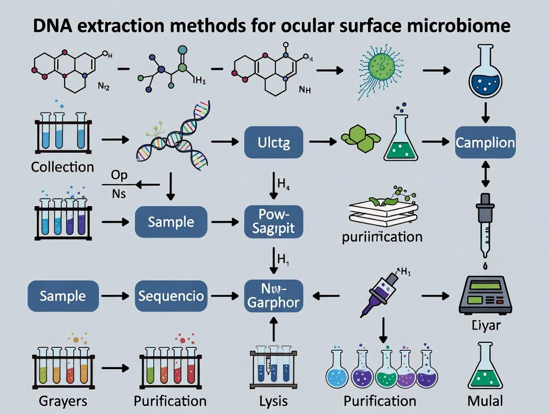
Optimizing DNA Extraction for Low-Biomass Ocular Surface Microbiome: A Guide for Researchers & Drug Developers
This article provides a comprehensive, current guide to DNA extraction methodologies specifically designed for the challenging low-biomass environment of the ocular surface microbiome.
Optimizing DNA Extraction for Low-Biomass Ocular Surface Microbiome: A Guide for Researchers & Drug Developers
Abstract
This article provides a comprehensive, current guide to DNA extraction methodologies specifically designed for the challenging low-biomass environment of the ocular surface microbiome. Targeted at researchers, scientists, and drug development professionals, it covers foundational principles, detailed protocols for common and emerging methods, key optimization strategies to overcome contamination and bias, and a critical comparison of commercial kits and in-house techniques. The review synthesizes best practices to ensure accurate, reproducible microbial profiling, which is crucial for advancing our understanding of ocular surface health, disease mechanisms, and the development of microbiome-targeted therapeutics.
Understanding the Challenge: Why Low-Biomass Ocular Samples Demand Specialized DNA Extraction
Application Notes
The study of the ocular surface microbiome (OSM) presents a paradigm of low-biomass niche research, characterized by low microbial density, high host DNA contamination, and vulnerability to environmental contamination. Accurate definition requires stringent controls and optimized DNA extraction protocols that maximize microbial lysis while minimizing bias and exogenous contamination. This note details methodologies framed within the thesis that robust DNA extraction is the most critical determinant for generating reliable, reproducible OSM profiles for research and therapeutic development.
Key Challenges & Solutions:
- Low Microbial Load: The ocular surface harbors approximately 0.5-2.0 CFU/µl in culture-based studies, with total bacterial DNA estimates often below 1 pg/µl in elution volume.
- High Host-to-Microbial DNA Ratio: Can exceed 10,000:1, necessitating methods that deplete host nucleic acids or selectively enrich microbial DNA.
- Contamination Mitigation: Negative extraction controls (NECs) and sterile collection controls (SCCs) are non-negotiable. Protocols must include steps to subtract contaminating operational taxonomic units (OTUs) present in controls from sample data.
Table 1: Comparison of DNA Extraction Kits for Low-Biomass Ocular Surface Samples
| Kit Name | Mechanical Lysis Step | Host DNA Depletion | Avg. DNA Yield (16S rRNA Gene Copies/µl) | Key Advantage for OSM | Primary Limitation |
|---|---|---|---|---|---|
| PowerSoil Pro Kit | Bead beating (0.1mm garnet beads) | No | 1.2 x 10³ - 5.0 x 10³ | Excellent for Gram-positive bacteria (e.g., Staphylococci) | Co-elution of PCR inhibitors common. |
| NucleoSpin Microbiome | Bead beating + enzymatic lysis | Yes (selective degradation) | 2.5 x 10³ - 8.0 x 10³ | Significantly reduces human DNA background (~50-70% reduction) | Higher cost per sample; potential loss of some Gram-negatives. |
| MasterPure Complete | Proteinase K + shaking | No | 0.8 x 10³ - 3.0 x 10³ | High DNA fragment size; good for shotgun metagenomics | Lower efficiency on tough Gram-positive cell walls. |
| MoBio Ultraclean | Bead beating (0.7mm silica beads) | No | 0.5 x 10³ - 2.0 x 10³ | Designed for low biomass environments; includes carrier RNA | Yield can be highly variable. |
Experimental Protocols
Protocol 1: Standardized Ocular Surface Sample Collection for Microbiome Analysis Objective: To collect microbial biomass from the conjunctival fornix without inducing inflammation or introducing contamination. Materials: Sterile swab (e.g., Puritan HydraFlock), sterile saline (0.9% NaCl), sterile tube with stabilization buffer (e.g., DNA/RNA Shield), NECs, SCCs. Procedure:
- Instill one drop of sterile saline onto the inferior palpebral conjunctiva.
- Gently retract the lower eyelid and rotate a pre-moistened sterile swab along the inferior conjunctival fornix from medial to lateral canthus for 10 seconds per eye.
- Immediately place the swab tip into a sterile tube containing 500 µl of stabilization buffer. Break the shaft to seal the tube.
- Process SCC by exposing a swab to the collection environment without touching the eye.
- Store all samples at -80°C until DNA extraction.
Protocol 2: Optimized DNA Extraction for Ocular Surface Low-Biomass Using a Modified PowerSoil Pro Protocol Objective: To extract total genomic DNA with high efficiency for both Gram-positive and Gram-negative ocular commensals. Materials: PowerSoil Pro Kit, 0.1mm garnet beads, sterile phosphate-buffered saline (PBS), heating block, microcentrifuge, NEC (reagents only). Procedure:
- Thaw sample on ice. Vortex for 1 minute. Aseptically transfer 200 µl of the sample buffer (containing swab eluate) to a PowerSoil Pro bead tube.
- Critical Modification: Add 50 µl of sterile PBS to increase fluid volume for optimal bead beating kinetics.
- Add Solution CD1. Secure tubes horizontally on a vortex adapter.
- Vortex at maximum speed for 15 minutes at 4°C.
- Incubate at 65°C for 10 minutes on a heating block.
- Centrifuge at 10,000 x g for 1 minute. Transfer supernatant to a clean tube.
- Continue with the manufacturer's standard protocol for inhibitor removal and DNA binding/washing (Steps 5-15).
- Elute DNA in 30 µl of Solution CE. Use a low-retention pipette tip.
- Quantify using a high-sensitivity qPCR assay targeting the V4 region of the 16S rRNA gene (e.g., 515F/806R primers) rather than fluorometric assays.
Mandatory Visualization
Title: OSM Research Workflow with Critical Controls
Title: Low-Biomass Challenges & DNA Extraction Solutions
The Scientist's Toolkit: Research Reagent Solutions
Table 2: Essential Materials for Ocular Surface Microbiome DNA Studies
| Item | Function in OSM Research | Key Consideration |
|---|---|---|
| DNA/RNA Shield Stabilization Buffer | Immediately lyses cells and inactivates nucleases, preserving the in situ microbial profile from collection to extraction. | Critical for preventing shifts in community representation during storage. |
| 0.1mm Garnet Beads | Provides aggressive mechanical lysis essential for breaking tough cell walls of ocular Staphylococci and Corynebacteria. | Superior to larger beads for low-biomass, small-cell-volume bacteria. |
| Mock Microbial Community (e.g., ZymoBIOMICS) | Serves as a positive process control to evaluate extraction kit bias, lysis efficiency, and sequencing accuracy. | Allows quantification of protocol-induced taxonomic bias. |
| Molecular Grade Water (PCR Clean) | Used as the negative extraction control (NEC). Identifies reagent- and kit-derived contaminating DNA sequences. | Must be from a dedicated, unopened bottle for each extraction batch. |
| Human DNA Depletion Cocktail | Enzymatically degrades unprotected human DNA (e.g., host epithelial cells) while protecting prokaryotic DNA. | Increases sequencing depth on microbial targets, improving detection sensitivity. |
| Carrier RNA (e.g., Poly-A) | Improves DNA binding to silica membranes during low-input extractions, increasing yield and reproducibility. | Reduces stochastic loss common in sub-nanogram extractions. |
The ocular surface, comprising the cornea and conjunctiva, is a low-biomass environment. Accurate characterization of its resident microbiome is critical for understanding ocular health, diseases like dry eye syndrome and blepharitis, and for developing targeted therapeutics. However, standard DNA extraction methods are confounded by three primary challenges:
- Host DNA Dominance: Human epithelial and immune cell DNA vastly outweighs microbial DNA, obscuring detection of low-abundance bacteria, viruses, and fungi.
- Contamination: Reagents (kits, water, tubes) and collection procedures introduce exogenous microbial DNA, leading to false positives.
- Inhibitor Presence: Tear components (lysozyme, lactoferrin), mucins, and preservatives in clinical samples can inhibit downstream PCR and sequencing.
This application note details protocols and solutions to mitigate these challenges, enabling robust metagenomic analysis.
Table 1: Comparative Performance of Host DNA Depletion Methods
| Method | Principle | Avg. Host DNA Reduction | Microbial DNA Recovery | Key Limitations |
|---|---|---|---|---|
| Selective Lysis | Differential lysis of human cells with mild detergents, followed by enzymatic degradation of released host DNA. | 70-85% | Moderate (30-50% loss) | Incomplete for robust human cells; co-loss of gram-positive bacteria. |
| DNase Treatment | Post-lysis treatment with DNase that selectively degrades eukaryotic DNA (e.g., Benzonase). | 90-95% | Low (High loss of microbial DNA if not protected) | Requires careful optimization to protect intracellular microbial DNA. |
| Methylation-Based Capture (sWGA) | Use of phage polymerases (Φ29) with primers biased against human CpG-methylated DNA. | 99% | Highly Variable (Can favor specific taxa) | Amplification bias; may miss underrepresented genera; cost. |
| Propidium Monoazide (PMA) | Photoactive dye penetrates dead cells, cross-linking DNA to preclude amplification. | N/A (Targets dead cells) | High for viable cells | Does not reduce host DNA from live human cells; requires light source. |
Table 2: Common Inhibitors in Ocular Samples and Neutralization Strategies
| Inhibitor (Source) | Effect on Downstream Assays | Neutralization Method |
|---|---|---|
| Lysozyme (Tears) | Degrades bacterial cell walls, reducing yield. | Addition of inhibitors (e.g., EDTA), rapid processing. |
| Mucins (Mucus) | Binds to DNA, impedes extraction. | Pre-treatment with mucolytic agents (e.g., DTT, N-acetylcysteine). |
| Polysaccharides | Co-precipitate with DNA, inhibit polymerases. | Use of high-salt buffers, specific column-based purification. |
| Heavy Metals | Interfere with enzymatic reactions. | Chelating agents (e.g., Chelex resin). |
Detailed Experimental Protocols
Protocol A: Optimized DNA Extraction with Host Depletion for Conjunctival Swabs
This protocol integrates selective lysis and enzymatic host DNA depletion.
Materials:
- Sterile conjunctival swabs (e.g., polyester-tipped)
- Negative control swab (exposed to air during collection)
- PBS + 0.1% Tween 20
- Lysozyme (20 mg/mL)
- Lysostaphin (for Staphylococci)
- Mutanolysin (for Streptococci)
- Proteinase K
- AL Buffer (Qiagen) or similar lysis buffer
- Benzonase (25 U/µL)
- Commercial DNA purification kit (e.g., Qiagen DNeasy PowerLyzer)
Procedure:
- Sample Elution: Vortex swab in 500 µL PBS-Tween for 1 min. Snap the swab shaft and retain liquid.
- Microbial Enrichment: Centrifuge at 14,000 x g, 4°C for 10 min. Discard supernatant (removes soluble inhibitors).
- Selective Host Lysis: Resuspend pellet in 200 µL of pre-warmed (37°C) TE buffer with 0.1% SDS. Incubate 10 min at 37°C.
- Bacterial Lysis: Add 20 µL lysozyme, 5 µL lysostaphin, 5 µL mutanolysin. Incubate 30 min at 37°C.
- Host DNA Digestion: Add 2 µL Benzonase and 5 µL MgCl2 (25 mM). Incubate 15 min at 37°C.
- Complete Lysis: Add 200 µL AL Buffer and 20 µL Proteinase K. Incubate at 56°C for 30 min.
- DNA Purification: Follow manufacturer's protocol for bead-beating (for gram-positives) and column-based purification. Elute in 30-50 µL.
Protocol B: Systematic Contamination Tracking Workflow
Implements a rigorous negative control strategy.
Procedure:
- Reagent Blank: Include a tube with only extraction reagents processed in parallel.
- Collection Control: Process an unused swab from the same lot opened during patient sampling.
- Sequencing: Sequence all controls (extraction, library prep, PCR) on the same flow cell as samples.
- Bioinformatic Subtraction: Use tools like
decontam(R package) with frequency and prevalence methods to identify and remove contaminant ASVs/OTUs present in controls from sample data.
Visualization Diagrams
Title: Optimized DNA Extraction Workflow for Ocular Microbiome
Title: Key Challenges & Mitigation Strategies in Ocular Microbiome Analysis
The Scientist's Toolkit: Research Reagent Solutions
Table 3: Essential Materials for Low-Biomass Ocular DNA Studies
| Item | Function | Example/Note |
|---|---|---|
| Polyester-tipped Swabs | Sample collection. Minimize DNA binding vs. cotton. | Puritan PurFlock Ultra. |
| DNA/RNA Shield | Immediate sample preservation, stabilizes nucleic acids, inhibits nucleases. | Zymo Research DNA/RNA Shield. |
| PMA Dye | Differentiates viable vs. dead microbial cells via DNA intercalation. | Biotium PMA dye. Requires blue light photolysis device. |
| Phi29 Polymerase Kit | For selective whole genome amplification (sWGA) to enrich microbial DNA. | REPLI-g Single Cell Kit (Qiagen). Risk of amplification bias. |
| Benzonase Nuclease | Degrades linear host DNA post-lysis while protected microbial DNA is intact. | MilliporeSigma. Requires optimization of Mg2+ and incubation time. |
| Mock Microbial Community | Defined mix of microbial genomes. Positive control for extraction efficiency and bias. | ATCC MSA-1000 (10 bacterial strains). |
| UltraPure Water | PCR-grade, sterile water for all reagents to minimize background DNA. | Invitrogen UltraPure. |
| Internal Spike-in DNA | Synthetic, non-native DNA sequence added pre-extraction to quantify loss. | Spike-in controls from ZymoBIOMICS or custom sequences. |
| Mucolytic Agent | Breaks down mucin polymers to release trapped microbes and DNA. | 1,4-Dithiothreitol (DTT) or N-Acetylcysteine. |
| High-Efficiency Library Prep Kit | For constructing sequencing libraries from low-input DNA. | Illumina DNA Prep, or Nextera XT. |
Within the specific challenges of low-biomass ocular surface microbiome research, DNA extraction is the primary gatekeeper of data fidelity. Extraction bias—the differential lysis of microbial cells and preferential recovery of genetic material based on cell wall structure, gram-status, or DNA integrity—systematically distorts microbial community profiles. This bias propagates through all downstream analyses, including 16S rRNA gene sequencing (targeting hypervariable regions) and shotgun metagenomic sequencing. For ocular samples, where biomass is minimal and contaminants are a significant concern, the choice of extraction method fundamentally determines whether observed taxa are biological signals or methodological artifacts. These biases impact diversity metrics (alpha and beta), relative abundance estimates, functional potential inference, and the identification of potential biomarkers for ocular disease or therapeutic response. The following protocols and data are framed to guide researchers in selecting, validating, and controlling for extraction bias in ocular microbiome studies.
Comparative Data on Extraction Bias
Table 1: Impact of Four Commercial Kits on Mock Community Analysis (Low Biomass Simulated) Mock Community: Defined mix of Gram-positive (e.g., *S. aureus, S. epidermidis), Gram-negative (e.g., P. aeruginosa, E. coli), and a yeast (e.g., C. albicans), spiked into a sterile tear-like matrix.*
| Kit Name (Typical Use) | Lysis Principle | Gram+ Bias (Rel. to Expected) | Gram- Bias (Rel. to Expected) | Fungal Bias (Rel. to Expected) | Human DNA % in Low-Biomass Simulant | Mean DNA Yield (pg/µL) |
|---|---|---|---|---|---|---|
| Kit A (Mechanical + Chemical) | Bead-beating, Guanidine salts | +5% to +15% | -10% to -5% | +20% to +30% | 5-15% | 45 |
| Kit B (Enzymatic + Chemical) | Lysozyme, Proteinase K, SDS | -25% to -40% | +20% to +35% | -50% to -70% | 40-70% | 120 |
| Kit C (Mild Chemical Lysis) | AL Buffer, thermal shock | -40% to -60% | +50% to +80% | -80% to -90% | 60-85% | 95 |
| Kit D (Optimized Mechanical) | Intensive bead-beating, Inhibitor removal | -2% to +8% | -5% to +3% | -10% to +5% | 1-10% | 25 |
Table 2: Downstream Analytical Consequences of Bias Analysis of the same low-biomass ocular swab sample extracted with different methods.
| Downstream Metric | Kit A (Mech+Chem) | Kit B (Enzymatic) | Kit C (Mild) | Kit D (Opt. Mech) |
|---|---|---|---|---|
| 16S: Observed Richness | 45 species | 18 species | 12 species | 52 species |
| 16S: Shannon Diversity Index | 2.8 | 1.5 | 0.9 | 3.1 |
| Shotgun: % Microbial Reads | 82% | 30% | 15% | 90% |
| Shotgun: Functional Pathway Coverage | High | Low | Very Low | Highest |
| False Positive Contaminants | Low | Moderate | High | Very Low |
Detailed Experimental Protocols
Protocol 1: Standardized Low-Biomass Ocular Surface Sample Collection Objective: To collect microbiome samples from the conjunctival fornix with minimal contamination.
- Preparation: Instill topical anesthetic (e.g., proparacaine 0.5%) if required by protocol.
- Positioning: Ask the participant to look up.
- Collection: Gently retract the lower eyelid. Using a pre-moistened (with sterile saline) synthetic tipped swab (e.g., FLOQSwab), rub the swab tip along the inferior conjunctival fornix from nasal to temporal, applying gentle pressure and rotating the swab.
- Storage: Immediately place the swab into a sterile, DNA-free, labeled collection tube containing a stabilization buffer (e.g., DNA/RNA Shield). Store at 4°C for <24 hours, then transfer to -80°C.
Protocol 2: Side-by-Side Extraction Bias Assessment Using a Mock Community Objective: To empirically quantify the bias introduced by different DNA extraction methods.
- Mock Community Preparation: Create a defined, low-concentration (10^3-10^4 CFU/mL) microbial mixture in a sterile artificial tear solution. Include Gram-positive (e.g., Staphylococcus epidermidis ATCC 12228), Gram-negative (e.g., Pseudomonas aeruginosa ATCC 27853), and fungal (e.g., Candida albicans SC5314) representatives.
- Spike and Extract: Aliquot 100 µL of the mock community into 10 replicate tubes per extraction method to be tested. Include negative control aliquots (artificial tears only).
- Parallel Extraction: Perform extractions strictly according to each kit's manufacturer protocol. Key variable steps to note: incubation times, temperature, bead-beating intensity/duration, and elution volume.
- Quantification & QC: Quantify total DNA yield using a fluorescence-based assay (e.g., Qubit dsDNA HS Assay). Assess DNA quality via spectrophotometry (A260/A280, A260/A230) and fragment analyzer.
- Downstream Analysis: a. 16S rRNA qPCR: Perform absolute quantification of total bacterial load using universal 16S primers (e.g., 341F/518R). b. Shotgun Sequencing Library Prep: Use equal input DNA masses (e.g., 1ng) from each extract to prepare sequencing libraries (e.g., Illumina DNA Prep). Include unique dual indices. c. Bioinformatic Analysis: Map shotgun reads to the reference genomes of the mock community members. Calculate recovery ratios (Observed/Expected read counts) for each organism.
Protocol 3: Protocol for Optimized, Low-Biomass Ocular Sample Extraction Objective: To maximize microbial DNA recovery while minimizing host DNA and bias.
- Sample Lysis: Transfer the swab or its stabilizing buffer to a PowerBead Pro tube. Add:
- 20 µL of Proteinase K (100 mg/mL)
- 200 µL of pre-heated (70°C) lysis buffer (e.g., from Kit D)
- Mechanical Disruption: Secure tubes in a vortex adapter or bead beater. Process at maximum speed for 10 minutes at room temperature. Pulse-centrifuge briefly.
- Inhibitor Removal: Add 250 µL of inhibitor removal solution. Vortex for 5 minutes. Centrifuge at 13,000 x g for 5 minutes.
- DNA Binding: Transfer the supernatant to a new tube. Add 1.5 volumes of binding buffer and mix. Load onto a silica spin column.
- Washes: Wash the column twice with 700 µL of wash buffer (with ethanol). Centrifuge at full speed for 2 minutes to dry the membrane.
- Elution: Elute DNA in 25-50 µL of pre-heated (55°C) nuclease-free water or TE buffer. Incubate on the column for 2 minutes before centrifugation.
Visualizations
Diagram 1 Title: From Extraction to Downstream Bias
Diagram 2 Title: Optimized Low-Biomass Extraction Workflow
The Scientist's Toolkit: Research Reagent Solutions
Table 3: Essential Materials for Low-Biomass Ocular Microbiome DNA Studies
| Item | Function & Rationale |
|---|---|
| FLOQSwabs (Synthetic Tip) | Minimizes background DNA retention and elutes efficiently compared to cotton. Critical for low biomass. |
| DNA/RNA Shield (or similar) | Immediate nucleic acid stabilization buffer. Inactivates nucleases and preserves microbial community structure at point of collection. |
| PowerBead Pro Tubes | Contain a mixture of ceramic and silica beads for rigorous mechanical lysis of tough Gram-positive and fungal cell walls. |
| Proteinase K (Molecular Grade) | Digests proteins and peptides, crucial for breaking down host cells and microbial proteins, improving yield. |
| Guanidine Thiocyanate (GuSCN) | Chaotropic agent that denatures proteins, inactivates nucleases, and promotes binding of DNA to silica. |
| Silica Spin Columns | Selective binding of DNA based on size and salt conditions, allowing for purification from inhibitors common in ocular samples (e.g., salts, mucins). |
| Qubit dsDNA HS Assay Kit | Fluorometric quantification essential for accurately measuring the very low concentrations of DNA from ocular samples; superior to UV spectrophotometry for purity. |
| Mock Microbial Community (Even/Staggered) | Defined mix of known microbes. The gold standard for quantifying extraction bias and benchmarking kit performance. |
| Human DNA Depletion Kit (e.g., NEBNext Microbiome) | Optional post-extraction step to enrich microbial sequences by selectively removing host-derived DNA, increasing sequencing depth on target. |
In low-biomass ocular surface microbiome research (e.g., conjunctiva, cornea), the DNA extraction process is the critical gatekeeper. The inherent challenges—extremely low microbial load, high human: bacterial DNA ratio, and delicate microbial communities—mean that extraction performance directly dictates downstream sequencing reliability. This note details the application and protocols for assessing the four foundational pillars of nucleic acid quality, framed within the specific demands of ocular surface studies.
Quantitative Benchmarks for Ocular Surface DNA
The following table summarizes target benchmarks and typical challenges for low-biomass ocular samples.
Table 1: Foundational Requirement Benchmarks & Challenges for Ocular Surface DNA
| Requirement | Target Benchmark | Measurement Method | Ocular Surface Specific Challenge |
|---|---|---|---|
| Yield | >0.5 ng/µL (total >5 ng) | Fluorometry (Qubit dsDNA HS) | Swab collection yields picogram to low nanogram totals. Inhibitor carryover from tears/lysozyme. |
| Purity | A260/A280: 1.8-2.0 A260/A230: >2.0 | Spectrophotometry (NanoDrop) | Low yield amplifies contamination signals. Keratins, salts, and phenol from swabs affect ratios. |
| Integrity | DIN/DQN >7.0 (if applicable) | Fragment Analyzer, TapeStation | Not always measured for microbial 16S; critical for shotgun metagenomics. Human DNA often intact. |
| Representativeness | Maximum microbial diversity recovery; Minimal bias. | qPCR for 16S rRNA genes vs. total DNA; Community analysis controls. | Host DNA depletion can co-deplete Gram+ bacteria. Lysis bias against hardy organisms (e.g., Corynebacterium). |
Detailed Experimental Protocols
Protocol: Integrated Extraction & QC for Low-Biomass Ocular Swabs
Sample: Flocked swab of inferior fornix, stored in 200 µL of DNA/RNA Shield. Objective: Extract total DNA while maximizing microbial yield and representativeness.
Materials (The Scientist's Toolkit):
| Item | Function & Rationale |
|---|---|
| Flocked nylon swab | Maximizes cell elution into buffer compared to wound fiber swabs. |
| DNA/RNA Shield (e.g., Zymo) | Preserves nucleic acids immediately, inhibits nucleases. |
| Enzymatic Lysis Cocktail (Lysozyme, Mutanolysin, Lysostaphin) | Breaks down diverse bacterial cell walls (Gram+, Gram-) critical for representativeness. |
| Mechanical Lysis Beads (0.1mm zirconia/silica) | Enhances disruption of tough bacterial and fungal cells. |
| Kit with Inhibitor Removal (e.g., Qiagen PowerSoil Pro, Zymo BIOMICS) | Specifically designed for difficult samples and inhibitor removal. |
| Carrier RNA (e.g., for Qiagen kits) | Improves nucleic acid binding to silica in low-concentration samples, boosting yield. |
| qPCR reagents (16S rRNA gene primers, SYBR Green) | Quantifies bacterial load independently of host DNA. |
Workflow:
- Sample Preparation: Vortex swab vial for 1 min. Transfer all liquid to a sterile tube. Centrifuge swab at 10,000 x g for 2 min to collect residual fluid; pool.
- Enzymatic Pre-treatment: Add 25 µL of freshly prepared lysis cocktail (20 mg/mL lysozyme, 5 U/µL mutanolysin, 1 U/µL lysostaphin in TE buffer). Incubate at 37°C for 60 min with gentle agitation.
- Mechanical Lysis: Transfer to a bead-beating tube. Add 0.1mm beads. Securely cap and bead-beat at 5.5 m/s for 45 sec (e.g., FastPrep-24).
- Binding & Wash: Follow manufacturer's protocol for the chosen inhibitor-removal kit. Critical Step: Add 2 µL of carrier RNA (1 µg/µL) to the binding solution if yield is expected to be <10 ng total.
- Elution: Elute in 20-30 µL of nuclease-free water or TE buffer. Do not heat elution buffer (>65°C) prior to use if carrier RNA was added.
- QC Analysis:
- Yield & Purity: Use 1 µL for Qubit (HS assay) and 1 µL for NanoDrop. Record concentrations and ratios.
- Integrity: If sufficient yield (>2 ng/µL), run 1 µL on High Sensitivity Genomic DNA TapeStation assay.
- Representativeness (qPCR): Perform triplicate qPCR with universal 16S rRNA gene primers (e.g., 341F/534R) against a standard curve of a known bacterial genomic DNA. Calculate 16S copy number/µL.
Protocol: Assessing Lysis Efficiency and Bias (Spike-in Control)
Objective: Quantify extraction bias by using a known quantity of an exotic, hard-to-lyse bacterial cell (e.g., Bacillus subtilis spores) as an internal spike-in control. Procedure:
- Prior to extraction, spike sample with 10^4 cells of an exogenous control (e.g., Pseudomonas chlororaphis, not found on human ocular surface).
- Proceed with extraction as in 3.1.
- Perform species-specific qPCR for the spike-in organism.
- Calculate recovery efficiency: (Observed spike-in copies / Expected spike-in copies) x 100%. Efficiency <50% indicates significant lysis bias.
Data Interpretation and Pathway Visualization
Title: Foundational DNA QC Metrics for Ocular Microbiome Research
Title: Optimized DNA Extraction Workflow for Low-Biomass Samples
Ethical and Practical Considerations in Sample Collection (Swabs, Brushes, Lavage)
Within the context of a broader thesis on DNA extraction methods for low biomass ocular surface microbiome research, sample collection represents a critical initial step. The choice of collection method directly impacts downstream DNA yield, microbial community representation, and the validity of subsequent analyses. This document outlines the ethical framework and provides detailed protocols for common ocular surface collection techniques.
Ethical Framework for Ocular Surface Sampling
All sampling must be conducted under an Institutional Review Board (IRB) or Ethics Committee-approved protocol. Key principles include:
- Informed Consent: Participants must be fully informed of the procedure's purpose, risks (minimal but including potential for minor discomfort or transient corneal abrasion), benefits, and data usage.
- Minimization of Risk: The least invasive effective method should be chosen. Proper training in technique is mandatory to prevent injury.
- Privacy and Confidentiality: Participant data must be de-identified and stored securely, with genomic data treated as sensitive information.
Comparative Analysis of Collection Methods
The following table summarizes key quantitative data from recent studies on ocular surface collection methods for low-biomass microbiome analysis.
Table 1: Comparison of Ocular Surface Sample Collection Methods
| Method | Typical DNA Yield (Range) | Key Microbial Taxa Recovered (Representative) | Primary Advantages | Primary Limitations & Practical Considerations |
|---|---|---|---|---|
| Swab (e.g., polyester, rayon) | 0.1 - 2.5 ng/µL | Corynebacterium, Propionibacterium, Staphylococcus, Streptococcus | Non-invasive, low-cost, rapid, well-tolerated. Easy to standardize. | Low biomass yield. Material can inhibit PCR. Risk of contamination from skin/lids. Variable pressure application. |
| Micro-brush (e.g., cytobrush) | 0.5 - 4.0 ng/µL | As above, with potential for deeper epithelial cells. | Can yield higher cellular material than some swabs. Standardized surface area. | Slightly more invasive. Requires careful, gentle rotation to avoid injury. Cost higher than swabs. |
| Conjunctival Lavage/Wash | 0.01 - 1.0 ng/µL (in large vol.) | Often higher proportion of environmental/transient taxa. | Samples a larger surface area. Less risk of cross-contamination from lid margin. | Very dilute sample, requiring concentration. Significant dilution effect. More cumbersome for subject. Risk of washout to nasolacrimal duct. |
| Filter Paper Imprint | 0.05 - 1.0 ng/µL | Similar to swab. | Minimal equipment needed. Can be stored dry. | Inconsistent pressure and contact area. Very low biomass. |
| Corneal Epithelial Scrape | 2.0 - 10.0 ng/µL (when clinically indicated) | Full spectrum of adherent microbiota. | High yield from specific corneal region. | Invasive: Only permissible during clinically necessary procedures (e.g., for infection). Not for healthy volunteer research. |
Detailed Experimental Protocols
Protocol 1: Sterile Synthetic Tip Swab Collection
Objective: To consistently collect microbiome samples from the inferior fornix and bulbar conjunctiva. Materials: Pre-sterilized synthetic (e.g., polyester) swabs, sterile saline (0.9% NaCl) vial for moistening (optional), sterile scissors or forceps, labeled collection tube with DNA stabilization buffer or lysis buffer. Procedure:
- Don appropriate personal protective equipment.
- Moistening (Optional): Aseptically remove swab from packaging. Briefly dip tip into sterile saline to moisten. Gently tap to remove excess droplet.
- Positioning: Instruct the participant to look up. Gently pull down the lower eyelid to expose the inferior palpebral conjunctiva and fornix.
- Swabbing: Gently roll the swab across the conjunctival surface from the nasal to the temporal side, applying minimal pressure. Avoid contact with eyelashes or skin.
- Transfer: Immediately place the swab tip into the prepared collection tube.
- Processing: Using sterile scissors, cut the swab shaft so the tip remains submerged in buffer. Close tube securely.
- Storage: Store tubes at -80°C immediately or within 4 hours if using stabilization buffer.
Protocol 2: Micro-Brush Collection
Objective: To collect a standardized area of ocular surface epithelium. Materials: Sterile single-use micro-brushes (e.g., cytobrush), slit lamp or stable chin rest, collection tube as in Protocol 1. Procedure:
- Prepare materials and position participant at the slit lamp or with head stabilized.
- Instruct participant to look in a specified direction (e.g., up and out) to expose the target bulbar conjunctiva.
- Under direct visualization, gently touch the micro-brush to the conjunctiva and rotate the brush handle 180 degrees once.
- Immediately retract the brush and place it into the collection tube. Snap the handle at the score mark to seal the tube.
- Store at -80°C.
Protocol 3: Membrane Filtration-Concentrated Lavage
Objective: To collect microbiome from a broad ocular surface area via lavage with sample concentration. Materials: Sterile saline (1-5 mL syringe), sterile collection vial, 0.22 µm polycarbonate membrane filter unit, vacuum manifold, sterile forceps. Procedure:
- Instill 200-500 µL of sterile saline onto the ocular surface while the participant's eye is gently held open.
- Have the participant blink several times, then collect the fluid from the lateral canthus using a sterile micro-pipette or by tilting the head into the collection vial.
- Immediate Processing: Assemble the sterile filter unit on the vacuum manifold. Transfer the lavage fluid to the unit.
- Apply gentle vacuum to draw fluid through the membrane, trapping microorganisms and cells.
- Using sterile forceps, aseptically transfer the membrane to a bead-beating tube containing lysis buffer.
- Proceed directly to DNA extraction or store tube at -80°C.
The Scientist's Toolkit: Research Reagent Solutions
Table 2: Essential Materials for Ocular Surface Microbiome Collection & Stabilization
| Item | Function in Research | Example Product/Brand |
|---|---|---|
| Sterile Polyester-tipped Swabs | Low-binding, PCR-inhibitor free material for gentle sample collection. | Puritan HydraFlock, BD Falcon |
| Single-use Micro-brushes | Standardized surface area collection for increased cellular yield. | Cytosoft cytobrush, Copan FLOQSwabs (nylon) |
| DNA/RNA Shield Stabilization Buffer | Preserves nucleic acids at ambient temperature for transport/storage, inactivating nucleases and pathogens. | Zymo Research DNA/RNA Shield, Norgen Biotek Preservative Buffer |
| PBS, Molecular Biology Grade | Sterile, nuclease-free solution for moistening swabs or performing lavage. | Thermo Fisher, Corning |
| Low-Binding Microcentrifuge Tubes | Prevents adhesion of low-biomass material to tube walls during processing. | Eppendorf LoBind, Axygen Maxymum Recovery |
| 0.22 µm Polycarbonate Filters | For concentration of dilute lavage samples; minimal DNA binding. | Merck Millipore GTTP, Whatman Nuclepore |
| Bead-beating Tubes with Lysis Matrix | Mechanical disruption of tough bacterial cell walls (e.g., Gram-positive) for complete DNA extraction. | MP Biomedicals FastPrep tubes, Qiagen PowerBead Tubes |
Workflow and Pathway Visualizations
Title: Ocular Microbiome Study Workflow
Title: Collection Method Decision Pathway
Step-by-Step Protocols: From Sample Lysis to Elution for Ocular Microbiome DNA
This document, framed within a broader thesis on DNA extraction for low-biomass ocular surface microbiome research, details optimized mechanical lysis protocols. Successful metagenomic analysis hinges on efficient, unbiased cell disruption to release DNA, particularly from resilient ocular pathogens like Staphylococcus epidermidis, Propionibacterium acnes, Corynebacterium spp., and fungi. Mechanical lysis via bead beating is critical but must be optimized to balance yield with DNA shearing, especially for low-input samples.
Key Research Reagent Solutions & Materials
Table 1: Essential Research Toolkit for Bead Beating Optimization
| Item | Function & Rationale |
|---|---|
| Silica/Zirconia Beads (0.1mm) | Primary lysis agents for rigid Gram-positive bacteria and fungal spores. Small size increases collision frequency. |
| Garnet Beads (0.5mm) | Alternative for tougher cell walls; provides heterogeneous grinding action. |
| Lysis Buffer (e.g., with GuHCl/SDS) | Disrupts membranes post-mechanical fracture, inhibits nucleases, and stabilizes released DNA. |
| Inhibitor Removal Technology (IRT) Beads | Magnetic or silica-based beads to co-purify and sequester PCR inhibitors (e.g., lysozyme, mucins) common in ocular samples. |
| Bench-top Vortex Adapter | Provides consistent, high-speed horizontal agitation for reproducible lysis across multiple samples. |
| High-Throughput Bead Mill Homogenizer | For standardized, parallel processing with precise control over time and frequency. |
| Microcentrifuge Tubes, 2mL (Tough-Tube style) | Withstand high mechanical stress during beating to prevent sample loss and aerosol generation. |
| Carrier RNA (e.g., from MS2 phage) | Added to lysis buffer to improve nucleic acid recovery by preventing adsorption to surfaces in low-biomass samples. |
| Proteinase K | Enzyme used post-bead beating to digest proteins and degrade nucleases, enhancing yield and purity. |
Quantitative Optimization Data
Table 2: Bead Beating Parameter Optimization for Ocular Pathogen Lysis
| Pathogen Type | Bead Material & Size | Beating Duration | Beating Speed | DNA Yield (ng/µL) | Fragment Size (bp) | Inhibition Rate (qPCR Ct Shift) |
|---|---|---|---|---|---|---|
| S. epidermidis (Gram+) | Zirconia, 0.1mm | 3 x 45s cycles | 6.5 m/s | 12.5 ± 1.8 | 5,000-10,000 | 0.5 |
| P. acnes (Anaerobic Gram+) | Silica, 0.1mm | 2 x 60s cycles | 5.0 m/s | 8.2 ± 1.2 | 8,000-15,000 | 0.3 |
| C. albicans (Fungal) | Zirconia, 0.1mm & 0.5mm mix | 1 x 90s cycle | 7.0 m/s | 15.1 ± 2.5 | 3,000-7,000 | 1.2 |
| Mixed Ocular Community | Garnet, 0.5mm | 3 x 30s cycles | 6.0 m/s | 18.7 ± 3.1* | 2,000-8,000 | 0.8 |
*Represents total community DNA yield. Protocols include a 5-minute Proteinase K (20 mg/mL) digestion step post-bead beating. All protocols use a 60-second rest on ice between cycles to prevent overheating.
Table 3: Impact of Lysis Adjuncts on Low-Biomass Swab Eluate Recovery
| Adjunct in Lysis Buffer | Concentration | Mean Yield Increase (%) | Inhibition Reduction (ΔCt) |
|---|---|---|---|
| Carrier RNA | 1 µg/µL | +45% | -0.7 |
| IRT Beads (co-processing) | 10 µL bead slurry | +22% | -2.1 |
| DTT (for mucus disruption) | 40 mM | +15% | +0.5* |
| Bovine Serum Albumin (BSA) | 0.1% w/v | +10% | -0.4 |
*DTT can increase inhibition if not thoroughly removed; use with optimized clean-up.
Detailed Experimental Protocols
Protocol 4.1: Optimized Bead Beating for Rigid Ocular Pathogens
Objective: To maximally lyse rigid Gram-positive bacteria and fungi from a low-biomass ocular swab sample while preserving DNA integrity for downstream NGS.
Materials:
- Sample: Dry or eluted ocular swab in 100µL sterile PBS.
- Lysis Buffer: 500µL of buffer ATL (Qiagen) or equivalent (GuHCl-based), supplemented with 1µg/µL Carrier RNA.
- Beads: ~100mg of 0.1mm zirconia/silica beads in a 2mL reinforced tube.
- Proteinase K (20 mg/mL).
- Bench-top vortex with tube adapter or bead mill homogenizer (e.g., MagNA Lyser, TissueLyser II).
- Microcentrifuge.
Procedure:
- Sample Preparation: Transfer the entire 100µL sample eluate into the 2mL bead tube containing lysis buffer. Add 20µL of Proteinase K. Pipette mix.
- Primary Bead Beating: Secure tubes in a pre-chilled (4°C) bead mill homogenizer. Process at 6.5 m/s for 3 cycles of 45 seconds each, with a 60-second pause on ice between cycles.
- Incubation: Incubate the lysate at 56°C for 10 minutes to allow Proteinase K digestion.
- Bead Separation: Centrifuge tubes at 14,000 x g for 1 minute to pellet beads and debris.
- Supernatant Transfer: Carefully transfer up to 500µL of the clarified supernatant to a new 1.5mL tube. Avoid transferring any beads.
- Inhibitor Removal: Add 10µL of IRT bead slurry to the supernatant. Vortex briefly and incubate at room temp for 5 minutes. Pellet beads magnetically or by brief centrifugation and transfer cleaned lysate to a new tube.
- Proceed to standard DNA purification (e.g., column-based, SPRI beads).
Protocol 4.2: Validation via qPCR and Fragment Analysis
Objective: To quantify lysis efficiency and assess DNA fragmentation.
Part A: Quantitative PCR (qPCR) for Bacterial Load
- Target: Amplify a ~150bp region of the 16S rRNA gene (e.g., V4).
- Reaction Setup: Use a master mix (e.g., SYBR Green), 2µL of 1:10 diluted template DNA, in 20µL reactions.
- Cycling Conditions: 95°C for 3 min; 40 cycles of 95°C for 15s, 60°C for 60s.
- Analysis: Compare Cycle Threshold (Ct) values to a standard curve of known genomic DNA. A lower Ct from the same input sample indicates more efficient lysis.
Part B: DNA Fragment Analysis (TapeStation/Bioanalyzer)
- Sample Prep: Use 1µL of extracted DNA per well on a High Sensitivity DNA assay chip.
- Run: According to manufacturer's instructions.
- Analysis: Assess the fragment distribution profile. Optimal bead beating shows a broad smear centered >2000bp, not a sharp peak <500bp.
Visualized Workflows & Relationships
Diagram 1: Ocular Microbiome DNA Extraction Workflow (100 chars)
Diagram 2: Bead Beating Optimization Trade-Off (100 chars)
Application Notes
Within ocular surface microbiome research, DNA extraction from low-biomass samples presents a unique challenge. The delicate balance between achieving sufficient cell lysis for adequate DNA yield and preserving DNA integrity for downstream analyses (e.g., 16S rRNA sequencing, shotgun metagenomics) is critical. Enzymatic lysis offers gentle, targeted degradation of cell walls, while chemical lysis provides robust, rapid disruption but risks DNA shearing. The optimal strategy often involves a synergistic combination tailored to the diverse microbial community (bacteria, fungi, viruses) and scant sample volume typical of corneal swabs or conjunctival scrapings. Contaminant removal and inhibition mitigation are paramount considerations.
Protocols for Ocular Surface Microbiome DNA Extraction
Protocol 1: Enzymatic Lysis (Gram-Positive & Fungal Bias)
This protocol is optimized for breaking tough cell walls prevalent in low-biomass ocular samples.
- Sample Resuspension: Vortex the clinical swab in 200 µL of sterile, molecular-grade phosphate-buffered saline (PBS) in a 2 mL Lysing Matrix E tube.
- Enzymatic Treatment: Add:
- 20 µL of lysozyme (50 mg/mL)
- 10 µL of lysostaphin (1 mg/mL) [for Staphylococci]
- 15 µL of mutanolysin (5 U/µL) [for Streptococci]
- 25 µL of lyticase (10 U/µL) [for fungal cells]
- Incubation: Incubate at 37°C for 60 minutes with gentle agitation (300 rpm).
- Proteinase K & SDS Addition: Add 20 µL of Proteinase K (20 mg/mL) and 40 µL of 10% SDS. Mix by inversion.
- Secondary Incubation: Incubate at 56°C for 30 minutes.
- Proceed to Purification: The lysate is now ready for standard phenol-chloroform or silica-membrane purification.
Protocol 2: Chemical Lysis (Broad-Spectrum, Rapid)
Designed for maximal, rapid lysis with awareness of potential DNA shear.
- Sample Preparation: Place the swab head directly into a tube containing 180 µL of ATL buffer from the DNeasy PowerLyzer Kit.
- Bead Beating: Add 0.1 mm glass beads. Homogenize in a bead mill for 2 x 45 seconds at 4.5 m/s, with a 2-minute pause on ice between cycles.
- Guanidine Thiocyanate Addition: Add 200 µL of a buffered guanidine thiocyanate solution (4 M final concentration) to denature proteins and inhibit nucleases.
- Detergent Boost: Add 40 µL of 10% Sarkosyl (N-Lauroylsarcosine). Vortex thoroughly.
- Heat Lysis: Incubate at 70°C for 10 minutes.
- Cool & Clarify: Centrifuge at 13,000 x g for 1 minute. Transfer supernatant to a new tube.
- Proceed to Purification: Combine supernatant with an equal volume of binding buffer for column-based purification.
Protocol 3: Hybrid Enzymatic-Chemical Lysis (Recommended for Maximal Yield/Integrity)
This integrated protocol balances efficiency and integrity for optimal NGS results.
- Perform Protocol 1, Steps 1-3 (Enzymatic Treatment).
- Chemical Lysis Integration: Without removing the enzymatic mix, add:
- 200 µL of AL buffer (Qiagen) or equivalent guanidine HCl-based lysis buffer.
- 20 µL of Proteinase K (20 mg/mL).
- Vortex & Incubate: Vortex vigorously for 15 seconds. Incubate at 56°C for 30 minutes.
- Bead Beating (Optional but Recommended for Diversity): Add 0.1 mm glass beads. Perform a short bead-beating step: 1 x 30 seconds at 4.0 m/s. Place immediately on ice.
- Inhibition Removal: Add 5 µL of Carrier RNA (1 µg/µL) to the lysate to improve low-DNA yield recovery. Add 200 µL of absolute ethanol and mix by pulse-vortexing.
- Purification: Transfer the entire mixture to a silica spin column and proceed with wash steps per manufacturer's instructions. Elute in 30-50 µL of TE buffer or nuclease-free water.
Table 1: Comparative Performance of Lysis Methods on Synthetic Low-Biomask Ocular Community
| Lysis Method | Avg. DNA Yield (ng) | DNA Fragment Size (avg. bp) | 16S α-Diversity (Shannon Index) | Inhibition Rate (qPCR Delay) | Process Time (min) |
|---|---|---|---|---|---|
| Enzymatic Only | 1.5 ± 0.4 | >15,000 | 2.8 ± 0.3 | Low | 120 |
| Chemical Only | 2.3 ± 0.6 | 3,000 - 5,000 | 2.1 ± 0.4 | High | 25 |
| Hybrid (Enz+Chem) | 3.1 ± 0.5 | 8,000 - 12,000 | 3.2 ± 0.2 | Medium | 90 |
| Commercial Kit (Ref) | 1.8 ± 0.3 | 10,000 - 15,000 | 2.9 ± 0.3 | Low | 65 |
Table 2: Reagent Solutions for Ocular Surface Microbiome Lysis
| Research Reagent Solution | Function in Lysis | Key Consideration for Low-Biomass |
|---|---|---|
| Lysing Matrix E Tubes | Contains a mixture of silica/zirconia beads for mechanical disruption of tough cell walls. | Essential for breaking Gram-positive and fungal cells; minimizes DNA shear with optimized bead sizes. |
| Lysozyme & Specialty Enzymes | Hydrolyzes peptidoglycan in bacterial cell walls. | Cocktail (lysozyme, lysostaphin, mutanolysin) is critical for diverse ocular bacteria. Must be molecular grade. |
| Lyticase | Degrades β-glucans in fungal cell walls. | Important for capturing fungal elements (e.g., Candida, Aspergillus) on the ocular surface. |
| Guanidine Thiocyanate / HCl | Chaotropic agent that denatures proteins, inhibits RNases/DNases, and aids nucleic acid binding to silica. | Primary chemical lysing agent; crucial for immediate nuclease inhibition in complex samples. |
| Sarkosyl (N-Lauroylsarcosine) | Anionic detergent that solubilizes membranes and disrupts protein complexes. | More effective than SDS on difficult membranes and less inhibitory in downstream steps. |
| Carrier RNA | Co-precipitates with and "carries" minute amounts of DNA through purification, increasing yield. | Vital for low-biomass recovery. Prevents adsorption losses to tube walls and columns. |
| Inhibitor Removal Technology (IRT) | Specific resins or buffers to sequester humic acids, ionic detergents, and ocular pigments. | Critical for samples containing trace amounts of melanin or host debris that inhibit PCR. |
Visualizations
Lysis Method Decision Pathway
Hybrid Lysis Workflow for Ocular Samples
Within low biomass ocular surface microbiome research, DNA extraction efficiency is a critical determinant of downstream analytical success. The paucity of microbial biomass, combined with high host DNA background and potential inhibitory substances from tear fluid, presents a unique challenge. This application note provides a detailed comparative analysis and protocols for three commercial kits specifically designed or adapted for challenging microbiome samples: the QIAamp DNA Microbiome Kit, the DNeasy PowerSoil Pro Kit, and the NEBNext Microbiome DNA Enrichment Kit.
Core Challenges in Ocular Surface DNA Extraction
Low total microbial load, variable lysis efficiency across diverse taxa (e.g., Gram-positive bacteria on the conjunctiva), and contamination risks from collection swabs or reagents are primary concerns. The ideal protocol maximizes microbial DNA yield, minimizes host DNA, and produces inhibitor-free, amplifiable DNA suitable for 16S rRNA gene sequencing and shotgun metagenomics.
The following table summarizes key performance metrics based on current literature and manufacturer data, contextualized for low-biomass ocular samples.
Table 1: Comparative Analysis of Microbiome DNA Extraction Kits
| Feature | QIAamp DNA Microbiome Kit | DNeasy PowerSoil Pro Kit | NEBNext Microbiome DNA Enrichment Kit |
|---|---|---|---|
| Primary Mechanism | Enzymatic & chemical lysis followed by selective host DNA depletion via methyl-CpG binding. | Mechanical (bead-beating) and chemical lysis optimized for tough-to-lyse cells. | Post-extraction enzymatic depletion of host (human) DNA via differential methylation. |
| Input Sample Type | Swabs, body fluids, tissues. | Soil, stool, biofilms, swabs (high inhibitors). | Purified DNA from any extraction method. |
| Host DNA Depletion | Integrated: Pre-extraction binding and removal of methylated host DNA. | Not a primary feature; focuses on total microbial DNA yield. | Specialized: Post-extraction depletion of 5mC-methylated eukaryotic DNA. |
| Hands-on Time | ~1.5 hours | ~30 minutes | ~2 hours (post-DNA extraction) |
| Total Processing Time | 4-5 hours | 1-1.5 hours | 3.5-4 hours (including incubation) |
| Optimal for Ocular Low-Biomass? | High suitability due to integrated host depletion. | High suitability for robust lysis, but co-extracts host DNA. | High suitability as a complementary step to enrich microbial DNA from any extract. |
| Typical Microbial Yield* (16S copies/µl) | 10^2 - 10^4 | 10^3 - 10^5 | Enrichment fold: 5-50x (depends on input) |
| Inhibitor Removal | Moderate (spin-column based) | High (PowerBead technology with inhibitor removal solution) | Not applicable (input is purified DNA). |
*Yields are highly variable and depend on sample biomass; ocular surface yields are typically at the lower end of these ranges.
Detailed Experimental Protocols
Protocol 1: Ocular Surface Sampling & DNA Extraction with QIAamp DNA Microbiome Kit
For conjunctival or corneal swab/brush samples collected in sterile saline or collection buffer.
Materials:
- Sterile corneal sponge or polyester-tipped swab
- Sterile phosphate-buffered saline (PBS)
- Microcentrifuge tubes (1.5 mL, DNA-free)
- QIAamp DNA Microbiome Kit (Qiagen, Cat No. 51704)
- Thermal shaker/heating block
- Ethanol (96-100%)
- Nuclease-free water
Procedure:
- Sample Collection: Gently sample the inferior fornix or corneal surface with a pre-moistened swab. Immediately place the swab head into a 1.5 mL tube containing 200 µL of Microbial Lysis Buffer (MLB).
- Transport/Storage: Vortex briefly and store at -80°C until processing.
- Host DNA Depletion & Lysis:
- Thaw sample on ice. Incubate at 56°C for 30 min in a thermal shaker (900 rpm).
- Add 15 µL of Proteinase K and 4 µL of Carrier RNA to the sample. Mix by vortexing.
- Add 200 µL of Lysis Solution MB, mix, and incubate at 56°C for 30 min with shaking.
- Briefly centrifuge to collect condensation.
- Binding & Washing:
- Add 350 µL of ethanol (96-100%) to the lysate. Mix by pipetting.
- Apply the entire mixture to a QIAamp UCP Mini column. Centrifuge at 17,000 x g for 1 min. Discard flow-through.
- Add 500 µL of Buffer AW1. Centrifuge at 17,000 x g for 1 min. Discard flow-through.
- Add 500 µL of Buffer AW2. Centrifuge at 17,000 x g for 1 min. Discard flow-through.
- Centrifuge at 17,000 x g for 2 min to dry the membrane.
- Elution: Place column in a clean 1.5 mL tube. Apply 30-50 µL of ATE buffer (nuclease-free water preferred for low biomass) to the center of the membrane. Incubate at room temperature for 3-5 min. Centrifuge at 17,000 x g for 1 min to elute DNA. Store at -80°C.
Protocol 2: Mechanical Lysis-Focused Extraction with DNeasy PowerSoil Pro Kit
Ideal for ocular samples where robust lysis of diverse communities (e.g., including potential Gram-positives) is paramount.
Materials:
- DNeasy PowerSoil Pro Kit (Qiagen, Cat No. 47014)
- Vortex adapter for 2 mL tubes
- Microcentrifuge
- Bead-beater (optional, for enhanced lysis)
Procedure:
- Sample Preparation: Place the ocular swab directly into a PowerBead Pro Tube provided. Add 200 µL of sterile PBS used for collection if needed to ensure the swab is moist.
- Lysis:
- Add 60 µL of Solution CD1 to the PowerBead Pro Tube.
- Secure the tube horizontally on a vortex adapter. Vortex at maximum speed for 10-15 minutes. Alternatively, use a bead-beater for 2 x 1 min cycles with cooling on ice between cycles.
- Centrifuge the tube at 15,000 x g for 1 minute at room temperature.
- Inhibitor Removal & Binding:
- Transfer up to 300 µL of supernatant to a clean 2 mL collection tube.
- Add 100 µL of Solution CD2. Vortex for 5 seconds. Incubate at 4°C for 5 minutes.
- Centrifuge at 15,000 x g for 1 minute.
- Transfer up to 300 µL of supernatant to a new 2 mL tube, avoiding the pellet.
- Add 450 µL of Solution CD3 and 50 µL of EDR Solution. Vortex for 5 seconds.
- Column Purification:
- Load 700 µL of the mixture onto an MB Spin Column and centrifuge at 15,000 x g for 1 min. Discard flow-through. Repeat with the remaining mixture.
- Add 500 µL of Solution EA. Centrifuge at 15,000 x g for 1 min. Discard flow-through.
- Add 500 µL of Solution EB. Centrifuge at 15,000 x g for 1 min. Discard flow-through.
- Centrifuge at 15,000 x g for 2 min to dry.
- Elution: Transfer column to a clean 1.5 mL tube. Apply 30-50 µL of Solution C6 (10 mM Tris, pH 8.5) to the membrane center. Incubate for 5 min. Centrifuge at 15,000 x g for 1 min. Store at -80°C.
Protocol 3: Post-Extraction Host DNA Depletion with NEBNext Microbiome DNA Enrichment Kit
To be used following a primary extraction (e.g., from Protocol 1 or 2) to further enhance microbial signal.
Materials:
- NEBNext Microbiome DNA Enrichment Kit (NEB, Cat No. E2612S/L)
- Magnetic rack for PCR tubes
- Thermal cycler or heating block
- Nuclease-free water
Procedure:
- DNA Input & Denaturation: Dilute 5-50 ng of total extracted DNA (host + microbial) to 25 µL with nuclease-free water in a PCR tube. Note: Input mass is often limiting for ocular samples; use maximum available eluate volume.
- MBD2-Fc Binding Reaction:
- Add 5 µL of 10X Bind Buffer 1 and 20 µL of MBD2-Fc Protein to the DNA. Mix thoroughly by pipetting.
- Incubate at room temperature for 10 minutes.
- Magnetic Bead Capture:
- Add 30 µL of well-resuspended Magnetic Beads to the reaction. Mix by gentle pipetting.
- Incubate at room temperature for 5 minutes.
- Place the tube on a magnetic rack until the supernatant is clear (2-5 minutes). Carefully transfer the supernatant (enriched microbial DNA) to a new PCR tube.
- Bead Wash (Optional Stringency):
- For high-host DNA samples, wash beads with 100 µL of 1X Bind Buffer 1 while on the magnet, then discard wash. Combine this wash with the initial supernatant for maximum microbial recovery in low-biomass contexts.
- Concentration & Clean-up: Purify the combined supernatant/enriched DNA using a standard ethanol precipitation or a clean-up kit (e.g., AMPure XP beads) to remove salts and concentrate the DNA. Elute in 20 µL.
Visualized Workflows
Host Depletion & Lysis Workflow (QIAamp)
Mechanical Lysis & Purification Workflow (PowerSoil)
Post-Extraction Host DNA Depletion (NEBNext)
The Scientist's Toolkit: Essential Research Reagents & Materials
Table 2: Key Reagents and Materials for Ocular Microbiome DNA Studies
| Item | Function/Benefit | Example/Catalog |
|---|---|---|
| Polyester-tipped Swabs | Minimize background DNA; no inhibitory wood/glue. | Puritan 25-806 1PD |
| DNA/RNA Shield Solution | Preserves sample integrity at room temp; inactivates nucleases. | Zymo Research R1100 |
| PCR Inhibitor Removal Solution | Critical for downstream success from swab/saline samples. | Zymo Research D6030 (in kits) |
| Carrier RNA | Enhances binding of low-concentration DNA to silica membranes. | Qiagen 1017645 |
| Magnetic Stand for PCR Tubes | Essential for NEBNext and SPRI bead clean-up protocols. | Thermo Fisher Scientific AM10027 |
| AMPure XP or SPRIselect Beads | Size-selective clean-up and concentration of DNA libraries. | Beckman Coulter A63881 |
| Human DNA Depletion Spike-in Control | Assesses host depletion efficiency in low-biomass samples. | Zymo Research D6320 |
| Mock Microbial Community (Low Biomass) | Validates extraction bias and kit performance. | ATCC MSA-1006 |
| Broad-Range 16S rRNA PCR Primers | Amplifies variable regions from low-abundance templates. | 27F (5'-AGRGTTTGATYMTGGCTCAG-3') / 1492R |
| Next-Gen Sequencing Library Prep Kit | Compatible with low DNA input (<1 ng). | Illumina DNA Prep |
In the context of ocular surface microbiome research, where biomass is exceptionally low and samples are precious, maximizing DNA yield and purity is paramount. This application note evaluates manual phenol-chloroform extraction as a potential gold standard against modern commercial kits. While labor-intensive, its unparalleled recovery efficiency for complex, challenging samples makes it a critical benchmark method for foundational studies in drug development and microbial discovery.
The ocular surface presents a unique, low-biomass environment. DNA extraction methods must efficiently lyse resilient bacterial cell walls, sequester inhibitors like lysozyme and mucins, and recover minute quantities of nucleic acid. Phenol-chloroform extraction, a classic protein denaturation and liquid-phase separation technique, is often cited for high yield and purity. This protocol assesses its applicability as a gold standard for maximum yield in ocular microbiome research.
Comparative Yield Data: Phenol-Chloroform vs. Commercial Kits
Table 1: Summary of Quantitative Yield and Purity Comparisons from Recent Studies.
| Method | Average Yield (ng/µL) from Low-Biomass Mock Community | A260/A280 Purity Ratio | Inhibitor Removal Efficiency (qPCR CT Shift) | Bacterial Community Bias (vs. Known Mock Profile) |
|---|---|---|---|---|
| Manual Phenol-Chloroform-IAA | 1.8 ± 0.3 | 1.80 ± 0.05 | Minimal (+0.5 cycles) | Lowest (Bray-Curtis Dissimilarity: 0.08) |
| Silica Column Kit (Kit A) | 1.2 ± 0.4 | 1.90 ± 0.10 | Moderate (+2.1 cycles) | Moderate (Bray-Curtis Dissimilarity: 0.15) |
| Magnetic Bead Kit (Kit B) | 0.9 ± 0.2 | 1.85 ± 0.08 | Low (+3.5 cycles) | High (Bray-Curtis Dissimilarity: 0.22) |
| Enzymatic Lysis + PCI | 2.1 ± 0.4 | 1.78 ± 0.07 | Minimal (+0.7 cycles) | Low (Bray-Curtis Dissimilarity: 0.10) |
Data synthesized from recent (2023-2024) methodological comparisons in microbiome literature. Mock community yield based on sample input equivalent to 10⁴ bacterial cells.
Detailed Protocol: Phenol-Chloroform-Isoamyl Alcohol (PCI) Extraction for Ocular Swabs
Reagent Preparation
- Lysis Buffer: 20 mM Tris-Cl (pH 8.0), 2 mM EDTA, 1.2% Triton X-100, 20 mg/mL Lysozyme. Add lysozyme fresh before use.
- Phenol:Chloroform:Isoamyl Alcohol (25:24:1): pH stabilized at 7.8-8.0. Store at 4°C in amber glass.
- 3M Sodium Acetate (NaOAc): pH 5.2.
- Absolute Ethanol & 70% Ethanol: Pre-chill to -20°C.
- Nuclease-Free TE Buffer: 10 mM Tris-Cl, 1 mM EDTA, pH 8.0.
Step-by-Step Procedure
- Sample Input: Place ocular swab (e.g., sterile polyester-tipped) directly into a 1.5 mL microcentrifuge tube containing 200 µL of Lysis Buffer.
- Mechanical Lysis: Vortex vigorously for 1 minute. Incubate at 37°C for 60 minutes with gentle agitation.
- Proteinase K Digestion: Add 20 µL of 20 mg/mL Proteinase K and 20 µL of 20% SDS. Mix by inversion. Incubate at 56°C for 2 hours.
- Phenol-Chloroform Extraction: a. Add 240 µL of PCI (equal volume to aqueous phase). Vortex for 30 seconds. b. Centrifuge at 16,000 x g for 10 minutes at 4°C. Three phases will form: lower organic (phenol-chloroform), interphase (denatured proteins), upper aqueous (DNA). c. Carefully transfer the upper aqueous phase to a new tube using a fine-tip pipette. Avoid the interphase.
- Chloroform Wash: Add an equal volume of chloroform only. Vortex for 30 seconds. Centrifuge at 16,000 x g for 5 minutes at 4°C. Transfer the upper aqueous phase to a new tube.
- DNA Precipitation: a. Add 0.1 volumes of 3M NaOAc (pH 5.2) and mix. b. Add 2.5 volumes of ice-cold absolute ethanol. Mix by inversion. c. Precipitate at -80°C for 1 hour or -20°C overnight.
- DNA Pellet Wash: Centrifuge at 16,000 x g for 30 minutes at 4°C. Carefully decant supernatant. Wash pellet with 500 µL of ice-cold 70% ethanol. Centrifuge at 16,000 x g for 5 minutes. Carefully aspirate ethanol.
- DNA Resuspension: Air-dry pellet for 5-10 minutes. Resuspend in 30-50 µL of TE Buffer. Incubate at 55°C for 10 minutes to aid dissolution.
- QC: Quantify yield via fluorometry (e.g., Qubit) and assess purity via A260/A280 ratio.
Workflow Diagram: Phenol-Chloroform DNA Extraction Steps
The Scientist's Toolkit: Essential Reagents & Materials
Table 2: Key Research Reagent Solutions for Manual PCI Extraction.
| Item | Function & Critical Note |
|---|---|
| pH-Balanced Phenol:Chloroform:IAA (25:24:1) | Organic solvent mixture denatures proteins, separating them from nucleic acids. pH 7.8-8.0 is critical to keep DNA in the aqueous phase. |
| Lysozyme (High Purity) | Enzymatically digests the peptidoglycan layer of Gram-positive bacteria, crucial for ocular surface microbiome. Must be nuclease-free. |
| Proteinase K | Broad-spectrum protease degrades cellular proteins and nucleases, enhancing yield and preventing DNA degradation. |
| RNase A (Optional) | Degrades RNA to increase DNA purity. May be added during lysis if DNA-only yield is desired. |
| Triton X-100/SDS | Detergents disrupt lipid membranes and aid in cell lysis and protein solubilization. |
| 3M Sodium Acetate (pH 5.2) | Provides monovalent cations (Na+) necessary for ethanol precipitation of DNA. Acidic pH optimizes precipitation. |
| Glycogen or tRNA Carrier | Essential for low biomass. Co-precipitates with DNA to visualize pellet and dramatically improve recovery from dilute solutions. |
| Phase Lock Gel (Heavy) Tubes | Optional but recommended. Forms a barrier during centrifugation, simplifying aqueous phase recovery and preventing interphase carryover. |
| Nuclease-Free TE Buffer | Resuspension buffer; EDTA chelates Mg2+ to inhibit nuclease activity, ensuring long-term DNA stability. |
Method Comparison Logic Diagram
For ocular surface microbiome research, where maximizing yield from minimal input is the primary objective, manual phenol-chloroform extraction remains a gold standard benchmark. Its superior recovery efficiency, minimal bias, and effective inhibitor removal justify its use despite the increased hands-on time and safety considerations. It is highly recommended for pilot studies, method validation, and processing irreplaceable low-biomass clinical samples where every fragment of DNA counts.
Within low biomass ocular surface microbiome research, host DNA contamination poses a significant challenge, often overwhelming microbial signals. This application note details protocols for host depletion using saponin, benzonase, and selective lysis, critical for obtaining high-fidelity microbial genomic data for downstream sequencing and analysis in drug development pipelines.
Key Research Reagent Solutions
| Reagent/Material | Function in Host Depletion | Key Considerations |
|---|---|---|
| Saponin | Selective detergent that permeabilizes mammalian cell membranes (e.g., epithelial cells) without lysing bacterial cells, allowing host DNA release for subsequent degradation. | Concentration and incubation time are critical to avoid co-lysing Gram-negative bacteria. |
| Benzonase Nuclease | Engineered endonuclease that digests all forms of DNA and RNA (linear, circular, chromosomal). Degrades released host DNA fragments post-lysis. | Requires Mg²⁺ as a cofactor. Must be thoroughly inactivated prior to microbial DNA extraction. |
| Selective Lysis Buffer | Typically contains lysozyme for enzymatic digestion of Gram-positive bacterial cell walls. Applied after host DNA removal to enrich for microbial DNA. | Effectiveness varies with bacterial community composition; may require optimization for ocular taxa. |
| Proteinase K | Broad-spectrum serine protease. Inactivates nucleases and digests proteins, often used after selective lysis to complete microbial cell wall breakdown. | Requires incubation at 56°C. Essential for digesting ocular surface mucins. |
| PBS (Phosphate-Buffered Saline) | Isotonic solution used for washing ocular surface samples (e.g., swabs, washes) to remove inhibitors and standardize sample milieu. | Calcium/Magnesium-free PBS is often recommended to prevent benzonase inhibition. |
| Magnetic Beads (SPRI) | Used for size-selective cleanup post-digestion to remove small host DNA fragments and enzyme reagents. | Bead-to-sample ratio determines the size cutoff; crucial for retaining microbial DNA. |
Experimental Protocols
Protocol 1: Saponin-Based Host Cell Permeabilization
Objective: To selectively permeabilize host epithelial cells in an ocular swab sample.
- Sample Input: Resuspend a dry conjunctival or corneal swab in 500 µL of sterile, molecular-grade PBS.
- Centrifugation: Pellet cells at 5,000 x g for 5 minutes at 4°C. Discard supernatant.
- Saponin Treatment: Resuspend pellet in 200 µL of pre-optimized saponin solution (0.5-2% w/v in PBS).
- Incubation: Incubate on a rotator for 15 minutes at room temperature.
- Wash: Add 1 mL PBS, centrifuge at 10,000 x g for 10 min. Carefully transfer supernatant (containing released host DNA) to a waste tube. Retain the pellet (containing intact microbial cells).
- Repeat Wash: Wash pellet once more with 1 mL PBS to ensure saponin removal.
Protocol 2: Benzonase Digestion of Free Host DNA
Objective: To degrade host DNA released by saponin or other gentle lysis methods.
- Prepare Benzonase Master Mix: For the pellet from Protocol 1 (or a similar sample), prepare 100 µL of digestion buffer containing: 1X benzonase reaction buffer (e.g., 2mM MgCl₂, 50mM Tris-HCl, pH 8.0), and 50-100 U of benzonase.
- Resuspend and Digest: Resuspend the sample pellet thoroughly in the benzonase master mix.
- Incubate: Incubate at 37°C for 30-45 minutes with gentle agitation.
- Inactivate: Heat-inactivate at 75°C for 15 minutes OR add 5 µL of 0.5M EDTA (to chelate Mg²⁺) and place on ice.
- Cleanup: Proceed to microbial DNA extraction or a magnetic bead cleanup (0.8X ratio) to remove digestion products and salts.
Protocol 3: Integrated Workflow for Ocular Surface Samples
Objective: Sequential application of host depletion followed by robust microbial lysis.
- Collection: Collect ocular surface sample via validated swab or lavage method into sterile PBS.
- Host Cell Lysis: Apply Protocol 1 (Saponin Treatment) followed by Protocol 2 (Benzonase Digestion).
- Microbial Enrichment Lysis: Resuspend the final, treated pellet in 100 µL of a selective lysis buffer containing: 20 mg/mL lysozyme, 20 mM Tris-HCl (pH 8.0), 2 mM EDTA, 1.2% Triton X-100.
- Incubate: Incubate at 37°C for 60 minutes.
- Complete Lysis: Add 25 µL of Proteinase K and 100 µL of AL buffer (or equivalent chaotropic lysis buffer). Incubate at 56°C for 60 min.
- DNA Purification: Purify total DNA using a silica-column or magnetic bead-based method per manufacturer's instructions. Elute in 30-50 µL of TE buffer or nuclease-free water.
Table 1: Effect of Depletion Steps on DNA Yield and Host Fraction in Simulated Ocular Samples
| Condition | Total DNA Yield (ng) | % Human DNA (qPCR) | % Microbial DNA (qPCR) | 16S rRNA Gene Copies/µL |
|---|---|---|---|---|
| No Depletion (Standard Lysis) | 155.2 ± 45.6 | 98.7 ± 1.1 | 1.3 ± 1.1 | 1.2e2 ± 1.1e2 |
| Saponin + Benzonase Pre-Treatment | 28.4 ± 8.7 | 65.3 ± 12.4 | 34.7 ± 12.4 | 1.8e4 ± 0.9e4 |
| Selective Lysis (Lysozyme) Only | 41.2 ± 11.2 | 89.5 ± 5.8 | 10.5 ± 5.8 | 5.3e3 ± 2.1e3 |
| Full Integrated Workflow | 19.8 ± 6.1 | 22.1 ± 8.5 | 77.9 ± 8.5 | 3.4e4 ± 1.2e4 |
Table 2: Sequencing Metrics Post-Depletion (16S rRNA Gene Amplicon)
| Metric | No Depletion | Full Integrated Workflow |
|---|---|---|
| Passing Filter Reads | 80,000 | 75,000 |
| Reads Aligned to Human Genome | 78,400 (98%) | 8,250 (11%) |
| Reads in Microbial ASVs | 1,600 (2%) | 66,750 (89%) |
| Observed ASVs | 15 ± 6 | 142 ± 38 |
| Shannon Diversity Index | 0.8 ± 0.3 | 3.4 ± 0.7 |
Visualized Workflows
Host DNA Depletion Workflow
qPCR Efficiency Assessment
1.0 Application Note: Scaling Low-Biomass Ocular Microbiome DNA Extraction for Multi-Center Clinical Trials
The transition of ocular surface microbiome research from small-scale discovery studies to large-scale clinical trials presents significant challenges in standardization, reproducibility, and throughput. Manual DNA extraction methods, while effective for pilot studies, introduce user variability and are a bottleneck for processing hundreds to thousands of samples required for robust clinical validation. This application note details the integration of automated liquid handling systems with optimized, inhibitor-removal chemistries to standardize and scale the extraction of microbial DNA from low-biomass ocular swabs.
Table 1: Comparison of Manual vs. Automated Processing for Ocular Swab DNA Extraction
| Parameter | Manual Column-Based Protocol | Automated Magnetic Bead-Based Protocol |
|---|---|---|
| Samples per Batch | 12-24 | 96 |
| Hands-on Time | ~4 hours | ~1 hour |
| Total Processing Time | 5-6 hours | 3-4 hours |
| Average DNA Yield (Swab) | 1.2 ± 0.8 ng/µL | 1.5 ± 0.5 ng/µL |
| 16S rRNA Gene PCR Success Rate | 85% ± 10% | 98% ± 2% |
| Inter-Operator CV (Yield) | 25-40% | <10% |
| Key Contaminant Risk | Human genomic DNA, kitome | Consistent kitome, cross-contamination |
2.0 Detailed Experimental Protocols
Protocol 2.1: Automated High-Throughput DNA Extraction from Ocular Swabs
Objective: To isolate total microbial DNA from flocked nylon ocular surface swabs in a 96-well format suitable for downstream 16S rRNA gene sequencing and qPCR.
Materials:
- Samples: Ocular swabs stored in 500µL of DNA/RNA Shield solution.
- Automation Platform: Hamilton Microlab STAR or equivalent liquid handler with a 96-channel head.
- Extraction Kit: MagAttract PowerMicrobiome DNA/RNA Kit (Qiagen) or similar magnetic-bead based kit optimized for inhibitor removal.
- Consumables: 96-well deep-well plates (2mL), skirted 96-well PCR plates, magnetic plate holders, sterile filter pipette tips.
Procedure:
- Sample Lysis: Transfer 200µL of swab storage buffer from each collection tube to the corresponding well of a deep-well plate. Program the liquid handler to add 250µL of lysis buffer (with Proteinase K) to each well. Seal the plate and incubate off-deck at 56°C for 30 minutes with shaking (700 rpm).
- Magnetic Bead Binding: Following incubation, return the plate to the deck. The system adds 400µL of binding buffer and 50µL of well-resuspended magnetic beads to each lysate. The mixture is aspirated and dispensed 10 times to mix. The plate is then incubated at room temperature for 10 minutes without shaking to allow DNA binding.
- Bead Washing: Engage the onboard magnetic module for 5 minutes to pellet beads. The system aspirates and discards the supernatant. With the magnet engaged, it performs two sequential washes: first with 500µL of wash buffer AW1, then with 500µL of wash buffer AW2. After each wash, the system pauses for 1-minute incubation before supernatant removal.
- Elution: The plate is removed from the magnetic module. Beads are air-dried for 5 minutes. The system dispenses 52µL of pre-heated (65°C) nuclease-free water directly onto the bead pellet. The mixture is mixed by pipetting and incubated at room temperature for 5 minutes. The magnetic module is re-engaged for 5 minutes. The system finally transfers 50µL of the purified eluate to a clean 96-well PCR plate. Store at -80°C.
Protocol 2.2: High-Throughput 16S rRNA Gene Library Preparation and Quality Control
Objective: To amplify the V3-V4 hypervariable region and attach dual-index barcodes in a 96-well PCR plate format.
Materials:
- PCR Mix: 2X KAPA HiFi HotStart ReadyMix.
- Primers: Indexed Illumina Nextera XT v2 compatible primers (e.g., 341F/805R).
- Automation: ThermoCycler with 96-well block, plate centrifuge.
- QC Kit: Fragment Analyzer or Bioanalyzer High Sensitivity DNA kit.
Procedure:
- Amplification: Program the liquid handler to assemble 25µL reactions in a 96-well PCR plate: 12.5µL 2X master mix, 5µL template DNA (diluted 1:10 in nuclease-free water), 2.5µL of each forward and reverse indexed primer (1µM final). Seal and run PCR: 95°C for 3 min; 25 cycles of (95°C for 30s, 55°C for 30s, 72°C for 30s); 72°C for 5 min.
- Clean-up: Use an automated SPRI (solid-phase reversible immobilization) bead clean-up protocol on the liquid handler to purify amplicons from primers and primer dimers.
- Quantification & Pooling: Quantify each library using a PicoGreen fluorescence assay on the liquid handler. Normalize concentrations based on fluorescence and pool equal volumes of each barcoded library into a single tube.
- Final QC: Analyze 1µL of the pooled library on a Fragment Analyzer to confirm a single peak at ~550bp.
Workflow: Automated Ocular Microbiome DNA Extraction and Library Prep
3.0 The Scientist's Toolkit: Essential Research Reagent Solutions
Table 2: Key Reagents and Materials for High-Throughput Ocular Microbiome Workflows
| Item | Function & Rationale |
|---|---|
| DNA/RNA Shield Collection Tubes | Preserves nucleic acid integrity and inactivates pathogens immediately upon sample collection, critical for multi-center trial consistency. |
| Magnetic Bead-Based Extraction Kits | Enables automation, efficient removal of PCR inhibitors from swab matrices, and consistent binding of low-concentration DNA. |
| Low-Binding 96-Well Plates & Tips | Minimizes surface adsorption of precious low-biomass DNA, maximizing yield and reproducibility. |
| Bench-Stable PCR Master Mix | Essential for automated dispensing; reduces pipetting steps and variability in library preparation. |
| Unique Dual-Index Primers | Allows massive multiplexing of samples with minimal index hopping risk, required for pooling hundreds of clinical samples. |
| SPRI Magnetic Beads | The core reagent for automated PCR clean-up and library size selection, replacing manual column-based methods. |
| Fluorometric QC Reagents | Enable high-throughput, nanoscale quantification of DNA and libraries directly in plates, informing accurate pooling. |
Logic: Solving Clinical Trial Challenges with Integrated Automation
Solving Common Pitfalls: Optimization Strategies for Reliable Ocular Microbiome Data
Application Notes for Low Biomass Ocular Surface Microbiome Research
Contamination control is the foundational challenge in low-biomass microbiome studies, such as those of the ocular surface (conjunctiva, cornea). The target microbial signal is often dwarfed by contaminating DNA introduced during sampling, processing, and analysis. This document outlines an integrated contamination mitigation strategy framed within a thesis on optimizing DNA extraction for this delicate niche.
1. The Hierarchy of Contamination Control The most effective strategies are preventive. A tiered approach is essential:
- Primary (Most Critical): Dedicated spaces, rigorous aseptic technique, and UV sterilization.
- Secondary: Use of molecular-grade reagents, filtration, and aliquoting.
- Tertiary: In silico correction via bioinformatics, based on data from extensive control samples.
2. Key Quantitative Data from Recent Studies
Table 1: Contaminant DNA Load from Common Sources
| Source | Typical 16S rRNA Gene Copy Number | Mitigation Strategy |
|---|---|---|
| Molecular Grade Water (1 µL) | 10 - 100 copies | Use 0.1 µm filtered, aliquoted, UV-treated |
| DNA Extraction Kit Reagents (per kit) | 10^2 - 10^4 copies | Pre-treat with DNase, use "low-biomass" dedicated kits |
| Laboratory Air (per cubic meter) | 10^3 - 10^5 copies | Use HEPA-filtered, positive-pressure, UV-irradiated hoods |
| Researcher Skin (touch) | 10^5 - 10^7 copies | Wear gloves, mask, dedicated lab coat, frequent changing |
Table 2: Efficacy of Sterilization Methods on Reagents
| Method | Target | Reduction Factor | Limitations |
|---|---|---|---|
| UV-C Irradiation (254 nm, 30 min) | Free DNA in solution | 10^2 - 10^3 | May not penetrate particulates; can damage enzymes |
| 0.1 µm Filtration | Bacterial Cells & Particles | >10^3 | Does not remove free DNA or viruses |
| DNase I Treatment (followed by heat inactivation) | Free DNA | >10^4 | Must be thoroughly inactivated to avoid degrading sample DNA |
| Autoclaving (121°C, 20 min) | Microbial Cells | >10^6 | Degrades most free DNA but can alter chemical reagents |
3. Experimental Protocols
Protocol 1: Processing Reagent Blanks and Negative Controls Purpose: To characterize and subtract the contaminant background. Materials: DNA extraction kits, 0.1 µm filtered molecular water, sterile swabs (dry or moistened with sterile saline), UV workstation. Procedure:
- Environmental Blank: Place an open, sterile collection tube in the sampling hood during patient sampling.
- Reagent Blank (Extraction Blank): Subject a tube containing only the lysis buffer and all subsequent extraction reagents (no sample) through the entire DNA extraction protocol.
- Template-Free Blank (PCR Blank): Use molecular-grade water as the template in the PCR amplification step.
- Process: Extract all blanks in parallel with true biological samples (in the same batch).
- Analysis: Sequence all blanks. Use their aggregated contaminant profile for bioinformatic decontamination (e.g., using R packages like
decontam).
Protocol 2: UV Sterilization of Reagents and Workspace Purpose: To degrade ambient and reagent-borne contaminant DNA. Materials: UV-C crosslinker or cabinet (254 nm), sterile microcentrifuge tubes, aliquoted reagents. Procedure: A. For Reagents (compatible buffers, water): 1. Aliquot reagents into sterile, UV-transparent tubes (e.g., quartz or special plastic). 2. Expose open tubes to 0.5 - 1.0 J/cm² of UV-C (typically 10-30 minutes in a calibrated cabinet). 3. Close tubes and use immediately or store frozen. B. For Workspace: 1. Prior to use, irradiate the interior of PCR workstations, laminar flow hoods, and benchtops with UV-C for a minimum of 15 minutes. 2. Ensure all consumables (pipettes, racks) are exposed. 3. Allow a 5-minute pause after UV turns off before entering to allow ozone dissipation.
Protocol 3: Establishing a Dedicated Low-Biomass Workflow Purpose: To spatially separate high-DNA and low-DNA activities. Materials: Separate rooms or enclosed cabinets, dedicated equipment, single-use consumables. Procedure:
- Physical Separation: Establish three distinct zones:
- Zone 1 (Pre-PCR, Low-Biomass): For sample handling, extraction, and PCR setup. Equipped with HEPA filtration, positive air pressure, and UV light. Restricted access.
- Zone 2 (Pre-PCR, High-Biomass): For culture work, plasmid prep, and high-DNA sample processing. Never enter Zone 1 after working in Zone 2 on the same day.
- Zone 3 (Post-PCR): For gel electrophoresis, sequencing library purification, and analysis. Contains amplified DNA.
- Unidirectional Workflow: Reagents and equipment flow from Zone 1 to 3, never backward.
- Dedicated Equipment: Assign pipettes, centrifuges, and vortexers exclusively to Zone 1. Use aerosol-filtered tips and single-use plasticware.
4. Visualized Workflows and Relationships
Title: Integrated Contaminant Control Workflow for Ocular Microbiome
Title: Root Causes and Solutions for Contamination
5. The Scientist's Toolkit: Essential Research Reagent Solutions
Table 3: Key Reagents and Materials for Contamination Control
| Item | Function & Rationale |
|---|---|
| 0.1 µm Filtered, Molecular Biology Grade Water | The solvent for all reagents; filtration removes bacterial cells, minimizing background DNA. |
| DNase I, RNase-free | Pre-treatment of non-enzymatic reagents (e.g., buffers) to degrade contaminating free DNA before use. |
| UV-C Crosslinker/Cabinet | Degrades nucleic acids via thymine dimer formation; for sterilizing surfaces, tools, and compatible reagents. |
| Aerosol-Barrier Pipette Tips | Prevents carryover of sample or aerosolized amplicons into pipette shafts, a major contamination vector. |
| "Low-Biomass" or "Microbiome" Dedicated DNA Extraction Kits | Often include enhanced bead-beating for Gram-positives and reagents processed for low DNA background. |
| Sterile, Individually Wrapped Swabs | For sample collection; pre-sterilized and DNA-free to avoid introducing contamination at the first step. |
| Phosphate-Buffered Saline (PBS), 0.1 µm Filtered & Autoclaved | For moistening swabs or sample resuspension; dual-treated to eliminate living cells and free DNA. |
| DNA LoBind Tubes | Reduce adsorption of low-concentration DNA to tube walls, maximizing recovery and reducing cross-talk between samples. |
Within the study of the low biomass ocular surface microbiome, DNA extraction yields are often minuscule and co-extracted with potent PCR inhibitors, including salts, proteins, mucins, and lysozyme from tear fluid. Effective management of PCR inhibition is therefore a critical step post-extraction to ensure accurate microbial community profiling. This application note details practical strategies centered on dilution, additive supplementation, and the use of inhibitor-resistant polymerases.
Table 1: Efficacy of Common PCR Inhibitor Mitigation Strategies
| Strategy | Typical Use Case | Key Advantage | Key Limitation | Estimated Recovery* |
|---|---|---|---|---|
| Simple Dilution | Mild to moderate inhibition from salts, humic acids. | Reduces all inhibitor concentrations; simple, low cost. | Dilutes target DNA; not suitable for very low biomass. | 40-70% |
| BSA (Bovine Serum Albumin) | Inhibition by polyphenols, humic acids, ionic detergents. | Binds inhibitors; stabilizes polymerase; inexpensive. | May interfere with downstream steps. | 60-80% |
| Betaine (1-1.5 M) | Inhibition from GC-rich secondary structures, some salts. | Reduces DNA secondary structure; uniformizes melting. | Can be inhibitory at high concentrations. | 50-75% |
| Polyvinylpyrrolidone (PVP) | Polyphenol inhibition (common in plant/soil extracts). | Binds polyphenols competitively. | Requires optimization; can inhibit if overused. | 55-75% |
| Inhibitor-Resistant Polymerase | Broad-spectrum inhibition (e.g., from ocular mucins). | Engineered tolerance; often requires no other additives. | Higher cost per reaction; may have fidelity trade-offs. | 70-95% |
| Combined (e.g., BSA + Betaine + Dilution) | Severe or complex inhibition profiles. | Synergistic effect; addresses multiple mechanisms. | Increased optimization complexity. | 75-90% |
*Recovery refers to the relative PCR amplification efficiency compared to an uninhibited control.
Table 2: Comparison of Selected Inhibitor-Tolerant DNA Polymerases
| Polymerase | Common Brand Names | Key Inhibitor Resistance | Recommended for Ocular Samples? | Hot-Start? |
|---|---|---|---|---|
| Taq DNA Pol | Standard Taq | Low | No (baseline control) | Optional |
| rTth Pol | Thermostable Pol from T. thermophilus | Moderate (to salts, blood) | Possible for mild inhibition | No |
| Tgo Pol | Expand High Fidelity System | Moderate | Possible | No |
| KAPA2G Robust | KAPA2G Robust HotStart | High (to blood, humic acid, heparin) | Yes | Yes |
| Phusion U | Phusion U Hot Start | Very High (to blood, humic acid, tannins) | Yes, for severe inhibition | Yes |
| OmniKlentaq | Omni Klentag LA | High (to urine, feces, plant extracts) | Yes | Configurable |
Detailed Experimental Protocols
Protocol 1: Systematic Dilution Series to Overcome Inhibition
Purpose: To identify the optimal dilution factor that reduces inhibitor concentration while retaining sufficient target DNA for amplification from ocular surface eluates. Materials: Purified DNA from ocular swab, sterile molecular-grade water, PCR master mix, target-specific primers (e.g., 16S rRNA gene V4 region). Procedure:
- Prepare a stock of your extracted DNA sample. Note the original elution volume (typically 50-100 µL).
- Create a dilution series in sterile PCR tubes:
- Tube 1: 2 µL DNA + 0 µL H₂O (1:1, or no dilution)
- Tube 2: 2 µL DNA + 2 µL H₂O (1:2 dilution)
- Tube 3: 2 µL DNA + 6 µL H₂O (1:4 dilution)
- Tube 4: 2 µL DNA + 14 µL H₂O (1:8 dilution)
- Tube 5: 2 µL DNA + 30 µL H₂O (1:16 dilution)
- Prepare a standardized PCR master mix for all reactions. Aliquot equal volumes to new reaction tubes.
- Add 2 µL from each dilution tube to its corresponding PCR reaction. Include a positive control (known DNA) and negative control (water).
- Run PCR. Analyze results via gel electrophoresis or qPCR Cq values.
- Analysis: The optimal dilution yields the lowest Cq (or strongest band) without significant signal loss at higher dilutions.
Protocol 2: Additive Supplementation with BSA and Betaine
Purpose: To enhance PCR amplification from inhibited ocular samples by supplementing reactions with chemical additives. Reagent Preparation:
- 10 mg/mL BSA Stock: Dissolve molecular-grade BSA (Fraction V) in sterile water. Store at -20°C.
- 5M Betaine Stock: Dissolve betaine monohydrate in sterile water. Filter sterilize. Store at -20°C. Procedure:
- Prepare a base PCR master mix, omitting additives.
- For BSA supplementation, add BSA stock to a final concentration of 0.1-0.5 µg/µL (e.g., 0.5-2.5 µL of 10 mg/mL stock in a 50 µL reaction).
- For Betaine supplementation, add betaine stock to a final concentration of 0.5-1.5 M (e.g., 5-15 µL of 5M stock in a 50 µL reaction).
- For Combined supplementation, add both agents at their mid-range concentrations.
- Aliquot the master mixes, add template DNA (undiluted or slightly diluted), and perform PCR.
- Compare amplification yields (gel band intensity or qPCR Cq) to a no-additive control.
Protocol 3: Validation with an Inhibitor-Resistant Polymerase
Purpose: To directly compare the performance of a standard polymerase versus an inhibitor-resistant polymerase on ocular surface DNA extracts. Procedure:
- Select two polymerases: a standard Taq (control) and an inhibitor-resistant enzyme (e.g., KAPA2G Robust or Phusion U).
- Prepare two separate master mixes according to the manufacturers' specific instructions for high-GC or inhibited samples. Use identical primer and template concentrations.
- Use the same DNA template(s): a dilution series of an inhibited ocular sample and a clean, positive control DNA.
- Run PCR in parallel using the optimal cycling conditions for each polymerase.
- Analyze products. The inhibitor-resistant polymerase should show improved amplification (lower Cq, stronger band) on the inhibited samples compared to standard Taq, with similar efficiency on the clean control.
Visualizations
Title: Decision Workflow for Overcoming PCR Inhibition
Title: Molecular Mechanisms of Inhibition and Mitigation
The Scientist's Toolkit: Research Reagent Solutions
Table 3: Essential Materials for Inhibitor Management in Ocular Microbiome PCR
| Item | Function & Rationale | Example Product/Brand |
|---|---|---|
| Molecular-Grade BSA (Fraction V) | Competitively binds ionic detergents and polyphenols; stabilizes enzymes. | Sigma-Aldrich A7906 |
| Betaine Monohydrate | A chemical chaperone that reduces DNA secondary structure, improving primer binding and polymerase processivity. | Sigma-Aldrich 61962 |
| Inhibitor-Resistant HotStart Polymerase | Engineered for tolerance to a wide range of inhibitors; HotStart reduces non-specific amplification. | KAPA Biosystems KAPA2G Robust HotStart |
| Polyvinylpyrrolidone (PVP-40) | Binds phenolic compounds through hydrogen bonding, preventing inhibitor interaction with polymerase. | Sigma-Aldrich PVP40 |
| Non-Acetylated BSA | Lacks fatty acids, preferred for some enzymatic reactions to prevent unintended inhibition. | NEB B9000S |
| PCR Enhancer/"Rescue" Buffers | Proprietary mixes often containing carrier proteins, osmoprotectants, and stabilizing agents. | Thermo Fisher Scientific PCR Enhancer Kit |
| Synthetic DNA Spike-In Controls | Quantifies inhibition level by comparing Cq values to expected results in clean vs. sample background. | Zymo Research SIPC1 |
| Magnetic Bead Clean-Up Kits | Post-extraction cleanup to remove residual salts and small organics prior to PCR. | Beckman Coulter AMPure XP |
| High Purity, Nuclease-Free Water | Critical for making dilutions and master mixes; contaminants can introduce inhibition. | Invitrogen UltraPure DNase/RNase-Free Water |
Within the context of low-biomass ocular surface microbiome research, optimizing DNA extraction is critical to overcoming inherent challenges such as low microbial load and high host DNA contamination. This application note details three synergistic strategies—carrier RNA supplementation, extended enzymatic incubation, and mechanical lysis bead size variation—to maximize microbial DNA recovery and representation for downstream genomic analyses.
Table 1: Impact of Carrier RNA on DNA Yield from Low-Biomass Ocular Swabs
| Carrier RNA Type | Concentration (ng/µL) | Mean DNA Yield (pg/µL) ± SD | % Increase vs. No Carrier | Inhibition in qPCR (Ct Shift) |
|---|---|---|---|---|
| None (Control) | 0 | 12.5 ± 3.2 | 0% | N/A |
| Poly-A RNA | 2.5 | 28.7 ± 5.1 | 130% | -0.3 |
| MS2 Bacteriophage RNA | 2.5 | 31.2 ± 4.8 | 150% | -0.5 |
| Glycogen | 50 | 15.1 ± 2.9 | 21% | +1.2 |
Table 2: Effect of Extended Proteinase K Incubation on Microbial Community Representation
| Incubation Time (hr) | Total DNA Yield (ng) | Bacterial DNA (16S Copies/µL) | Fungal DNA (ITS Copies/µL) | Host DNA (%) |
|---|---|---|---|---|
| 1 (Standard) | 1.8 ± 0.3 | 1.2 x 10^3 ± 210 | 45 ± 12 | 92.5 |
| 3 | 2.9 ± 0.4 | 2.8 x 10^3 ± 430 | 98 ± 21 | 89.1 |
| 6 | 3.5 ± 0.5 | 3.5 x 10^3 ± 510 | 155 ± 34 | 84.7 |
| Overnight (~16) | 4.1 ± 0.6 | 4.1 x 10^3 ± 605 | 210 ± 45 | 80.3 |
Table 3: Influence of Bead Size on Lysis Efficiency and Microbial Profile
| Bead Composition (mm) | Lysis Efficiency (%)* | Gram-positive Recovery (vs. Gram-negative) | DNA Fragment Size (avg bp) | Inhibition Index |
|---|---|---|---|---|
| 0.1 (Homogeneous) | 45 ± 8 | 0.65:1 | 850 ± 120 | 1.05 |
| 0.5 (Homogeneous) | 78 ± 9 | 0.82:1 | 620 ± 95 | 1.12 |
| 2.0 (Homogeneous) | 85 ± 7 | 0.91:1 | 350 ± 75 | 1.45 |
| 0.1 + 0.5 (Mixed) | 92 ± 6 | 0.95:1 | 580 ± 110 | 1.20 |
| 0.1 + 2.0 (Mixed) | 88 ± 8 | 0.93:1 | 450 ± 85 | 1.38 |
*Measured via qPCR of a spiked-in synthetic control.
Detailed Experimental Protocols
Protocol 3.1: Optimized DNA Extraction from Ocular Surface Swabs
Principle: This protocol integrates carrier RNA, extended enzymatic incubation, and optimized bead beating for maximal recovery of microbial DNA from low-biomass samples like conjunctival or corneal swabs, while mitigating host DNA dominance.
Materials:
- Sample: Dry or preserved (e.g., in RNA/DNA Shield) ocular swab.
- Lysis Buffer: Commercial kit lysis buffer (e.g., from DNeasy PowerSoil Pro Kit, QIAamp DNA Microbiome Kit) supplemented with 2.5 ng/µL MS2 carrier RNA.
- Proteinase K (20 mg/mL).
- Bead Beating Tubes: Tubes containing a mixture of 0.1 mm and 0.5 mm silica/zirconia beads.
- Binding Buffers & Wash Buffers: As per chosen silica-membrane kit.
- Elution Buffer: 10 mM Tris-HCl, pH 8.5.
- Equipment: Vortex adapter for bead beating, microcentrifuge, heating block (56°C), vacuum manifold or centrifuge for spin columns.
Procedure:
- Sample Lysis: Transfer the swab head or its storage solution to a bead-beating tube containing the supplemented lysis buffer. Add 20 µL of Proteinase K. Vortex briefly.
- Extended Enzymatic Incubation: Incubate the sample at 56°C for 6 hours in a heating block with occasional vortexing (every 60-90 minutes).
- Mechanical Disruption: Secure tubes in a vortex adapter and bead-beat at maximum speed for 10 minutes.
- Inhibition Reduction: Centrifuge tubes at 13,000 x g for 1 minute. Transfer the supernatant to a new collection tube, avoiding the pellet of debris and beads.
- DNA Binding & Washing: Follow the standard protocol for your chosen silica-membrane kit. Typically, this involves adding a binding buffer, applying the lysate to a column, washing twice with wash buffers, and drying the membrane by centrifugation.
- Elution: Elute DNA in 30-50 µL of pre-warmed (56°C) Elution Buffer. Incubate the loaded column at room temperature for 2 minutes before centrifugation.
Validation: Quantify total DNA yield via fluorometry (e.g., Qubit dsDNA HS Assay). Assess bacterial load via qPCR targeting the V4 region of the 16S rRNA gene.
Protocol 3.2: Comparative Bead Beating Efficiency Test
Principle: To empirically determine the optimal bead size(s) for ocular sample lysis, evaluating both total DNA yield and the recovery efficiency of hard-to-lyse Gram-positive bacteria.
Procedure:
- Sample Standardization: Aliquot identical volumes of a homogenized, pooled ocular wash sample into 5 separate bead-beating tubes, each pre-loaded with a different bead composition from Table 3.
- Parallel Processing: Add identical volumes of lysis buffer and Proteinase K to each tube. Perform lysis and bead beating under identical conditions (time, speed).
- DNA Extraction: Continue with the DNA purification steps as per Protocol 3.1, steps 4-6, processing all samples in parallel.
- Analysis: Quantify total DNA yield. Perform qPCR with pan-bacterial (16S), Staphylococcus-specific, and Propionibacterium-specific primers to compare lysis efficiency across microbial cell wall types.
Visualizations
Diagram 1: Workflow for Optimized Ocular Microbiome DNA Extraction
Diagram 2: Impact of Optimizations on Downstream Analysis
The Scientist's Toolkit: Research Reagent Solutions
Table 4: Essential Materials for Optimized Low-Biomass DNA Extraction
| Item | Function & Rationale | Example Product(s) |
|---|---|---|
| Carrier RNA | Co-precipitates with trace nucleic acids during binding/isopropanol steps, drastically improving recovery efficiency from dilute solutions. Critical for low-biomass samples. | MS2 Bacteriophage RNA, Poly-A RNA |
| Bead Beating Tubes (Mixed Silica/Zirconia Beads) | Different bead sizes target different cell structures. Small beads (0.1mm) disrupt tight aggregates and biofilms; larger beads (0.5-2.0mm) provide impact force for tough cell walls. | Tubes with 0.1mm & 0.5mm bead mix |
| Proteinase K (High Purity) | Digests proteins, inactivates nucleases, and helps lyse cells. Extended incubation digests host epithelial cells and tough microbial cell walls more completely. | Molecular biology-grade Proteinase K |
| Inhibitor Removal Technology Buffers | Specialized lysis/binding buffers designed to adsorb humic acids, pigments, and other PCR inhibitors common in clinical/environmental samples. | PowerSoil Lysis Buffer, InhibitorEX Tablets |
| Low-Binding Microcentrifuge Tubes & Tips | Minimizes adsorption of precious low-concentration DNA to plastic surfaces during processing. | DNA LoBind tubes, RNase/DNase-free aerosol barrier tips |
| Fluorometric DNA Quantification Kit (HS) | Accurate quantification of double-stranded DNA in the low-concentration range (pg/µL to ng/µL). More reliable for microbiome samples than UV absorbance. | Qubit dsDNA HS Assay, Quant-iT PicoGreen |
In low-biomass ocular surface microbiome (OSM) research, inherent sample variability—from low microbial yields to high human DNA contamination—poses a significant challenge for reproducible and biologically meaningful DNA extraction. Effective normalization is critical to distinguish true biological variation from technical artifacts. This document details application notes and protocols for handling sample variability, framed within a thesis exploring optimized DNA extraction methods for low-biomass OSM studies.
Normalization can be applied pre-extraction (input mass) or post-extraction (output DNA). The choice depends on research goals: absolute quantification versus comparative community profiling.
Table 1: Normalization Strategies for Low-Biomass Ocular Surface Samples
| Strategy | Stage | Method | Pros | Cons | Best For |
|---|---|---|---|---|---|
| Input Normalization | Pre-extraction | Standardizing sampling area/time (e.g., uniform swab rotation), cell count from wash buffer. | Preserves absolute abundance data; mimics clinical reality. | Practically difficult on ocular surface; host cell count highly variable. | Studies requiring absolute microbial load (e.g., infection). |
| Output Normalization | Post-extraction | Standardizing total DNA concentration for library prep. | Simple, ensures equal sequencing depth. | Skews true proportions if host DNA fraction varies. | 16S rRNA gene sequencing for relative abundance. |
| Spike-In Normalization | Pre-extraction | Adding known quantity of exogenous control (e.g., synthetic cells, DNA) to sample pre-lysis. | Enables absolute quantification; controls for extraction efficiency variance. | Risk of contamination; requires separate bioinformatics removal. | Rigorous cross-study comparisons; absolute quantification. |
| Host DNA Depletion | Pre/post-lysis | Selective host cell lysis, methylation-based depletion, or probes (e.g., NEBNext Microbiome Enrichment). | Increases microbial sequencing depth; reduces variability from host DNA. | Potential bias against microbes similar to host; additional cost/steps. | Shotgun metagenomics where host DNA >90%. |
Protocols for Key Experiments
Protocol: Evaluation of Spike-In Controls for Input Normalization
Objective: To assess the use of an exogenous synthetic DNA spike-in (e.g., Salmonella enterica serotype typhimurium LT2 gene fragment) to normalize for extraction efficiency and enable absolute quantification. Materials: Synthetic spike-in DNA (e.g., ZymoBIOMICS Spike-in Control I), OSM sample collection swabs, DNA extraction kit (e.g., Qiagen DNeasy PowerLyzer), qPCR system. Procedure:
- Spike-in Addition: Prior to lysis, add a fixed volume (e.g., 2 µL) of the synthetic spike-in control (10⁴ copies/µL) to each OSM sample lysate.
- Co-extraction: Perform DNA extraction following the manufacturer's protocol for difficult samples, incorporating bead-beating.
- Quantitative PCR (qPCR):
- Perform dual qPCR assays: one targeting the 16S rRNA gene V3-V4 region (for total bacteria) and one targeting the unique spike-in sequence.
- Use standard curves for absolute quantification.
- Data Calculation:
- Calculate extraction efficiency: (Measured spike-in copies / Added spike-in copies) * 100.
- Normalize measured bacterial 16S copies: (Measured 16S copies / Extraction Efficiency) * 100.
Protocol: Standardized Ocular Surface Swabbing for Input Mass Consistency
Objective: To minimize pre-analytical variability through a standardized sampling protocol. Materials: Sterile polyester-tipped swabs (e.g., Puritan 25-806 1PD), 1.5mL sterile SCF-1 buffer, metronome/timer. Procedure:
- Sampling: After topical anesthetic (if used), swab the inferior and superior palpebral conjunctival fornices.
- Standardization: Apply uniform pressure and rotate the swab slowly for a count of 10 seconds per fornix, guided by a metronome (1 rotation/sec).
- Elution: Immediately place the swab tip in a tube containing 500 µL of SCF-1 buffer. Vortex for 30 seconds, then press the swab against the tube wall to express liquid.
- Processing: Proceed to DNA extraction or store at -80°C. Record sampling time and any clinical parameters (e.g., Schirmer's test score).
Visualizations
Title: Normalization Decision Workflow for OSM DNA
The Scientist's Toolkit: Research Reagent Solutions
Table 2: Essential Materials for Handling OSM Sample Variability
| Item | Supplier Example | Function in OSM Research |
|---|---|---|
| Polyester-tipped Swabs | Puritan 25-806 1PD | Minimal DNA retention and inhibitor release compared to cotton/calcium alginate. |
| Synthetic Spike-in Control | ZymoBIOMICS Spike-in Control I | Exogenous DNA added pre-extraction to calibrate for extraction efficiency and enable absolute quantification. |
| Host Depletion Kit | NEBNext Microbiome DNA Enrichment Kit | Uses methylation-based binding to selectively remove human/mammalian DNA, enriching microbial DNA. |
| Inhibitor Removal Beads | Sera-Mag Carboxylate-Modified Beads | Efficient removal of PCR inhibitors (e.g., lysozyme, mucins) common in ocular samples post-lysis. |
| Mock Microbial Community | ZymoBIOMICS Microbial Community Standard | Defined mix of microbial genomes used as a positive control to assess bias in extraction and sequencing. |
| Low-Binding Microtubes | Eppendorf LoBind Tubes | Minimizes adhesion of low-concentration DNA to tube walls during purification steps. |
| High-Sensitivity DNA Assay | Qubit dsDNA HS Assay Kit | Accurate quantification of low-yield DNA (<10 ng/µL) without interference from RNA or contaminants. |
Within a thesis investigating DNA extraction methods for the low-biomass ocular surface microbiome, stringent quality control (QC) is paramount. The minimal microbial load, coupled with high host DNA background, necessitates validation of extraction success and the detection of potential contamination at multiple stages. This application note details three essential QC checkpoints—fluorometry, quantitative PCR (qPCR) targeting the 16S rRNA gene, and gel electrophoresis—to assess DNA yield, quality, and suitability for downstream sequencing.
Fluorometry for DNA Quantification
Fluorometric assays using dyes like PicoGreen provide sensitive, double-stranded DNA (dsDNA)-specific quantification, superior to absorbance (A260) for low-concentration samples.
Protocol: dsDNA Quantification using PicoGreen
- Reagents: Quant-iT PicoGreen dsDNA Assay Kit, TE buffer (10 mM Tris-HCl, 1 mM EDTA, pH 7.5), lambda DNA standard.
- Procedure:
- Prepare DNA standards (0 to 1000 pg/µL) from the stock in TE buffer.
- Dilute samples 1:5 to 1:10 in TE buffer to mitigate assay inhibitors.
- Prepare 1X PicoGreen working solution in TE.
- In a black 96-well plate, mix 100 µL of PicoGreen solution with 100 µL of each standard or sample (in duplicate).
- Incubate at room temperature for 5 minutes, protected from light.
- Measure fluorescence (excitation ~480 nm, emission ~520 nm).
- Data Analysis: Generate a standard curve (fluorescence vs. concentration) and calculate sample concentrations, applying the dilution factor.
Table 1: Fluorometry QC Interpretation for Low-Biomass Ocular Samples
| DNA Yield (per sample) | Interpretation & Action |
|---|---|
| < 0.1 ng/µL | Very Low Yield. High risk of sequencing failure. Re-extract or pool replicates. Check inhibition. |
| 0.1 - 0.5 ng/µL | Low Yield. Typical for ocular samples. Proceed with low-input library prep protocols. |
| 0.5 - 10 ng/µL | Adequate Yield. Ideal range for most downstream applications. |
| > 10 ng/µL | High Yield. Potential host DNA contamination or sample carryover. Assess with gel/qPCR. |
Diagram 1: Fluorometric DNA Quantification Workflow
qPCR for 16S rRNA Gene Quantification & Contamination Screening
qPCR targeting the prokaryotic 16S rRNA gene quantifies bacterial DNA load and screens for extraction kit/ reagent contamination via no-template controls (NTCs).
Protocol: SYBR Green qPCR for Bacterial 16S rRNA Gene
- Primers: 341F (5'-CCTACGGGNGGCWGCAG-3'), 805R (5'-GACTACHVGGGTATCTAATCC-3') (Klindworth et al., 2013).
- Reaction Setup (20 µL):
- SYBR Green Master Mix: 10 µL
- Primer Mix (10 µM each): 0.8 µL
- Template DNA (sample, standard, NTC): 2 µL
- Nuclease-free H2O: 7.2 µL
- Thermocycling Conditions:
- Initial Denaturation: 95°C for 5 min.
- 40 Cycles: 95°C for 30 sec, 55°C for 30 sec, 72°C for 45 sec.
- Melting Curve: 65°C to 95°C, increment 0.5°C.
- Standards: Use a gBlock or purified PCR product of known copy number (e.g., 10^1 to 10^8 copies/µL).
- Analysis: Determine 16S copy number/µL for samples. NTCs should show Cq > 35 or be undetectable.
Table 2: qPCR 16S QC Metrics and Thresholds
| Metric | Target Range | Out-of-Range Implication |
|---|---|---|
| Standard Curve R² | > 0.990 | Assay efficiency and reliability. |
| Assay Efficiency | 90-110% | Accurate quantification. |
| NTC Cq Value | > 35 or undetectable | Contamination in reagents or process. |
| Sample 16S Copy # | Variable; used for normalization | Informs input for amplicon sequencing. |
Diagram 2: qPCR QC Data Decision Tree
Gel Electrophoresis for DNA Integrity & Contaminant Detection
Agarose gel electrophoresis visually assesses DNA fragment size, integrity, and detects RNA or sheared DNA contamination.
Protocol: Agarose Gel Electrophoresis for Extracted DNA
- Gel Preparation: Prepare a 1% agarose gel in 1X TAE buffer with a safe DNA stain (e.g., GelRed).
- Loading: Mix 5 µL of DNA sample with 1 µL of 6X loading dye. Load alongside a DNA ladder (e.g., 1 kb Plus).
- Electrophoresis: Run at 5-8 V/cm for 45-60 minutes in 1X TAE.
- Visualization: Image using a blue light or UV transilluminator.
- Interpretation: High-quality genomic DNA appears as a tight, high-molecular-weight band (>10 kb). Smearing indicates degradation. A low-molecular-weight band may indicate residual RNA or highly sheared DNA.
Table 3: Gel Electrophoresis Profile Interpretation
| Observed Profile | Interpretation | Recommended Action for Ocular Microbiome Study |
|---|---|---|
| Single, high MW band | High-quality, intact genomic DNA. | Optimal. Proceed to sequencing. |
| High MW band + smearing | Partial degradation. | Caution. May bias microbial composition. Re-extract if severe. |
| Low MW smear only | Severe degradation. | Unacceptable. Review extraction protocol; check sample handling. |
| Band at ~100-200 bp | Potential residual RNA. | Treat with RNase. Repeat fluorometry post-treatment. |
Diagram 3: Gel Electrophoresis QC Workflow
The Scientist's Toolkit: Research Reagent Solutions
| Item | Function in QC | Key Consideration for Low-Biomass |
|---|---|---|
| Quant-iT PicoGreen | Fluorescent dye for sensitive, dsDNA-specific quantification. | Critical for accurate measurement of sub-nanogram concentrations. Use black plates to reduce background. |
| SYBR Green Master Mix | For qPCR detection of 16S rRNA gene copies. | Enables quantification of bacterial load and screening for contamination. Low-DNA-binding tubes are recommended. |
| Universal 16S rRNA Primers (341F/805R) | Amplify hypervariable regions for bacterial quantification. | Choose a primer set with broad bacterial coverage to avoid bias. |
| GelRed / Safe DNA Stain | Fluorescent nucleic acid gel stain for visualization. | Safer alternative to ethidium bromide; compatible with blue light imaging. |
| Low DNA Mass Ladder | Provides precise size markers for gel electrophoresis. | Essential for confirming high molecular weight of extracted genomic DNA. |
| Molecular Biology Grade Water | Used in all reagent preparation and dilutions. | Must be certified nuclease-free and used as NTC in qPCR to monitor background. |
| RNase A (optional) | Degrades contaminating RNA. | Use if gel shows low molecular weight band; re-quantify post-treatment. |
Benchmarking Performance: A Critical Review of DNA Extraction Methods for Ocular Research
Application Notes
The study of the ocular surface microbiome (OSM) is a paradigm of low-biomass environmental sampling, where the choice of DNA extraction kit is a critical determinant of downstream microbial profiling results. Inconsistent findings across OSM studies can often be traced to methodological variability, particularly in the extraction phase. This application note presents a structured comparison of four commercially available DNA extraction kits, evaluating their performance in terms of microbial richness (alpha-diversity), community differentiation (beta-diversity), and taxonomic bias. The data underscores that kit selection is not neutral; it imposes a compositional signature that must be accounted for in cross-study comparisons and diagnostic assay development.
Experimental Protocol: Comparative Evaluation of DNA Extraction Kits for Ocular Surface Swabs
1. Sample Collection:
- Material: Sterile polyethylene terephthalate (PET) flocked swabs moistened with sterile 0.9% sodium chloride.
- Procedure: Under slit-lamp examination, a single trained clinician performs a standardized bilateral conjunctival sweep. Swabs from both eyes of a participant are pooled in a single 2mL sterile microcentrifuge tube containing 500µL of DNA/RNA Shield to immediately stabilize nucleic acids. Store at -80°C until processing.
2. DNA Extraction (Compared Kits): Process aliquots of the same homogenized sample across four kits in parallel.
- Kit A: Qiagen DNeasy PowerLyzer PowerSoil Pro Kit.
- Kit B: ZymoBIOMICS DNA Miniprep Kit.
- Kit C: MO BIO Laboratories (now Qiagen) DNeasy PowerSoil Kit.
- Kit D: Norgen Biotek Microbiome DNA Isolation Kit.
- Protocol Modifications: All kits are used according to manufacturer protocols for bacterial DNA isolation, with the following uniform modifications: (1) Include a pre-extraction bead-beating step (0.1mm silica/zirconia beads) at 5.5 m/s for 60s using a homogenizer. (2) Incorporate an external spike-in control (known concentration of Pseudomonas veronii cells, absent from human microbiota) into the lysis buffer to monitor extraction efficiency and identify kit-induced bias. (3) Elute in 50µL of provided elution buffer.
3. Library Preparation & Sequencing:
- Amplify the V4 region of the 16S rRNA gene using 515F/806R primers with attached Illumina adapters.
- Perform triplicate PCR reactions per sample to reduce stochastic bias.
- Purify amplicons, index, pool equimolarly, and sequence on an Illumina MiSeq platform (2x250 bp).
4. Bioinformatic Analysis:
- Process raw sequences through DADA2 pipeline (QIIME2 v.2024.5) to infer amplicon sequence variants (ASVs).
- Taxonomic assignment using SILVA v138 database.
- Alpha-diversity: Calculate observed ASVs and Shannon Index.
- Beta-diversity: Calculate weighted UniFrac distances and visualize via PCoA.
- Compositional Bias: Compare relative abundance of major phyla (e.g., Proteobacteria, Actinobacteria, Firmicutes) and the recovery efficiency of the spike-in control.
Results Summary Table
Table 1: Performance Metrics of DNA Extraction Kits for Low-Biomass Ocular Samples
| Metric | Kit A (PowerLyzer) | Kit B (ZymoBIOMICS) | Kit C (PowerSoil) | Kit D (Norgen) |
|---|---|---|---|---|
| Mean DNA Yield (pg/µL) | 145.2 ± 22.1 | 189.5 ± 18.7 | 132.8 ± 25.4 | 101.3 ± 30.5 |
| Spike-in Recovery % | 95.4 ± 5.2 | 102.1 ± 3.8 | 88.7 ± 7.1 | 72.3 ± 10.6 |
| Observed ASVs (Mean) | 85.4 | 112.7 | 78.9 | 64.2 |
| Shannon Index (Mean) | 2.45 | 3.01 | 2.32 | 1.98 |
| Dominant Phylum | Proteobacteria | Actinobacteria | Proteobacteria | Firmicutes |
| Relative Abundance of Actinobacteria (%) | 18.5 | 41.2 | 15.8 | 9.4 |
| PCR Inhibition Indicator (260/230) | 2.1 ± 0.1 | 2.3 ± 0.1 | 2.0 ± 0.2 | 1.7 ± 0.3 |
Table 2: Key Research Reagent Solutions
| Item | Function in OSM Research |
|---|---|
| DNA/RNA Shield (Zymo Research) | Preservative for immediate nucleic acid stabilization at point-of-collection, inhibiting nuclease activity and microbial growth. |
| External Spike-in Control (e.g., P. veronii) | Non-human microbe added pre-extraction to quantitatively assess kit efficiency, bias, and for data normalization. |
| PET Flocked Swabs | Maximize cell elution and sample recovery compared to traditional fiber-wound swabs. |
| Silica/Zirconia Beads (0.1mm) | Mechanical lysis of tough microbial cell walls (e.g., Gram-positives) during bead-beating step. |
| Mock Microbial Community (e.g., ZymoBIOMICS) | Defined mix of microbial genomes used as a positive control to assess fidelity of the entire workflow. |
| PCR Inhibitor Removal Beads | Often included in kits, critical for removing humic acids, salts, and ocular surface compounds that inhibit downstream amplification. |
Visualization of Experimental Workflow and Findings
Title: OSM DNA Extraction Kit Comparison Workflow
Title: Kit Properties Determine Output Metrics
Application Notes
The analysis of the ocular surface microbiome presents a significant low-biomass challenge, where DNA extraction method selection is critical. A major, often unaddressed, source of bias lies in the differential lysis efficiency for Gram-positive bacteria (with thick peptidoglycan layers), Gram-negative bacteria (with outer lipid membranes), and cyst-forming microbes (e.g., Acanthamoeba spp., with robust double-walled cysts). This bias directly impacts downstream 16S rRNA and metagenomic sequencing results, skewing community profiles and potentially obscuring pathogenic taxa relevant to conditions like infectious keratitis, blepharitis, and dry eye disease. The following notes and protocols are designed to quantify and mitigate this extraction bias within the constraints of ocular surface sampling (e.g., using swabs, corneal scrapings, or conjunctival washes).
Quantitative Data Summary
Table 1: Comparative Lysis Efficiency and DNA Yield of Common Extraction Methods on Model Taxa
| Extraction Method/Kit | Gram-Negative Model (P. aeruginosa) Yield (ng/µL) | Gram-Positive Model (S. epidermidis) Yield (ng/µL) | Cyst-Forming Model (Acanthamoeba castellanii) Yield (ng/µL) | Bias Index (G+/G- Ratio) |
|---|---|---|---|---|
| Pure Enzymatic Lysis (Lysozyme/Mutanolysin) | 5.2 ± 0.8 | 22.5 ± 3.1 | 1.1 ± 0.3 | 4.33 (High G+ Bias) |
| Bead Beating Only (0.1mm silica) | 18.3 ± 2.5 | 35.7 ± 4.2 | 15.8 ± 2.1 | 1.95 (Moderate G+ Bias) |
| Chemical Lysis Only (Kit A) | 25.1 ± 3.3 | 8.4 ± 1.2 | 3.2 ± 0.9 | 0.33 (High G- Bias) |
| Integrated Mechanical + Chemical (Kit B) | 32.6 ± 4.0 | 30.8 ± 3.8 | 5.5 ± 1.2 | 0.94 (Low Overall Bias) |
| Pre-lysis Physical Disruption (Freeze-Thaw + Beads) | 28.9 ± 3.5 | 33.1 ± 4.0 | 28.4 ± 3.7 | 1.15 (Low Bias, High Cyst Efficiency) |
Table 2: Impact of Extraction Bias on Downstream Sequencing Metrics (Mock Community)
| Observed Taxa (Expected=10) | Gram-Negative Abundance Deviation | Gram-Positive Abundance Deviation | Cyst-Forming Eukaryote Detection | |
|---|---|---|---|---|
| Chemical Lysis Only | 6 | +40% | -65% | False Negative |
| Bead Beating Only | 9 | -15% | +25% | False Negative |
| Integrated Mechanical + Chemical | 10 | ±5% | ±8% | False Negative |
| Enhanced Lysis Protocol | 10 | ±3% | ±5% | True Positive |
Experimental Protocols
Protocol 1: Benchmarking Lysis Efficiency for Ocular Surface Microbiome Analysis
Objective: To quantitatively compare the lysis efficiency of different DNA extraction methods against Gram-positive, Gram-negative, and cyst-forming microbes relevant to the ocular surface.
Materials: See "Research Reagent Solutions" below. Procedure:
- Mock Community Preparation: Create a standardized mock microbial community with equal CFU/mL (for bacteria) or cysts/mL (for Acanthamoeba) of Pseudomonas aeruginosa (Gram-negative), Staphylococcus epidermidis (Gram-positive), and Acanthamoeba castellanii cysts. Spike this community onto sterile synthetic swabs to mimic ocular sampling.
- Extraction Methods Tested:
- Group 1 (Enzymatic): Swabs eluted in 200µL TE buffer with 20mg/mL lysozyme and 200U/mL mutanolysin; incubate 1h at 37°C.
- Group 2 (Mechanical): Swabs placed in tubes with 0.1mm silica/zirconia beads and lysis buffer; bead beat at 6.5 m/s for 45s.
- Group 3 (Chemical): Process swabs using a common column-based kit relying primarily on guanidinium thiocyanate and detergent lysis.
- Group 4 (Integrated): Process swabs using a kit combining bead beating and chemical lysis.
- Group 5 (Enhanced): Swabs subjected to three cycles of freeze-thaw (-80°C/65°C) prior to bead beating (6.5 m/s, 60s) with 0.5mm and 0.1mm beads, followed by enzymatic treatment (lysozyme, proteinase K).
- DNA Purification: Complete purification per kit or standard phenol-chloroform protocol for enzymatic group.
- Quantification & Analysis: Quantify DNA yield via fluorometry (Qubit). Use taxon-specific qPCR (e.g., P. aeruginosa 16S, S. epidermidis tuf gene, Acanthamoeba 18S rDNA) to calculate absolute recovery and bias index (G+/G- DNA yield ratio).
Protocol 2: Assessing Methodological Bias in Low-Biomass Sequencing
Objective: To evaluate how extraction bias influences the apparent composition in a low-biomass 16S rRNA gene amplicon sequencing study.
Materials: As above, plus primers for 16S rRNA V4 region, sequencing platform. Procedure:
- Sample Processing: Apply the five extraction methods from Protocol 1 to replicate swabs spiked with an identical, defined mock community.
- Library Preparation: Amplify the 16S rRNA V4 region using dual-indexed primers. Use a high-fidelity polymerase and limit PCR cycles (<30) to reduce amplification bias.
- Sequencing & Bioinformatic Analysis: Perform sequencing on an Illumina MiSeq. Process reads through a standard pipeline (DADA2, QIIME 2). Compare observed relative abundances to the known input ratios.
- Bias Metric Calculation: For each method, calculate the Log2 Fold-Change for each taxon relative to its expected proportion. A method with low bias will show fold-changes close to 0 for all taxa.
Visualization Diagrams
Title: Source of Extraction Bias in Ocular Microbiome Analysis
Title: Enhanced Lysis Protocol for Balanced Recovery
Research Reagent Solutions
| Item/Category | Function & Relevance to Ocular Microbiome |
|---|---|
| Mechanical Lysis Beads (0.1mm & 0.5mm Zirconia/Silica) | Critical for disrupting robust cell walls. A mix of sizes is recommended: smaller beads (0.1mm) for bacterial clumps, larger (0.5mm) for cyst walls. Essential for Gram-positives and cysts. |
| Lysozyme & Mutanolysin | Enzymes that hydrolyze peptidoglycan. Mandatory pre-treatment step to weaken Gram-positive cell walls before chemical lysis, reducing bias. |
| Proteinase K | Broad-spectrum protease. Degrades proteins, inactivates nucleases, and aids in lysing tough structures, including components of microbial cysts. |
| Guanidine Thiocyanate (GuSCN) | Chaotropic salt. Denatures proteins, inactivates RNases/DNases, and aids in nucleic acid binding to silica membranes. Core component of chemical lysis. |
| Inhibitor Removal Technology (e.g., PTB) | Ocular samples contain PCR inhibitors (lysozyme, mucin). Specific additives like PTB (Patent Blue V) or alternative buffers are crucial for low-biomass eluates. |
| Mock Microbial Community | Defined mix of Gram-positive, Gram-negative, and eukaryotic cysts. The gold standard for benchmarking extraction bias in method development. |
| Fluorometric DNA Quantitation (Qubit) | Essential over spectrophotometry (Nanodrop) for accurate quantitation of low-concentration, potentially contaminated samples typical of ocular swabs. |
Within the critical context of optimizing DNA extraction methods for low-biomass ocular surface microbiome research, validation of methodological accuracy is paramount. The extreme environment of the eye, characterized by low microbial biomass and high human DNA background, exacerbates biases from contamination, DNA extraction efficiency, and PCR amplification. Mock microbial communities—synthetic, defined mixtures of known microorganisms—provide the gold standard for benchmarking these methods. They enable precise quantification of biases, allowing researchers to systematically compare extraction protocols and bioinformatic pipelines.
Core Principles of Mock Community Validation
A mock community should be representative of the expected sample environment. For ocular research, this includes typical commensals (e.g., Corynebacterium, Staphylococcus, Propionibacterium), potential pathogens (e.g., Pseudomonas aeruginosa), and fastidious bacteria. Validation involves spiking the mock community into a sterile matrix mimicking the sample (e.g., sterile saline or swab eluent) and processing it through the entire workflow—from extraction to sequencing and bioinformatic analysis. The observed microbial profile is then compared to the expected, known composition.
Key Metrics for Assessment
Quantitative data from mock community experiments should be analyzed using the following metrics, summarized in the table below.
Table 1: Key Performance Metrics for Extraction Method Validation Using Mock Communities
| Metric | Formula/Description | Ideal Value (Per Taxon) | What It Measures |
|---|---|---|---|
| Bias (Log2 Fold Change) | Log2(Observed Abundance / Expected Abundance) | 0 | Extraction & amplification efficiency bias. Positive = over-representation; Negative = under-representation. |
| Recall (Sensitivity) | (True Positives) / (True Positives + False Negatives) | 1.0 | Ability to detect all species present in the mock community. |
| Precision | (True Positives) / (True Positives + False Positives) | 1.0 | Specificity; absence of contamination or cross-talk. |
| Community Composition Correlation | Spearman’s rho between expected and observed relative abundances. | 1.0 | Fidelity in reconstructing the true community structure. |
| Limit of Detection (LoD) | Lowest input abundance at which a taxon is consistently detected. | Context-dependent | Sensitivity of the entire workflow for low-abundance members. |
Detailed Protocol: Validating Ocular Surface DNA Extraction Kits
Materials & Preparation
- Mock Community: Commercially available defined genomic DNA mix (e.g., ZymoBIOMICS Microbial Community Standard) or in-house assembled from cultured ocular-relevant strains.
- Negative Controls: Sterile elution buffer or nuclease-free water.
- Low-Biomass Matrix: Sterile phosphate-buffered saline (PBS) or synthetic tear solution.
- DNA Extraction Kits for Comparison: e.g., Qiagen DNeasy PowerLyzer, MO BIO PowerSoil, and a enzymatic lysis-based kit.
- qPCR Reagents: For 16S rRNA gene quantification.
- Sequencing Platform: e.g., Illumina MiSeq with 16S V3-V4 or shotgun metagenomics.
Experimental Workflow
Diagram Title: Mock Community Validation Workflow
Stepwise Procedure
- Normalization & Spiking: Dilute the mock community DNA (or cells) to a total concentration representative of low-biomass ocular samples (e.g., 10^3 - 10^4 bacterial genomes/µL). Spike equal aliquots into 200 µL of sterile matrix.
- DNA Extraction: Process each spiked sample in triplicate through each candidate extraction protocol. Strictly adhere to manufacturer's guidelines, but note any modifications (e.g., increased bead-beating time for tough ocular Gram-positives).
- Control Processing: Run extraction blanks (matrix only) and negative PCR controls alongside all samples.
- Quality Control: Measure DNA concentration (e.g., Qubit dsDNA HS Assay) and 16S rRNA gene copy number (via qPCR with universal primers) for each extract.
- Library Preparation & Sequencing: Prepare sequencing libraries using a standardized protocol (e.g., Illumina 16S Metagenomic Sequencing Library Preparation). Pool libraries equimolarly.
- Bioinformatics: Process raw reads through a pipeline (e.g., QIIME 2, DADA2). Use a standardized, curated database (e.g., SILVA, Greengenes) for taxonomy assignment. Do not pre-filter low-abundance reads at this stage to assess sensitivity.
- Data Analysis: For each taxon in the mock community, calculate Bias, Recall, and Precision (See Table 1). Perform principal coordinate analysis (PCoA) on Bray-Curtis distances to visualize separation between kits and controls.
The Scientist's Toolkit: Essential Research Reagents
Table 2: Key Research Reagent Solutions for Mock Community Validation
| Reagent/Material | Function & Role in Validation |
|---|---|
| ZymoBIOMICS Microbial Community Standard (DNA or Cell) | A commercially available, well-defined mock community with strain-level genomic validation. Serves as a cross-laboratory benchmarking standard. |
| MSA-1002: Microbial Standard for Ocular Research (Hypothetical) | A custom mock community comprising genomic DNA from Corynebacterium mastitidis, Staphylococcus epidermidis, Cutibacterium acnes, Pseudomonas aeruginosa, and Moraxella spp. Tailored for ocular relevance. |
| Benzonase Nuclease | Degrades contaminating host and free DNA in samples or reagents, crucial for improving specificity in low-biomass extracts. |
| PhiX Control v3 (Illumina) | Spiked into sequencing runs (1-5%) to monitor sequencing error rates and calibrate base calling, essential for accurate variant detection. |
| PCR Depletion Kit (e.g., MICROBEnrich) | Selectively depletes human DNA background, increasing microbial sequencing depth—critical for validating extraction efficiency in host-dominated samples. |
| Synthetic Tear Solution | A sterile, defined matrix mimicking the ionic and protein composition of tears. Provides a biologically relevant suspension for spiking mock communities. |
Interpretation & Integration into the Thesis
The results from mock community validation provide decisive, quantitative criteria for selecting an optimal DNA extraction method. A protocol demonstrating minimal bias across taxa, high recall for low-abundance members, and zero signal in negative controls should be selected for subsequent research on true human ocular surface samples. This step is non-negotiable; it transforms the thesis from a descriptive comparison to a rigorously validated methodological framework, ensuring that subsequent findings on dysbiosis and disease associations are grounded in technical accuracy.
Application Notes
In low-biomass ocular surface microbiome (OSM) research, distinguishing true biological signals from technical noise is paramount. A high-quality DNA extraction that maximizes yield while minimizing contamination is the foundational step for generating data with biological relevance. The subsequent correlation of microbiome features (e.g., taxonomic abundance, diversity indices, functional gene predictions) with precise clinical phenotypes is what transforms technical data into actionable biological insight. These application notes detail protocols and analytical frameworks to achieve this critical linkage, framed within a thesis investigating optimized DNA extraction methods for OSM studies.
Core Protocol: Integrated Workflow from Sample to Correlation
I. Pre-Analysis Phase: Clinical Phenotype Quantification
- Objective: Systematically capture and categorize clinical data to create robust variables for correlation.
- Protocol:
- Standardized Clinical Grading: For each participant, record clinical scores (e.g., OSDI, SPEED for symptoms; TFOS DEWS II grading, fluorescein/rose bengal staining for signs). Use slit-lamp photography with standardized lighting.
- Quantitative Molecular Phenotyping: Collect tear fluid via capillary tube or Schirmer’s strip. Assay for key inflammatory mediators (e.g., MMP-9, IL-6, IL-17A, Lactoferrin) using multiplex ELISA or Luminex assays.
- Data Structuring: Compile all phenotypic data into a structured matrix (samples x variables) with continuous (e.g., cytokine pg/μL) and categorical (e.g., disease severity stage) variables.
II. DNA Extraction & Sequencing (Thesis Core Focus)
- Objective: Obtain microbial DNA representative of the in vivo state, minimizing bias.
- Protocol (Optimized for Low Biomass):
- Sample Collection: Collect conjunctival swabs from inferior fornix using validated sterile synthetic swabs. Immediately place in a sterile tube containing a lysis buffer with proteinase K.
- Negative Controls: Include extraction blanks (lysis buffer only) and collection controls (sterilized swab waved near collection site) in every batch.
- Extraction Method (Benchmarking): Following the thesis hypothesis, compare performance of:
- Kit A: Commercial kit with bead-beating for mechanical lysis.
- Kit B: Commercial kit with enzymatic lysis only.
- Protocol C: In-house phenol-chloroform-isoamyl alcohol (PCIA) extraction with carrier RNA.
- Criteria for Evaluation: Quantify DNA yield (Qubit dsDNA HS Assay), assess inhibitor presence (qPCR amplification efficiency), and evaluate bacterial DNA proportion (16S rRNA gene qPCR vs. total human β-actin qPCR).
- Library Preparation & Sequencing: Amplify the V3-V4 region of the 16S rRNA gene using primers 341F/806R with dual-index barcodes. Use a minimum of 25 PCR cycles. Clean amplicons and sequence on an Illumina MiSeq (2x300 bp). Include a positive control (mock microbial community) and a no-template PCR control.
III. Bioinformatics & Statistical Correlation
- Objective: Derive microbiome features and test associations with clinical phenotypes.
- Protocol:
- Bioinformatics Processing: Process raw sequences through DADA2 or QIIME2 for denoising, chimera removal, and Amplicon Sequence Variant (ASV) generation. Assign taxonomy using a curated database (e.g., SILVA). Rigorous contamination subtraction using the R package
decontam(prevalence method) based on negative controls is mandatory. - Feature Generation: Calculate alpha-diversity (Shannon, Faith's PD) and beta-diversity (weighted/unweighted UniFrac, Bray-Curtis) matrices.
- Correlation Analysis:
- For continuous phenotypes (e.g., cytokine levels): Perform Spearman correlation tests between ASV relative abundance/alpha-diversity and the phenotype. Correct for multiple testing (Benjamini-Hochberg FDR).
- For categorical phenotypes (e.g., disease vs. control): Perform PERMANOVA on beta-distance matrices. Use Linear Discriminant Analysis Effect Size (LEfSe) to identify discriminatory taxa.
- Bioinformatics Processing: Process raw sequences through DADA2 or QIIME2 for denoising, chimera removal, and Amplicon Sequence Variant (ASV) generation. Assign taxonomy using a curated database (e.g., SILVA). Rigorous contamination subtraction using the R package
Key Research Reagent Solutions
| Item | Function in OSM Research |
|---|---|
| Flocked Nylon Swabs | Low retention, consistent sample release. Minimizes host cell collection. |
| DNA/RNA Shield Solution | Immediate chemical lysis and stabilization of sample at collection, preserving the microbial profile. |
| Carrier RNA | Added during low-biomass extraction to improve nucleic acid recovery by preventing non-specific adsorption to surfaces. |
| Mock Microbial Community | Defined mix of bacterial genomes. Serves as a positive control to assess extraction bias, PCR efficiency, and sequencing accuracy. |
| Human DNA Depletion Kit | Selectively degrades host DNA post-extraction to increase the relative proportion of microbial sequence data. |
| Microbial DNA Standard | Known quantity of microbial DNA for standard curve generation in qPCR, enabling absolute quantification. |
Data Summary: DNA Extraction Method Benchmarking
Table 1: Comparison of DNA Extraction Methods for Low-Biomass Ocular Surface Samples (Hypothetical Data Based on Current Literature)
| Method | Mean Total DNA Yield (fmol) | Mean Bacterial DNA % (16S qPCR) | Inhibition Rate (qPCR) | Mock Community Recovery Fidelity (Bray-Curtis) | Key Advantage | Key Limitation |
|---|---|---|---|---|---|---|
| Kit A (Bead-beating) | 150.2 ± 45.6 | 12.5% ± 4.2 | Low | 0.92 | Lyses tough Gram+ cells; high yield. | Potential for higher human DNA co-extraction and contamination from beads. |
| Kit B (Enzymatic) | 85.7 ± 32.1 | 18.3% ± 5.1 | Very Low | 0.87 | Gentle; low inhibition; simpler protocol. | May under-represent Gram+ bacteria. |
| Protocol C (PCIA + Carrier) | 102.4 ± 38.9 | 22.7% ± 6.8 | Moderate | 0.95 | High purity; efficient removal of inhibitors and host DNA. | Hazardous chemicals; requires expertise; more variable. |
Visualizations
Title: End-to-End Workflow for Phenotype Correlation
Title: DNA Extraction Method Benchmarking Strategy
1. Introduction: Context in Ocular Surface Microbiome Research Low biomass samples, such as those from the ocular surface (conjunctiva, cornea), present unique challenges for DNA extraction. The minimal microbial load heightens contamination risks and biases introduced during nucleic acid isolation. This application note provides a structured cost-benefit analysis for selecting a DNA extraction workflow, contrasting high-flexibility research laboratory approaches with high-throughput clinical laboratory protocols, specifically for ocular microbiome studies.
2. Comparative Quantitative Analysis: Research vs. Clinical Lab DNA Extraction
Table 1: Core Parameter Comparison for Ocular Swab DNA Extraction
| Parameter | Research Laboratory (e.g., Manual Column/Phenol-Chloroform) | Clinical Laboratory (e.g., Automated Platform) |
|---|---|---|
| Throughput (Samples/Technician/Day) | Low (20-40) | High (96-384+) |
| Hands-on Time (Per 24 Samples) | High (4-6 hours) | Low (1-2 hours) |
| Start-up Cost | Low ($5K - $15K) | Very High ($50K - $150K+) |
| Cost Per Sample (Reagents/Consumables) | $5 - $15 | $8 - $25 |
| Protocol Flexibility | Very High (easily modified) | Very Low (locked protocols) |
| Contamination Risk | Higher (manual handling) | Lower (closed system) |
| Extraction Efficiency (Yield) | Variable, can be optimized for low biomass | Consistent, may be suboptimal for low biomass |
| Data Quality (For Low Biomass) | Potentially higher with tailored lysis | Risk of low yield; consistent but potentially biased |
Table 2: Data Quality Metrics in Ocular Microbiome Context
| Metric | Research Lab (Tailored Protocol) | Clinical Lab (Standard IVD Kit) |
|---|---|---|
| Host DNA Depletion | Can integrate specific steps (e.g., saponin) | Rarely available on automated platforms |
| Inhibition Removal | Can be optimized (multi-step purification) | Standardized, may be insufficient for PCR inhibitors in tears |
| Bias in Community Profile | Lower with rigorous mechanical lysis (bead-beating) | Higher if lysis is gentle/biased towards easy-to-lyse cells |
| Reproducibility (Inter-lab) | Lower | Higher |
| Suitability for Metagenomics | Higher with sufficient yield | May fail due to inadequate DNA yield/quality |
3. Experimental Protocols for Ocular Surface Microbiome DNA Extraction
Protocol 3.1: Research-Grade, Low-Biomass Optimized Manual Extraction Title: Enhanced Lysis and Purification for Ocular Microbiome. Principle: Maximize microbial cell wall lysis while mitigating contamination and PCR inhibitors. Materials: See Scientist's Toolkit below. Procedure:
- Sample Collection: Collect ocular surface sample using validated moistened synthetic swab. Snap swab into 500 µL of PowerBead Solution (Qiagen) with 1% saponin in a 2 mL screw-cap tube.
- Host Cell Depletion (Optional): Incubate at 37°C for 15 min to lyse human epithelial cells. Centrifuge at 500 x g for 5 min. Transfer supernatant to a new PowerBead tube.
- Microbial Lysis: Add 50 µL of lysozyme (100 mg/mL). Incubate 37°C for 30 min. Add 25 µL proteinase K and 300 µL AL buffer (Qiagen). Vortex.
- Mechanical Disruption: Secure tubes in a bead-beater homogenizer (e.g., MP Biomedicals FastPrep-24). Process at 6.0 m/s for 45 seconds. Place on ice for 2 min. Repeat bead-beating once.
- Centrifugation: Centrifuge at 13,000 x g for 5 min. Transfer supernatant (~700 µL) to a new 2 mL tube.
- Binding & Washing: Add 700 µL of binding buffer (e.g., Agencourt AMPure XP or column-based buffer). Follow manufacturer's protocol for silica-membrane column binding. Perform two wash steps with 80% ethanol.
- Elution: Elute DNA in 30-50 µL of nuclease-free water or 10 mM Tris-HCl (pH 8.5). Store at -80°C.
Protocol 3.2: Clinical-Grade, Automated High-Throughput Extraction Title: Standardized Extraction on a Clinical Automation Platform. Principle: Reproducible, hands-off nucleic acid isolation using magnetic bead technology. Materials: Approved IVD extraction kit (e.g., MagMAX Microbiome Ultra Kit - Thermo Fisher), Liquid handling robot (e.g., KingFisher Flex - Thermo Fisher), 96-well deep-well plates, pre-filled reagent plates. Procedure:
- Sample Preparation: In a 96-well deep-well plate, aliquot 200 µL of ocular transport medium containing the swab sample into each well.
- Lysis Setup: Add 300 µL of lysis buffer containing carrier RNA and proteinase K to each sample. Seal and vortex the plate briefly.
- Automated Run: Load the sample plate, magnetic bead plate, wash buffer plates, and elution plate onto the KingFisher Flex deck according to manufacturer's layout.
- Program Selection: Initiate the pre-programmed "Microbiome_DNA" protocol. The system automatically performs: a. Binding (15 min incubation, bead mixing). b. Magnetic bead capture and transfer through two wash buffers. c. Final elution into a 96-well elution plate.
- Recovery: Retrieve the elution plate containing DNA in 50-100 µL of elution buffer. Seal and store at -20°C or proceed to downstream qPCR/NGS.
4. Visualized Workflows and Decision Pathways
Title: Research Lab Manual DNA Extraction Workflow
Title: Clinical Lab Automated DNA Extraction Workflow
Title: DNA Extraction Method Selection Decision Tree
5. The Scientist's Toolkit: Research Reagent Solutions
Table 3: Key Reagents for Low-Biomass Ocular Microbiome Research
| Reagent/Material | Function in Protocol | Key Consideration for Low Biomass |
|---|---|---|
| Synthetic Tip Swabs (e.g., FloqSwabs) | Sample collection. | Minimal microbial adhesion, no inherent bacterial DNA. |
| PowerBead Solution (Qiagen) | Lysis matrix. | Heterogeneous beads (e.g., ceramic, silica) mechanically disrupt tough cell walls. |
| Molecular Grade Saponin | Host cell depletion agent. | Selectively lyses human epithelial cells, reducing host DNA contamination. |
| Lysozyme | Enzymatic lysis. | Digests peptidoglycan in Gram-positive bacteria common on ocular surface. |
| Proteinase K | Protein digestion. | Inactivates nucleases and digests proteins, improving yield. |
| Carrier RNA (e.g., from kits) | Enhancement of binding. | Improves nucleic acid binding to silica/magnetic beads at low concentrations. |
| Agencourt AMPure XP Beads | SPRI-based size selection. | Removes short fragments (e.g., primer dimers) and concentrates DNA. |
| PCR Inhibitor Removal Reagents | Clean-up. | Critical for samples containing tear film inhibitors (lysozyme, lactoferrin). |
Conclusion
Effective DNA extraction is the critical first step in unlocking the secrets of the ocular surface microbiome. This review synthesizes that no single method is universally perfect; the choice depends on specific research goals, whether prioritizing depth of coverage (shotgun metagenomics) or broad community profiling (16S rRNA sequencing). Successful strategies invariably combine rigorous contamination control, optimized mechanical lysis, and careful validation against mock communities. For drug development, standardized, high-throughput protocols are essential for biomarker discovery and clinical trial analysis. Future directions point toward integrated microfluidic extraction, improved host DNA depletion, and standardized benchmarking panels to enable cross-study comparisons. Mastering these low-biomass techniques will accelerate our understanding of ocular diseases like dry eye, blepharitis, and infection, paving the way for novel microbiome-modulating therapies and personalized medicine approaches in ophthalmology.