Strategies to Minimize Host DNA Contamination: Optimized Extraction Methods for Pathogen and Microbiome Research
Host DNA contamination remains a critical challenge in pathogen detection, metagenomics, and microbiome studies, often obscuring target signals and compromising data quality.
Strategies to Minimize Host DNA Contamination: Optimized Extraction Methods for Pathogen and Microbiome Research
Abstract
Host DNA contamination remains a critical challenge in pathogen detection, metagenomics, and microbiome studies, often obscuring target signals and compromising data quality. This article provides a comprehensive guide for researchers and drug development professionals on current methodologies to reduce host DNA during nucleic acid extraction. We explore the foundational sources and impacts of contamination, detail practical wet-lab and bioinformatic techniques for host DNA depletion, offer troubleshooting for common protocol failures, and present a comparative analysis of commercial kits and emerging technologies. The goal is to equip scientists with the knowledge to select, optimize, and validate extraction protocols that maximize target DNA yield and purity for downstream applications like next-generation sequencing and diagnostic assays.
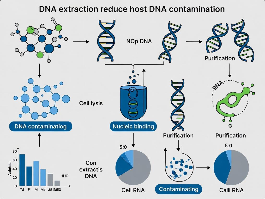
The Host DNA Problem: Understanding Sources, Impacts, and the Need for Purification
Troubleshooting & FAQs
Q1: My NGS library from a sputum sample shows >99% human reads. What are the primary causes and immediate steps? A: This indicates severe host DNA overrepresentation. Primary causes: 1) Lysis protocol too harsh, rupturing human cells; 2) Inefficient pathogen/enrichment steps; 3) Sample with very low pathogen load. Immediate Steps: 1) Quantify host DNA removal efficiency after extraction using qPCR for a human-specific gene (e.g., RPP30). 2) For future extractions, incorporate a differential lysis step (gentle for eukaryotic cells, harsh for microbes) or use a commercial host DNA depletion kit.
Q2: When using enzymatic host DNA depletion (e.g., kits using CpG methylation recognition), my yield is extremely low. How can I optimize? A: Low yield post-depletion often stems from over-digestion or insufficient input DNA. Optimization Protocol:
- Titrate Enzyme: Perform a digestion time course (5 min to 1 hr) and enzyme volume gradient (0.5x to 2x recommended).
- DNA Input: Ensure input DNA is within the kit's optimal range (typically 50ng-1µg). Excessive input can inhibit the enzyme.
- Inhibition Check: Spike a control, non-host DNA (e.g., lambda phage) into the reaction to confirm the enzyme is active and not inhibited by sample contaminants.
Q3: My microbiome sequencing from tissue biopsies has high inter-sample variability in host:microbe ratio. How do I standardize this? A: Variability often arises from inconsistent tissue homogenization and initial cell lysis. Standardization Method:
- Mechanical Homogenization: Use a bead-beater with fixed parameters (speed, time, bead type/size) for all samples.
- Initial Fixation: Consider brief ethanol fixation of tissue pieces prior to homogenization to stabilize human cells and reduce shearing.
- Post-Homogenization Filter: Pass homogenate through a 5µm filter to capture large human cell/debris fragments before proceeding with microbial DNA extraction.
Q4: For low biomass samples (e.g., plasma for cell-free pathogen DNA), how do I distinguish true low signal from host DNA background? A: This requires rigorous controls and bioinformatics. Experimental Controls:
- Negative Extraction Controls: Include multiple "blank" extraction controls (no sample added) to identify kit/reagent contaminants.
- Spike-In Controls: Use a synthetic, non-natural DNA sequence (e.g., from Arabidopsis thaliana) at a known, low copy number as an internal process control to assess recovery efficiency.
- Bioinformatics Threshold: Set a minimum read threshold for pathogen identification above the highest level seen in any negative control.
Key Experimental Protocols
Protocol 1: Differential Lysis for Selective Host Cell Removal
Objective: Gently lyse mammalian cells to release host DNA for degradation, while keeping microbial cells intact.
- Suspend sample (e.g., bronchoalveolar lavage) in 1 mL of Gentle Lysis Buffer (10 mM Tris-HCl pH 8.0, 1 mM EDTA, 0.1% Triton X-100).
- Incubate on ice for 30 minutes with gentle inversion every 10 minutes.
- Centrifuge at 500 x g for 10 min at 4°C. The pellet contains mostly intact microbes and host nuclei.
- Discard the supernatant (contains solubilized host cytoplasmic DNA).
- Resuspend pellet in robust microbial lysis buffer (e.g., with proteinase K and bead beating) to extract microbial DNA.
Protocol 2: qPCR-Based Quantification of Host DNA Depletion Efficiency
Objective: Quantitatively measure the amount of host DNA before and after a depletion step.
- Design Primers: Target a multi-copy human-specific gene (e.g., ALU or RPP30).
- DNA Standard: Prepare a serial dilution of human genomic DNA (10^6 to 10^1 copies/µL) for a standard curve.
- Sample Prep: Split your extracted DNA sample pre- and post-host depletion. Dilute to fall within the standard curve.
- Run qPCR: Perform qPCR in triplicate for standards and samples.
- Calculation:
% Host DNA Removal = [1 - (Host DNA copies post-depletion / Host DNA copies pre-depletion)] * 100
Research Reagent Solutions Toolkit
| Reagent/Material | Function & Rationale |
|---|---|
| Selective Lysing Buffers (e.g., with low [SDS] or mild detergents) | Gently disrupt mammalian cell membranes without lysing robust microbial (e.g., Gram-positive bacterial, fungal) cell walls. |
| Benzonase Nuclease | Degrades linear DNA in lysates without Mg²⁺ requirement; can be used post-differential lysis to digest released host DNA before microbial lysis. |
| Methylation-Dependent Restriction Enzymes (e.g., McrBC) | Cuts methylated (mammalian) DNA; key component in enzymatic depletion kits. Requires high-quality, input DNA. |
| Phosphothioate-Modified Probes (for ALU qPCR) | Resist nuclease degradation, providing robust quantification of host DNA in complex, nuclease-rich lysates. |
| Synthetic Spike-in DNA (e.g., A. thaliana sequences) | Non-biological internal control to monitor DNA recovery and sequencing efficiency across samples with variable host DNA content. |
| Size Selection Beads (e.g., AMPure XP at specific ratios) | Can be used to selectively remove large DNA fragments (>1-2 kbp) often associated with sheared host genomic DNA, enriching for smaller microbial DNA. |
| Density Gradient Media (e.g., Percoll) | For physical separation of host cells (e.g., leukocytes) from smaller microbes or free DNA in blood/plasma samples. |
Table 1: Comparison of Host DNA Depletion Methods
| Method | Principle | Typical Host DNA Reduction* | Key Limitation | Best For |
|---|---|---|---|---|
| Density Gradient Centrifugation | Physical separation by cell size/density. | 10-50% | Low recovery of microbes that clump or adhere to host cells. | Blood, BAL fluid. |
| Differential Lysis | Sequential chemical lysis. | 40-70% | Optimization required for each sample type. | Sputum, tissue homogenates. |
| Enzymatic Depletion (Methylation) | Digestion of methylated host DNA. | 90-99% | Requires high DNA input; inefficient on unmethylated or degraded DNA. | High biomass samples (stool, tissue). |
| Probe-Based Hybridization | Biotinylated probes pull out host sequences. | 95-99.9% | High cost, requires known host genome sequence. | Any sample with sufficient DNA. |
*Reduction values are sample-dependent estimates from recent literature.
Table 2: Impact of Host DNA on NGS Metrics in a Simulated Bronchial Sample
| Host DNA in Library | Pathogen (MTB) Reads Mapped | Microbial Alpha Diversity (Shannon Index) Estimated | Required Sequencing Depth for 10x Pathogen Coverage |
|---|---|---|---|
| 99% (No Depletion) | ~10,000 | Severely Underestimated | 100 Million reads |
| 90% (After Depletion) | ~100,000 | Significantly Improved | 10 Million reads |
| 50% (After Depletion) | ~500,000 | Near True Value | 2 Million reads |
Assumptions: Sample contains 0.1% *M. tuberculosis (MTB) DNA; Total library DNA = 1µg. Simulation based on 2023-2024 benchmarking studies.
Diagrams
Technical Support Center: Troubleshooting Host DNA Contamination
Troubleshooting Guides
Guide 1: Excessive Host DNA in Microbial Pellet Post-Differential Lysis
- Problem: After differential lysis (gentle lysis of human cells followed by harsh lysis of microbial cells), the resulting microbial pellet still contains a high percentage of host DNA, overwhelming the microbial signal in downstream sequencing.
- Diagnosis: Inefficient separation of host cell debris from intact microbial cells during the initial gentle lysis step. The host DNA is physically trapped or co-sedimenting with the microbes.
- Solution:
- Optimize Centrifugation: Reduce the centrifugation speed and time for the initial gentle lysis spin. Empirical data suggests 500 x g for 5-10 minutes is more effective for pelleting large human cell debris while leaving most bacteria in suspension, compared to standard 10,000 x g protocols.
- Introduce a Filtration Step: Pass the supernatant from the gentle lysis step through a 5.0 µm polycarbonate filter to capture remaining host debris before pelleting microbes.
- Add a DNase Step: Treat the microbial pellet with a benzonase-like enzyme that degrades linear DNA (released host DNA) but not DNA within intact microbial cell walls. Wash thoroughly afterwards.
Guide 2: Inconsistent Host DNA Depletion Across Sample Types
- Problem: A host depletion protocol works well for blood but fails for sputum or tissue biopsies, yielding highly variable host DNA percentages.
- Diagnosis: Sample collection and initial stabilization methods introduce artifacts that alter cell wall integrity and lysis susceptibility.
- Solution:
- Standardize Collection: For tissues, use immediate snap-freezing in liquid nitrogen or placement in a dedicated DNA/RNA stabilization reagent to prevent autolysis.
- Pre-process Mucosal Samples: For sputum or bronchoalveolar lavage (BAL), use a mucolytic agent (e.g., DTT) and a series of washes in PBS or saline to break down mucus trapping host cells before the lysis steps.
- Titrate Lysis Conditions: For each sample matrix, empirically determine the optimal concentration of a gentle detergent (e.g., 0.1-0.5% SDS) and incubation time for host cell lysis.
Frequently Asked Questions (FAQs)
Q1: During differential centrifugation, I can't find a speed that pellets human cells but leaves all bacteria in suspension. Some of my target bacteria are large (e.g., Helicobacter pylori). What can I do? A: You are correct that size overlap exists. Consider moving to a density gradient centrifugation approach. Using a medium like Percoll or Histodenz, create a gradient (e.g., 20%-80%) and layer your sample. Centrifuge at 2,500 x g for 15-30 min. Host cells and large microbes will pellet, while many bacteria will band at a specific density. This physically separates them based on buoyancy, not just size.
Q2: I'm using commercial host depletion kits, but they are very expensive for large-scale studies. Are there robust, published in-house protocols I can adapt? A: Yes. Two widely cited methods are the MO BIO (now QIAGEN) PowerMicrobiome protocol basis and the ‘Bleach Lysis’ method for tough spores. Key cost-saving, in-house adaptations involve:
- Using laboratory-prepared lysozyme/mutanolysin cocktails for Gram-positive cell walls.
- Replacing proprietary buffers with a defined TES Lysis Buffer (Tris, EDTA, Sucrose) with added lysozyme for gentle host lysis.
- Implementing a PMAP37 antimicrobial peptide treatment to selectively lyse human cells (requires careful optimization).
Q3: How do I definitively quantify the level of host DNA contamination in my sample before sequencing? A: Use a qPCR-based assay with taxon-specific primers. This provides a quantitative metric for protocol optimization.
- For Human DNA Contamination: Target the human Alu or LINE-1 repetitive elements. These provide high-sensitivity detection.
- For Bacterial Load: Target the conserved 16S rRNA gene. Calculate the Host DNA % = (Human DNA concentration) / (Human DNA concentration + Bacterial DNA concentration) * 100.
Table 1: Comparison of Host DNA Depletion Methods for Sputum Samples (n=5 per method)
| Method | Avg. Host DNA % Post-Depletion (±SD) | Avg. Microbial DNA Yield (ng) (±SD) | Cost per Sample | Key Limitation |
|---|---|---|---|---|
| Differential Centrifugation (500 x g) | 45.2% (±12.1) | 15.5 (±4.2) | Low | High variability |
| Density Gradient (Percoll) | 22.7% (±5.8) | 8.3 (±2.1) | Medium | Lower yield |
| Selective Lysis (saponin/DTAB) | 18.5% (±4.3) | 12.8 (±3.6) | Low-Medium | Inhibitor carryover |
| Commercial Kit (MICROBEnrich) | 9.8% (±2.5) | 25.1 (±5.7) | High | Cost-prohibitive |
Table 2: Impact of Sample Storage on Host Cell Lysis Efficiency
| Storage Condition | Time | Human Cell Viability Post-Thaw (%) | Host DNA in Supernatant After Gentle Lysis (ng/µL) |
|---|---|---|---|
| Fresh (Immediate Processing) | 0 hrs | 98% | 105.2 |
| -80°C (No Stabilizer) | 1 week | 15% | 32.1 |
| -80°C (With RNA/DNA Shield) | 1 week | 85% | 98.7 |
| 4°C in PBS | 24 hrs | 65% | 78.4 |
Experimental Protocol: Optimized Differential Lysis for Sputum
Objective: Maximize removal of human DNA from induced sputum samples for lung microbiome analysis.
Materials: See "The Scientist's Toolkit" below.
Procedure:
- Homogenization & Mucolysis: Mix 500µL of raw sputum with 500µL of Sputumolysin (0.1% DTT in PBS). Vortex for 30 sec, then incubate at 37°C for 15 min with intermittent vortexing.
- Filtration & Wash: Pass the homogenate through a 70 µm nylon cell strainer into a 50mL conical tube. Wash with 10mL PBS. Centrifuge filtrate at 500 x g for 10 min at 4°C.
- Gentle Host Cell Lysis: Discard supernatant. Resuspend pellet in 2 mL of Gentle Lysis Buffer. Incubate on ice for 30 min with gentle inversion every 10 min.
- Debris Removal: Centrifuge at 500 x g for 10 min at 4°C. Carefully transfer supernatant (containing microbes) to a new tube. Optionally, filter supernatant through a 5.0 µm filter.
- Microbial Pellet & Harsh Lysis: Centrifuge the supernatant/filtrate at 16,000 x g for 20 min at 4°C to pellet microbes. Discard supernatant. Proceed with mechanical (bead-beating) or enzymatic lysis of the microbial pellet for total DNA extraction.
- Optional DNase Treatment: Resuspend microbial pellet in 1 mL of PBS with 20 U/mL Benzonase. Incubate 15 min at 37°C. Centrifuge at 16,000 x g for 5 min. Wash pellet with PBS.
Visualizations
Title: Differential Lysis Workflow for Sputum Samples
Title: Troubleshooting Decision Tree for Host DNA Contamination
The Scientist's Toolkit: Key Research Reagent Solutions
| Item | Function in Host Depletion |
|---|---|
| Dithiothreitol (DTT) / Sputumolysin | Mucolytic agent. Breaks disulfide bonds in mucus glycoproteins to release trapped microbial and host cells for more effective separation. |
| Percoll / Histodenz | Density gradient media. Used to separate cells based on buoyant density, effectively partitioning human cells from many microbial species. |
| Saponin | Mild, cholesterol-targeting detergent. Selectively permeabilizes eukaryotic (host) cell membranes at low concentrations while leaving bacterial membranes intact. |
| Benzonase Nuclease | Endonuclease that degrades all forms of DNA and RNA. Used to digest free-floating host DNA released during gentle lysis before microbial pellet lysis. |
| Lysozyme & Mutanolysin | Enzymatic cell wall lysis agents. Target peptidoglycan; crucial for gentle lysis of Gram-positive host cells (e.g., neutrophils) and subsequent harsh lysis of Gram-positive bacteria. |
| TES Lysis Buffer (Tris-EDTA-Sucrose) | Isotonic, gentle lysis buffer. Sucrose maintains osmolarity to prevent premature bacterial lysis while EDTA chelates Mg2+ to weaken host cell membranes. |
| PMAP-37 Antimicrobial Peptide | Synthetic peptide derived from myeloid cells. Selectively lyses eukaryotic cells over prokaryotic membranes at specific concentrations. |
| Polycarbonate Filters (5.0 µm) | Size-exclusion filters. Capture large host cell debris and nuclei after gentle lysis, allowing smaller microbes to pass through. |
Technical Support Center
FAQs & Troubleshooting Guides
Q1: In our host DNA depletion study, despite using a validated depletion protocol, our NGS data shows poor sensitivity for low-abundance microbial targets. Is this a library prep or a sequencing issue? A: This is most commonly a sequencing depth (coverage) issue. Host depletion increases the proportion of microbial reads, but absolute microbial read count is king for detecting low-abundance taxa. If your sequencing depth is too low, you will lack sufficient microbial reads for statistically significant detection.
- Troubleshooting Step: Calculate your effective microbial sequencing depth.
- Determine total reads after QC (e.g., 50 million reads).
- Multiply by your post-depletion microbial DNA % (e.g., 15%): 50M * 0.15 = 7.5 million microbial reads.
- Compare this to recommended depths for your goal (see Table 1).
- Solution: Increase total sequencing depth or optimize the depletion protocol to further increase microbial DNA percentage.
Q2: We are sequencing host-depleted samples for pathogen detection. How do we balance the cost of ultra-deep sequencing with the need for high sensitivity? A: This requires a cost-benefit optimization based on your limit of detection (LOD) requirement. Use pilot studies to define the relationship.
- Troubleshooting Step: Perform a pilot study with spiked-in controls.
- Experimental Protocol:
- Spike-in Control: Introduce a known, low-abundance synthetic microbial genome or a characterized non-host DNA at defined fractions (e.g., 0.1%, 0.01%, 0.001%) into your host DNA background pre-depletion.
- Process Samples: Subject all spiked samples to your standard host DNA depletion and library prep protocol.
- Sequencing: Sequence the libraries at multiple depth tiers (e.g., 10M, 30M, 100M total reads).
- Analysis: Plot the detection probability of your spike-in against the effective microbial reads. This defines your empirical LOD curve.
- Solution: Use the data from the protocol above to choose the minimum depth (and thus cost) that meets your required LOD. See Table 1 for generalized guidance.
Q3: After implementing a new DNA extraction method designed to reduce host DNA, our NGS metrics show high duplicate read percentages. What does this mean for sensitivity and cost-efficiency? A: High duplication rates indicate low library complexity, often due to insufficient starting material (microbial DNA mass after depletion) or PCR over-amplification. This severely reduces cost-efficiency, as you pay for redundant sequences that do not improve coverage.
- Troubleshooting Step: Calculate the estimated non-duplicate microbial reads.
Effective Unique Microbial Reads = (Total Reads * (1 - Duplication Rate)) * (% Microbial Reads) - Solution: Optimize the DNA extraction and depletion protocol to maximize yield of microbial DNA. Increase input biomass if possible, or adjust PCR cycles during library prep to preserve complexity.
Data Presentation
Table 1: Sequencing Depth Impact on Downstream Analysis for Host-DNA-Depleted Samples
| Analysis Goal | Recommended Effective Microbial Reads | Typical Total Reads Required (at 20% Microbial DNA) | Impact of Insufficient Depth | Cost Consideration |
|---|---|---|---|---|
| Pathogen Detection (Abundant) | 1 - 5 million | 5 - 25 million | False negatives for low-viral-load samples. | Moderate. Balance with sample multiplexing. |
| Microbiome Profiling (16S rRNA) | 50,000 - 100,000 per sample | 0.5 - 1 million | Loss of rare taxa; skewed community diversity metrics. | Lower. High multiplexing is feasible. |
| Metagenomic Shotgun (Strain-level) | 20 - 50 million | 100 - 250 million | Incomplete genome assembly; inability to call rare genes/variants. | High. Requires premium flow cells or low-plex pools. |
| Host Transcriptome Co-analysis | Varies; 10-30% of total reads for host | 50 - 100 million (total) | Compromised power for both microbial detection and host gene expression. | Highest. Dual objectives demand maximum depth. |
Mandatory Visualization
Title: NGS Workflow from Extraction to Data: Key Variables
Title: Decision Logic for Optimizing Sequencing Depth
The Scientist's Toolkit: Research Reagent Solutions
| Item | Function in Host-DNA Depletion / NGS Workflow |
|---|---|
| Selective Lysis Buffers | Lyse host cells gently while preserving intact microbial cells (e.g., Gram+, Mycobacteria) for subsequent separation. |
| Nucleases (e.g., DNase I) | Digest free host DNA (e.g., from lysed human cells) prior to microbial cell lysis, enriching for intracellular microbial DNA. |
| Probe-Based Depletion Kits | Use oligonucleotide probes (e.g., methyl-CpG binding) to hybridize and remove host DNA post-extraction. Critical for high-host-content samples. |
| Spike-in Synthetic Controls | Defined, non-host DNA sequences added pre-extraction to monitor depletion efficiency, extraction yield, and LOD. |
| PCR-Free Library Prep Kits | Minimize amplification bias and duplicate reads, crucial for maintaining complexity in low-microbial-DNA samples post-depletion. |
| Size Selection Beads | Used to remove short fragments (often degraded host DNA) or select optimal library insert sizes, improving microbial read percentage. |
| Blocking Oligos | Suppress amplification of residual host DNA during library PCR, increasing the relative fraction of microbial sequences sequenced. |
Troubleshooting Guide & FAQs
Q1: During host DNA depletion from whole blood, my target pathogen DNA yield is extremely low. What could be the cause? A: This is often due to overly stringent lysis or depletion conditions. Human white blood cells are robust; if the initial hypotonic or gentle lysis step is too harsh, it can co-lyse fragile bacterial or fungal pathogens, releasing their DNA which is then degraded or inadvertently removed during host cell pelleting. Ensure a differential lysis protocol: use a mild detergent (e.g., 0.1% Triton X-100 in an isotonic buffer) to selectively lyse human cells, pellet intact host nuclei and debris, then apply a stronger lysis (e.g., with proteinase K and bead beating) to the supernatant/enriched pathogen pellet to liberate pathogen DNA.
Q2: My tissue samples show high human DNA background even after depletion protocols. How can I improve this? A: Tissue homogenization is critical. Incomplete homogenization leaves human cells intact, failing to expose them to depletion agents. Use optimized mechanical homogenization (e.g., gentleMACS Dissociator) followed by enzymatic treatment (collagenase/DNase-free RNase) for single-cell suspensions. Then apply a proven depletion method, such as selective lysis or saponin-based treatment (see protocol below). Also, consider targeting the human nucleus. For formalin-fixed paraffin-embedded (FFPE) tissue, deparaffinization must be complete before homogenization.
Q3: For sputum and BALF samples, how do I handle viscous mucus that impedes depletion efficiency? A: Mucolytic agents are essential. However, common agents like dithiothreitol (DTT) can inhibit downstream PCR. Use a two-step process: 1) Treat with recombinant mucolytic enzymes like Pulmozyme (dornase alfa) which cleaves DNA networks without inhibiting enzymes, followed by centrifugation to pellet cells. 2) Resuspend the pellet in a buffer containing saponin to selectively permeabilize human cells, allowing DNase treatment to degrade host DNA. Wash thoroughly before pathogen lysis.
Q4: When using enzymatic depletion (e.g., nucleases), how do I ensure complete enzyme inactivation to prevent degradation of my target DNA? A: Inactivation is paramount. For Benzonase or similar endonucleases, use EDTA (chelates Mg2+ cofactor) and a heat step (75°C for 15 min). For exonuclease-based host depletion (e.g., Selective Whole Genome Amplification kits), the enzyme is typically thermally labile and is inactivated by a simple 5-10 min heat step at 65°C. Always include a control with pure pathogen DNA spiked into the inactivation mix to confirm no loss of target.
Detailed Experimental Protocols
Protocol 1: Saponin-Based Host Cell Depletion for Blood and BALF
Principle: Saponin selectively permeabilizes eukaryotic (host) cell membranes, allowing diffusion of DNase I into the cytoplasm to degrade host genomic DNA, while leaving bacterial cells intact. Steps:
- Sample Prep: Lyse RBCs in blood with ACK buffer. For BALF, treat with 0.1% dornase alfa in PBS for 15 min at 37°C. Centrifuge at 500 x g for 10 min. Pellet contains human and pathogen cells.
- Permeabilization: Resuspend pellet in 1 mL of 0.1% saponin in PBS + 5mM MgCl2. Incubate 15 min at room temperature.
- DNase Treatment: Add 50 U of DNase I (RNase-free). Incubate 30 min at 37°C.
- Inactivation & Washing: Add EDTA to 10 mM (final concentration) to inactivate DNase. Centrifuge at 5000 x g for 5 min. Wash pellet 2x with PBS+EDTA.
- Pathogen Lysis: Proceed with mechanical (bead beating) or enzymatic lysis of the intact pathogen pellet.
Protocol 2: Differential Centrifugation & Lysis for Sputum
Principle: Uses density and differential lysis to separate and selectively deplete human cells. Steps:
- Mucolysis: Mix 1mL of sputum with 1mL of Sputasol (containing DTT) and 10µL of dornase alfa (1mg/mL). Vortex vigorously. Incubate at 37°C for 20 min.
- Clarification: Centrifuge at 300 x g for 5 min to pellet human cells and debris. Transfer supernatant (enriched in some pathogens) to a new tube.
- Host Cell Pellet Depletion: Resuspend the pellet in 1mL of 0.5% saponin/PBS. Incubate 10 min, then pellet at 5000 x g for 5 min. Discard supernatant (contains lysed host DNA). This pellet (P1) is retained.
- Supernatant Pathogen Concentration: Centrifuge the supernatant from step 2 at 16,000 x g for 15 min to pellet pathogens. Resuspend pellet (P2) in lysis buffer.
- Combine: Combine pellets P1 and P2 for total DNA extraction via bead beating and kit-based purification.
Table 1: Comparison of Host DNA Depletion Efficiency Across Sample Types
| Sample Type | Typical Total DNA Yield (Untreated) | Typical % Host DNA (Untreated) | Method | Post-Depletion % Host DNA | Key Challenge |
|---|---|---|---|---|---|
| Whole Blood | 2-5 µg/mL | >99.99% | Saponin+DNase I | 85-95% | Preserving low-titer bacteremia DNA |
| Lung Tissue | 10-50 µg/100mg | >99.9% | Mechanical Homogenization + Saponin | 70-90% | Complete homogenization |
| Sputum | 1-10 µg/mL | >99% | Dornase Alfa + Differential Lysis | 60-80% | Viscosity; diverse microbiota |
| Bronchoalveolar Lavage Fluid (BALF) | 0.5-5 µg/mL | ~99.5% | Dornase Alfa + Centrifugation + DNase | 50-75% | Low pathogen biomass |
Table 2: Performance of Commercial Host Depletion Kits
| Kit Name | Primary Mechanism | Best For Sample Type | Avg. Host Reduction | Cost per Sample |
|---|---|---|---|---|
| Microbiome Enrichment Kit (Molzym) | Selective lysis & DNase | Blood, BALF | 95-99% | High |
| NEBNext Microbiome DNA Enrichment Kit | Methylation-binding depletion | Stool, Saliva | >90% | Medium |
| QIAseq Host Depletion Kit | Probe-based capture/removal | Blood, Tissue | >99% | Very High |
Visualizations
Title: General Workflow for Host DNA Depletion from Complex Samples
Title: Troubleshooting Flow: Choosing a Depletion Strategy by Sample
The Scientist's Toolkit: Key Research Reagent Solutions
| Item | Function in Host Depletion |
|---|---|
| Saponin (from Quillaja bark) | Mild detergent that selectively permeabilizes cholesterol-rich eukaryotic (host) cell membranes, allowing DNase entry without lysing prokaryotic cells. |
| Recombinant Dornase Alfa (Pulmozyme) | Mucolytic enzyme that cleaves extracellular DNA networks in sputum/BALF, reducing viscosity and exposing cells without inhibiting downstream molecular assays. |
| Benzonase Nuclease | Potent endonuclease that degrades all linear and circular DNA/RNA. Used in kits to digest host DNA after non-selective lysis, requiring careful inactivation. |
| Selective Whole Genome Amplification (SWGA) Primers | Oligonucleotides designed with biased binding to pathogen genomes, enabling preferential amplification of microbial DNA in a background of host DNA. |
| MyOne Silane Dynabeads | Magnetic beads functionalized to bind nucleic acids. Used in conjunction with probe sets to selectively capture (and remove) human DNA sequences. |
| Collagenase Type IV | Enzyme for digesting collagen in tissue samples, crucial for creating single-cell suspensions from solid tissues prior to depletion steps. |
| ACK Lysing Buffer | Ammonium-Chloride-Potassium buffer for the gentle and effective osmotic lysis of red blood cells in whole blood samples, simplifying downstream white blood cell handling. |
Regulatory and Diagnostic Implications for Assay Development and Validation
Technical Support Center
Troubleshooting Guides & FAQs
Q1: During host DNA depletion experiments, my target pathogen DNA yield is unacceptably low post-extraction. What could be the cause? A: This is a common issue in differential lysis-based methods. The primary cause is often overly stringent lysis conditions for the host cells, which can co-damage or co-precipitate fragile pathogen (e.g., bacterial, viral) particles/nucleic acids. Validate by performing a spike-and-recovery experiment: spike a known quantity of pathogen control (e.g., synthetic DNA or cultured pathogen) into the sample post-host depletion and proceed with extraction. Recovery <70% indicates protocol issues. Solution: Titrate the host lysis reagent (e.g., concentration of detergent or enzyme like lysozyme for bacterial cells) and incubation time. A sequential, mild-to-harsh lysis approach is recommended.
Q2: My qPCR assay for pathogen detection shows high Ct values and poor reproducibility after implementing a new host DNA depletion kit. How should I investigate? A: This points to inhibition or inconsistent depletion efficiency. Follow this diagnostic workflow:
- Check for PCR Inhibitors: Perform the same qPCR on a 1:10 dilution of your extract. If the Ct decreases significantly, residual depletion reagents (e.g., alcohols, salts, polymers) are inhibitory. Solution: Include an additional wash step or change the wash buffer.
- Assess Depletion Consistency: Use a host-specific qPCR (e.g., for human β-actin or 18S rRNA gene) on replicates. High variability in host Ct values indicates uneven sample processing. Solution: Ensure homogeneous sample mixing before depletion and consistent incubation conditions.
- Review Validation Data: The kit's limit of detection (LoD) may have been established with different matrices. Re-establish the LoD and PCR efficiency in your specific sample type (e.g., sputum, blood) as part of assay re-validation.
Q3: For IVD development, what are the key regulatory validation parameters for an extraction method that includes host depletion, and how are they calculated? A: Per FDA/EMA/ISO 15189 guidelines, the extraction component must be validated as part of the complete test system. Key parameters include:
Table 1: Key Validation Parameters for Host Depletion-Integrated Extraction
| Parameter | Definition & Calculation | Target (Typical for IVD) |
|---|---|---|
| Efficiency (Yield) | % of target nucleic acid recovered. (QuantityOutput / QuantityInput) * 100. |
≥70% recovery for qualitative; ≥90% for quantitative assays. |
| Precision (Repeatability) | Intra-assay variability. Expressed as CV% of log10 copies/µL or Ct across ≥20 replicates. | CV% < 5% for Ct; <25% for copies. |
| Depletion Factor (DF) | Log10 reduction of host DNA. Log10(Host DNA concentration without depletion / Host DNA concentration with depletion). |
≥3-log10 reduction (99.9%) is often targeted. |
| Limit of Detection (LoD) | Lowest concentration detected in ≥95% of replicates. Determined via probit analysis on diluted spiked samples. | Must be established in the presence of expected maximum host background. |
| Carryover/Crosso ver Contamination | Rate of false positives in negative controls placed adjacent to high-positive samples. | <1% for high-throughput systems. |
Q4: Can you provide a detailed protocol for validating host DNA depletion efficiency? A: Protocol: Validation of Host DNA Depletion Factor.
- Sample Preparation: Create a contrived sample by spiking a known quantity of cultured pathogen (or its genomic DNA) into a matrix containing a high, quantified load of host DNA (e.g., 10^6 human leukocytes/mL).
- Experimental Arms: Process the sample in parallel with (Test) and without (Control) the host depletion step. Use n≥5 replicates per arm.
- Extraction & Quantification: Perform complete nucleic acid extraction. Use:
- Host-specific qPCR Assay: (e.g., RNase P, β-globin) to quantify host DNA in both Test and Control extracts.
- Pathogen-specific qPCR Assay: to quantify pathogen DNA in both extracts (to calculate yield loss).
- Calculation:
- Average the host DNA concentration (copies/µL) for Control ([Host]C) and Test ([Host]T).
- Depletion Factor (DF) = Log10( [Host]C / [Host]T ).
- Pathogen Yield = ( [Pathogen]T / [Pathogen]C ) * 100.
Q5: How does choice of sample type impact the regulatory strategy for assay validation? A: The sample type (matrix) is critical and dictates the scope of validation. Regulatory bodies require matrix-specific claims.
Table 2: Impact of Sample Type on Validation Strategy
| Sample Type | Key Considerations | Additional Validation Experiments Required |
|---|---|---|
| Whole Blood | High inhibitor load (heme, IgG), variable host cell count. | Inhibition testing with ICs, stability studies across anticoagulants (EDTA, heparin). |
| Formalin-Fixed Paraffin-Embedded (FFPE) | Nucleic acid fragmentation, cross-linking. | Demonstration of performance across a range of fixation times and block ages. |
| Respiratory (BAL, Sputum) | Viscosity, mucins, heterogeneous cellularity. | Homogenization procedure validation, LoD in each specific matrix. |
| Tissue Biopsies | Low pathogen load, high host background. | Minimum input mass validation, demonstration of depletion efficacy in fibrous/fatty tissues. |
Diagram 1: Diagnostic Assay Dev & Validation Workflow
Diagram 2: Host DNA Depletion Mechanisms
The Scientist's Toolkit: Research Reagent Solutions
Table 3: Essential Reagents for Host DNA Depletion Research
| Reagent/Material | Function in Research | Key Consideration for Validation |
|---|---|---|
| DNase I (Benzonase) | Degrades free host DNA post-host cell lysis, but not internalized pathogen DNA. | Must validate that pathogen particles/nucleic acids are protected (e.g., by capsid or membrane). |
| Selective Lysis Buffers | Mild detergents (e.g., saponin) lyse specific host cells (RBCs, WBCs) while leaving pathogens intact. | Requires precise titration for each sample matrix to balance host lysis vs. pathogen integrity. |
| Magnetic Beads (Functionalized) | Beads coated with antibodies (CD45 for leukocytes) or lectins bind and remove host cells. | Batch-to-batch consistency of coating is critical; validate binding capacity per sample volume. |
| Proteinase K | General protease for digesting proteins in tough samples (FFPE, sputum). | Source and activity can affect pathogen recovery; use a standardized, RNAse/DNase-free grade. |
| Internal Control (IC) | Non-target nucleic acid (e.g., phage RNA) spiked into sample pre-extraction. | Monitors extraction efficiency and identifies PCR inhibition. Must not cross-react with host/pathogen. |
| Inhibitor Removal Resins | (e.g., silica, charged polymers) bind PCR inhibitors (heme, humic acid) during wash steps. | Can also bind target DNA if not optimized; validate recovery with spiked targets. |
Practical Guide to Host DNA Depletion: Wet-Lab Techniques and Protocol Selection
Technical Support Center: Troubleshooting Guides & FAQs
FAQ Section
Q1: My selective lysis step for reducing human DNA in sputum samples is inconsistently effective. What could be causing this? A: Inconsistent lysis often stems from sample viscosity and heterogeneity. For sputum, a mandatory pre-treatment with dithiothreitol (DTT) or N-acetyl-L-cysteine (NAC) is required to homogenize the mucin matrix. Ensure the pre-treatment incubation is at 37°C for 15-30 minutes with vigorous vortexing. Post-homogenization, a centrifugation step (500 x g for 10 minutes) to pellet human cells can significantly improve selective bacterial lysis reagent performance.
Q2: When using enzymatic pre-treatment (e.g., lysozyme, mutanolysin) for Gram-positive bacteria, my final DNA yield is low. How can I optimize this? A: Low yield post-enzymatic treatment typically indicates incomplete lysis or inhibitor carryover. First, verify the enzyme activity buffer; many require Tris-HCl (pH 8.0) and do not tolerate chelating agents. Increase incubation time to 60 minutes at 37°C. For difficult-to-lyse cells like Mycobacterium, incorporate proteinase K and a brief bead-beating step post-enzymatic treatment. Refer to Table 1 for optimized reagent concentrations.
Q3: I am using a detergent-based selective lysis buffer for host cell depletion in blood cultures, but I'm still getting high human DNA contamination. How do I improve depletion? A: This indicates that lysis conditions are too harsh or too gentle. Use a mild detergent (e.g., 0.1% Triton X-100 or 0.5% Saponin) in an isotonic sucrose buffer to selectively lyse human cells while leaving bacterial cells intact. Critical parameters are osmotic support and incubation time. Incubate on ice for exactly 5-10 minutes, then immediately centrifuge (1000 x g, 5 min) to pellet intact bacteria. Discard the supernatant containing lysed host DNA. A second wash step is recommended.
Q4: Post pre-treatment, my sample volume has increased significantly, diluting my target pathogen. How do I manage this? A: Volume increase is common after homogenization buffers are added. Always include a concentration step post pre-treatment and prior to DNA extraction. For liquid samples, use a low-speed centrifugation (e.g., 5000 x g for 10 min) to pellet microbial cells. Resuspend the pellet in a minimal volume (e.g., 100-200 µL) of the selective lysis buffer or PBS. For filter-concentrated samples, perform enzymatic or mechanical lysis directly on the filter membrane.
Troubleshooting Guide
Issue: Complete Inhibition of Downstream PCR after Selective Lysis.
- Checkpoint 1: Reagent Compatibility. Ensure the selective lysis detergent or enzyme is compatible with your downstream DNA extraction kit. Ionic detergents like SDS can inhibit silica-column binding. Perform a buffer exchange or clean-up spin column step if needed.
- Checkpoint 2: Inhibitor Carryover. Physical pre-treatment (e.g., density gradient centrifugation) can co-pellet inhibitors like heme. Incorporate a wash step with a low-salt buffer (e.g., 1X PBS) before proceeding to main extraction.
- Checkpoint 3: Lysis Time. Over-incubation in a harsh lysis buffer can cause irreversible damage to pathogen cell walls, leading to DNA degradation. Strictly adhere to recommended times.
Issue: Poor Reproducibility Between Technical Replicates in Host DNA Depletion.
- Checkpoint 1: Sample Inhomogeneity. Solid tissues (e.g., biopsies) must be thoroughly homogenized using a mechanical homogenizer before aliquoting for pre-treatment. Powder samples under liquid nitrogen for best consistency.
- Checkpoint 2: Inaccurate Timing. The selective lysis step is often time-sensitive. Use a dedicated timer and process samples sequentially or in batches you can handle precisely.
- Checkpoint 3: Temperature Fluctuation. Perform incubations in a calibrated heat block or water bath, not on the lab bench.
Experimental Protocols & Data
Protocol: Selective Lysis of Human Cells in Bronchoalveolar Lavage (BAL) Fluid for Microbial DNA Enrichment
Objective: To deplete human eukaryotic cells prior to DNA extraction, enriching for bacterial and fungal pathogen DNA. Reagents: Saponin Lysis Buffer (0.25% w/v Saponin, 0.5 M Sucrose, 10 mM Tris-HCl pH 8.0), PBS. Procedure:
- Centrifuge 1-2 mL of fresh BAL fluid at 500 x g for 10 minutes at 4°C.
- Carefully decant supernatant (may be used for other assays). Resuspend pellet in 1 mL of ice-cold Saponin Lysis Buffer by gentle pipetting.
- Incubate on ice for 10 minutes, inverting tube every 2 minutes.
- Centrifuge at 1000 x g for 10 minutes at 4°C.
- Discard the supernatant (contains lysed host cell material). Wash pellet with 1 mL of ice-cold PBS.
- Centrifuge again at 1000 x g for 5 minutes at 4°C. Discard supernatant.
- Proceed to mechanical or enzymatic lysis of the microbial pellet for DNA extraction.
Protocol: Enzymatic Pre-treatment of Gram-Positive Bacterial Colonies
Objective: To weaken the peptidoglycan layer for efficient DNA extraction. Reagents: Lysozyme Solution (20 mg/mL in 10 mM Tris-HCl, pH 8.0), Mutanolysin Solution (5 U/µL in same buffer), TE Buffer. Procedure:
- Harvest 1-5 bacterial colonies and suspend in 100 µL of TE buffer in a microcentrifuge tube.
- Add 50 µL of Lysozyme Solution. Mix by vortexing briefly.
- Incubate at 37°C for 30 minutes.
- Add 10 µL of Mutanolysin Solution. Mix gently.
- Incubate at 37°C for an additional 30 minutes.
- Proceed immediately to a standard proteinase K/SDS or kit-based total lysis step.
Table 1: Efficacy of Common Selective Lysis Reagents on Host Cell Depletion in Sputum
| Pre-treatment Method | Concentration | Incubation Time | Avg. Host DNA Reduction | Avg. Pathogen DNA Recovery |
|---|---|---|---|---|
| Saponin (Isotonic) | 0.1% | 10 min on ice | 85-90% | 95% |
| Triton X-100 (Isotonic) | 0.1% | 15 min on ice | 80-85% | 90% |
| Water (Hypotonic) | N/A | 5 min RT | 95% | 40-60% (Variable due to co-lysis) |
| Commercial HostZap | 1X | 10 min RT | 70-80% | 98% |
Table 2: Impact of Enzymatic Pre-treatment on DNA Yield from Hard-to-Lyse Bacteria
| Bacterial Type | Enzymatic Pre-treatment | Subsequent Lysis Method | DNA Yield (ng/µL) ± SD | PCR Inhibition Rate |
|---|---|---|---|---|
| Staphylococcus aureus | Lysozyme (30 min) | Kit-based column | 45.2 ± 5.1 | 0% |
| Staphylococcus aureus | None | Kit-based column | 12.5 ± 3.8 | 0% |
| Mycobacterium tuberculosis | Lysozyme + Proteinase K (60 min) | Phenol-Chloroform | 65.7 ± 7.3 | High (requires purification) |
| Mycobacterium tuberculosis | Bead-beating only | Phenol-Chloroform | 30.1 ± 10.2 | Moderate |
Diagrams
Diagram 1: Workflow for Selective Host Cell Lysis in Blood Samples
Diagram 2: Decision Tree for Sample Pre-treatment Strategy
The Scientist's Toolkit: Research Reagent Solutions
| Reagent/Material | Function in Pre-Extraction | Key Consideration |
|---|---|---|
| Dithiothreitol (DTT) / N-Acetyl-L-Cysteine (NAC) | Mucolytic agent for sputum homogenization. Breaks disulfide bonds in mucin proteins. | Prepare fresh; can inhibit PCR if carried over. |
| Saponin (in Isotonic Sucrose) | Mild, non-ionic detergent for selective lysis of eukaryotic cell membranes. | Concentration and time-critical; avoids microbial lysis. |
| Lysozyme | Enzyme targeting β-1,4-glycosidic bonds in peptidoglycan of Gram-positive bacteria. | Activity is pH and buffer dependent; ineffective alone for Mycobacteria. |
| Mutanolysin | Enzyme hydrolyzing the glycan strands in peptidoglycan, effective on many Gram-positives. | Often used in combination with lysozyme for synergistic effect. |
| Proteinase K | Broad-spectrum serine protease. Degrades proteins and inactivates nucleases. | Requires SDS or other denaturants for full activity on cellular structures. |
| Percoll/Density Gradient Media | Forms density gradient for physical separation of host and microbial cells via centrifugation. | Useful for blood samples; preserves pathogen viability. |
| Silica/Zirconia Beads (0.1mm) | Used in bead-beating for mechanical disruption of tough cell walls and biofilms. | Can generate heat; samples must be kept cold during processing. |
Technical Support Center
Troubleshooting Guides & FAQs
Q1: During differential lysis for bacterial DNA extraction from blood, my host cell lysis buffer is also lysing the target bacterial cells. What can I do? A: This indicates overly harsh conditions. Troubleshoot by:
- Optimize Buffer Osmolarity: Ensure the host lysis buffer contains a non-ionic detergent (e.g., 0.1% Triton X-100 or SDS) in an isotonic solution (e.g., 0.25M sucrose). Avoid ionic detergents at this stage.
- Reduce Incubation Time/ Temperature: Perform the host lysis step on ice for 5-10 minutes only, with gentle vortexing.
- Validate Separately: Test your host lysis buffer on a pure bacterial pellet to confirm it leaves the cells intact for your specific species.
Q2: I used Benzonase to degrade host nucleic acids, but my final target DNA yield is unacceptably low. A: This is common and often due to co-degradation. Address with:
- Optimize Cation Dependence: Benzonase requires Mg²⁺. Ensure your buffer contains 1-2 mM MgCl₂. To stop digestion, add a chelator (e.g., 5 mM EDTA) precisely after the host lysis step before disrupting your target cells.
- Shield Target DNA: Bind target DNA to a carrier or solid phase (e.g., silica beads/columns) prior to Benzonase treatment if protocol allows.
- Titrate Enzyme: Use the minimal effective unit (e.g., 25-50 U/mL) for the shortest time (10-15 mins at 37°C).
Q3: My density gradient centrifugation (e.g., Percoll or sucrose) fails to separate host debris from my target organelles (e.g., mitochondria) or microbes. A: Poor separation arises from improper gradient formation or sample loading.
- Verify Gradient Integrity: Prepare a discontinuous gradient carefully by layering solutions of decreasing density. Allow a slow diffusion step (e.g., 1 hour at 4°C) to form a continuous gradient for smoother separations.
- Check Centrifugation Parameters: Use a swinging-bucket rotor, not a fixed-angle. Ensure k-factor and g-force are correct for your target particle. For bacterial cells, typical settings are 10,000-20,000 x g for 30 min. For organelles, 40,000-100,000 x g for 1 hour may be needed.
- Sample Density: Ensure your sample load is less dense than the top layer of the gradient. Dilute sample in isotonic buffer if needed.
Q4: After all steps, my qPCR shows high levels of residual host gDNA. Which step likely failed? A: Perform a diagnostic check.
- Assess Differential Lysis: Take an aliquot post-host lysis, centrifuge, and run supernatant on gel. A strong host DNA band indicates effective host lysis.
- Assess Benzonase: Take an aliquot from Step 1, add Benzonase/Mg²⁺, incubate, and check gel. The smear should be gone. If not, enzyme is inactive.
- Assess Gradient: Isolate the target band/zone carefully; cross-contamination occurs if bands are aspirated too broadly.
Q5: How do I scale down these protocols for small sample volumes (e.g., <1 mL of blood)? A: Scaling requires maintaining reagent-to-sample ratios.
- Direct Scalability: Reduce all volumes proportionally.
- Centrifugation: Use micro-ultracentrifuge tubes (e.g., 1.5 mL thick-walled tubes) and appropriate rotors. Relative Centrifugal Force (RCF) must remain constant; time can sometimes be reduced slightly for smaller path lengths.
- Enzyme Use: Maintain final concentration (U/µL), not total units.
Experimental Protocols in Thesis Context
Protocol 1: Differential Lysis for Bacterial DNA from Whole Blood
- Objective: Selectively lyse human cells while keeping bacterial cells intact.
- Method:
- Mix 1 mL of whole blood with 3 mL of Host Lysis Buffer (20 mM Tris-HCl pH 8.0, 0.25M Sucrose, 0.1% Triton X-100, 10 mM EDTA).
- Incubate on ice for 10 minutes with gentle inversion every 2 minutes.
- Centrifuge at 500 x g for 10 min at 4°C to pellet intact bacteria and nuclei.
- Discard supernatant (contains host cytoplasmic debris).
- Resuspend pellet in 1 mL of Target Lysis Buffer (20 mM Tris-HCl pH 8.0, 2% SDS, 2 mg/mL Lysozyme, 20 mg/mL Proteinase K). Incubate at 56°C for 1 hour.
Protocol 2: Benzonase Treatment to Reduce Host Nucleic Acid Contamination
- Objective: Degrade host gDNA/RNA released after initial lysis.
- Method:
- Following Protocol 1, Step 4, resuspend the pellet in 500 µL of Benzonase Buffer (20 mM Tris-HCl pH 8.0, 2 mM MgCl₂).
- Add Benzonase to a final concentration of 50 U/mL.
- Incubate at 37°C for 15 minutes.
- Immediately halt digestion by adding EDTA to a final concentration of 5 mM.
- Proceed to target cell lysis (Protocol 1, Step 5).
Protocol 3: Sucrose Density Gradient Centrifugation for Mitochondrial DNA Enrichment
- Objective: Separate mitochondria from nuclear debris.
- Method:
- Prepare a discontinuous sucrose gradient in an ultracentrifuge tube: 2 mL of 60% sucrose (bottom), 2 mL of 40% sucrose, 2 mL of 20% sucrose (top). All solutions in 10 mM Tris-HCl pH 7.4.
- Carefully load 1 mL of post-homogenization cell lysate (in isotonic buffer) on top.
- Centrifuge in a swinging-bucket rotor at 40,000 x g for 1 hour at 4°C.
- Mitochondria will band at the 40%/60% interface. Carefully aspirate with a pipette.
Table 1: Optimization of Benzonase Treatment for Host DNA Depletion
| Sample Type | Benzonase Conc. (U/mL) | Incubation Time (min) | % Host DNA Remaining (qPCR) | % Target DNA Recovery |
|---|---|---|---|---|
| Spiked Blood Lysate | 0 | 0 | 100% | 100% |
| Spiked Blood Lysate | 25 | 15 | 15% | 95% |
| Spiked Blood Lysate | 50 | 15 | 5% | 90% |
| Spiked Blood Lysate | 100 | 15 | 2% | 70% |
| Spiked Blood Lysate | 50 | 30 | 1% | 65% |
Table 2: Comparative Efficiency of Core Biochemical Approaches
| Method | Primary Mechanism | Avg. Host DNA Reduction | Avg. Target DNA Yield | Typical Processing Time |
|---|---|---|---|---|
| Differential Lysis Only | Selective membrane disruption | 10-50 fold | High | 1-2 hours |
| Differential Lysis + Benzonase | Selective lysis + enzymatic degradation | 100-1000 fold | Medium-High | 2-3 hours |
| Density Gradient Centrifugation | Physical separation by density | 50-200 fold | Low-Medium | 3-4 hours |
| Combined (Lysis + Gradient) | Biochemical & Physical | >1000 fold | Low-Medium | 4-5 hours |
Visualizations
Diagram 1: Combined Workflow for Host DNA Depletion
Diagram 2: Decision Tree for Contamination Troubleshooting
The Scientist's Toolkit: Research Reagent Solutions
| Item | Function in Host DNA Depletion | Key Consideration |
|---|---|---|
| Triton X-100 (Non-ionic detergent) | Selectively disrupts eukaryotic (host) cell membranes in isotonic buffers, leaving prokaryotic and organelle membranes intact. | Concentration is critical (0.1-0.5%); use ice-cold. |
| Benzonase Nuclease | Degrades all forms of DNA and RNA (linear, circular, chromosomal). Used to digest host nucleic acids post-lysis. | Absolutely requires Mg²⁺ (1-2 mM). Must be inactivated by EDTA post-digestion. |
| Sucrose (Optimal Density Media) | Forms density gradients for separating particles (bacteria, organelles) from host debris based on buoyant density. | Prepares iso-osmotic solutions; concentrations from 20-60% w/v common. |
| Percoll (Silica-based Media) | Colloidal silica coated with PVP for isosmotic gradient centrifugation. Separates live bacteria from dead cells/debris. | Low viscosity allows faster centrifugation times than sucrose. |
| Lysozyme | Hydrolyzes peptidoglycan layer of Gram-positive bacteria. Used in target lysis step after host DNA removal. | Ineffective alone on Gram-negatives; requires EDTA pretreatment. |
| Proteinase K | Broad-spectrum serine protease. Degrades nucleases and proteins during target cell lysis, increasing DNA yield/quality. | Requires SDS and elevated temperature (56°C) for full activity. |
| EDTA (Chelating Agent) | 1) Inhibits DNases by chelating Mg²⁺. 2) Halts Benzonase activity. 3) Helps disrupt Gram-negative walls with lysozyme. | Critical for protocol timing—added to stop digestion or as buffer component. |
Troubleshooting Guides & FAQs
Q1: I am using a PMAxx-based kit for selective host DNA depletion in stool samples. My pathogen signal remains low post-treatment, even with spiked controls. What could be wrong? A: This is commonly due to suboptimal PMA photoactivation. Ensure the light-emitting diode (LED) array delivers uniform 465-475 nm light at the recommended power (e.g., ≥40 W). Tube placement and ice bath use are critical; samples must be kept cold during the 15-minute exposure to prevent heat-induced cell damage. Verify dye concentration and incubation time in the dark (5-10 min) are per protocol. Incomplete light exposure leaves PMA unbound, failing to crosslink host DNA.
Q2: During saponin-based host cell lysis for blood samples, my target bacterial DNA yield has dropped precipitously. How can I troubleshoot? A: Over-lysed bacterial cells are likely. Saponin concentration and incubation time are highly sample-volume dependent. For a standard 1 mL blood sample:
- Confirm saponin concentration is ≤0.5% (w/v).
- Reduce incubation time on a rotating mixer from 15 min to 5-10 min at 4°C.
- Immediately after incubation, centrifuge at 500 x g for 5 min (not higher) to pellet only host nuclei and intact host cells, leaving bacteria in supernatant. Increase subsequent bacterial pellet centrifugation to 10,000 x g.
Q3: My magnetic bead-based pathogen DNA isolation post-PMA treatment results in low elution volumes and poor recovery. What steps should I check? A: Focus on bead handling and buffer conditions.
- Bead Pellet Integrity: Ensure the magnetic separation time is sufficient (≥2 min) for a clear supernatant. Do not disturb the bead pellet during wash steps.
- Ethanol Contamination: Incomplete removal of Wash Buffer 2 (often ethanol-based) inhibits elution. After the final wash, spin briefly, place back on magnet, and use a 10 µL pipette to remove all residual ethanol. Air-dry beads for 5-10 minutes until cracks appear.
- Elution Buffer: Elute with pre-warmed (55-70°C) nuclease-free water or TE buffer (10 mM Tris-HCl, pH 8.5). Incubate on a heat block for 5 min while mixing.
Q4: For a saponin+magnetic bead combined workflow, I'm getting high levels of human genomic DNA contamination. Where is the failure? A: The failure likely occurs at the initial differential lysis. After saponin treatment, the low-speed centrifugation step is crucial. If the speed is too high (>800 x g), it may pellet both host debris and pathogen cells together. Re-optimize the g-force and time. Additionally, after saponin lysis, the supernatant containing pathogens should be transferred to a new tube before adding proteinase K and proceeding with bead-based DNA extraction to avoid carry-over of host debris.
Q5: PMA treatment appears to also crosslink DNA from my target Gram-negative bacteria in culture. Is this possible? A: Yes, if bacterial membrane integrity is compromised. PMA can penetrate dead/damaged bacterial cells. Validate cell viability and PMA penetration controls. For a pure culture, include a sample treated with 70% ethanol for 30 min to kill cells, followed by PMA. If DNA from the ethanol-killed sample is significantly reduced compared to an untreated killed control, PMA is penetrating damaged targets. Optimize by ensuring healthy, mid-log phase cultures and confirm no mechanical damage occurred during sample preparation.
Table 1: Performance Comparison of Host DNA Depletion Technologies
| Parameter | PMA-Based Technology | Saponin-Based Lysis | Magnetic Bead Capture (Pathogen-Specific) |
|---|---|---|---|
| Primary Mechanism | Photocrosslinking of free DNA & compromised host cells | Selective lysis of mammalian cell membranes | Immobilized probes binding target pathogen DNA/RNA |
| Typical Host DNA Reduction | 2-4 log10 reduction (stool, saliva) | 1-3 log10 reduction (blood, BALF) | 3-6 log10 reduction (post-lysis) |
| Target Pathogen Integrity | Preserves intact cells (vital) | Preserves intact cells (vital) | Can capture from lysate; not viability-dependent |
| Key Limitation | Light penetration in dense samples; dye optimization | Over-lyses fragile pathogens (e.g., Neisseria) | Requires prior knowledge of target; probe design |
| Best Suited For | Complex microbiomes (stool, sputum) where host cells are dead/damaged | Liquid biopsies (blood, plasma) with intact host cells | Specific detection in high-host background (e.g., B. burgdorferi in blood) |
| Typical Process Time | 1.5 - 2 hours (incl. photoactivation) | 30 - 60 minutes | 2 - 3 hours (incl. hybridization) |
Table 2: Troubleshooting Common Issues & Solutions
| Problem | Likely Cause | Suggested Solution |
|---|---|---|
| Low pathogen yield post-PMA | Incomplete host DNA crosslinking | Verify light source spectral output; ensure sample is in thin-walled, clear tubes on ice. |
| Bacterial DNA loss with saponin | Non-selective lysis of pathogens | Titrate saponin (0.1%-0.5%); reduce incubation time and temperature. |
| Low eluate concentration (beads) | Beads not fully resuspended or dried | Ensure thorough bead resuspension during binding/washes. Do not over-dry beads (>10 min). |
| High human DNA in bead eluate | Non-specific binding to beads or carryover | Increase stringency of wash buffers (e.g., add 5-10% ethanol to Wash Buffer 1). |
| Inconsistent PMA results | Variable sample matrix effects | Include an internal control (spiked intact cells) and normalize PMA concentration per sample type. |
Experimental Protocols
Protocol 1: PMA Treatment for Selective Host DNA Depletion in Sputum Samples This protocol is designed within the thesis context to enrich for bacterial pathogen DNA from cystic fibrosis sputum.
- Sample Preparation: Homogenize 500 µL of sputum with 500 µL of 1X PBS containing 0.1% dithiothreitol (DTT). Centrifuge at 800 x g for 5 min to pellet host cells and debris.
- PMA Treatment: Resuspend pellet in 1 mL PBS. Add PMAxx dye to a final concentration of 50 µM. Mix thoroughly.
- Incubation & Photoactivation: Incubate in the dark for 10 minutes at room temperature. Place tubes horizontally on ice 15 cm from a PMA-Lite LED array (465-475 nm). Expose for 15 minutes, gently agitating tubes every 5 minutes.
- DNA Extraction: Proceed with mechanical lysis (e.g., bead beating) followed by a standard magnetic bead-based total DNA extraction kit.
- Analysis: Quantify total DNA and perform qPCR for a human single-copy gene (e.g., RNase P) and a universal bacterial 16S rRNA gene to assess depletion efficiency.
Protocol 2: Sequential Saponin-Magnetic Bead Workflow for Bacterial DNA from Whole Blood This protocol aims to isolate *Staphylococcus aureus DNA from septic blood with minimal host background.*
- Selective Host Lysis: Mix 1 mL of whole blood with 9 mL of 0.5% (w/v) saponin in 1X PBS. Rotate gently for 15 minutes at 4°C.
- Differential Centrifugation: Centrifuge at 500 x g for 10 minutes at 4°C. Carefully transfer the supernatant (containing bacteria) to a new 15 mL tube. Pellet bacteria at 10,000 x g for 10 minutes. Discard supernatant.
- Bacterial Lysis: Resuspend pellet in 200 µL enzymatic lysis buffer (20 mM Tris-HCl, pH 8.0, 2 mM EDTA, 1.2% Triton X-100, 20 mg/mL lysozyme). Incubate 30 min at 37°C.
- Pathogen-Specific Capture: Add magnetic beads conjugated with S. aureus-specific peptide nucleic acid (PNA) probes. Hybridize at 55°C for 30 min with shaking.
- Wash & Elute: Wash beads twice with 500 µL of stringent wash buffer (10 mM Tris, 1 M NaCl, 0.1% SDS). Elute DNA in 50 µL of 10 mM Tris-HCl, pH 8.5, at 80°C for 5 min.
Diagrams
Title: PMA-Based Selective Host DNA Depletion Workflow
Title: Technology Mechanism and Profile Comparison
The Scientist's Toolkit: Research Reagent Solutions
| Item | Function in Host DNA Depletion |
|---|---|
| PMAxx or EMA Dye | Membrane-impermeant DNA intercalator. Crosslinks DNA upon light exposure, preventing PCR amplification from dead/damaged host cells. |
| Saponin (from Quillaja bark) | Cholesterol-binding detergent. Selectively lyses eukaryotic (host) cell membranes while leaving many bacterial membranes intact. |
| Magnetic Beads (Streptavidin-coated) | Solid-phase support for immobilizing biotinylated probes (e.g., PNA, DNA) to capture specific pathogen nucleic acids via hybridization. |
| Peptide Nucleic Acid (PNA) Clamps/Probes | DNA mimics with a neutral backbone. Used to block amplification of host sequences (clamps) or as capture probes for bead-based isolation. |
| Lysozyme & Mutanolysin | Enzymatic lysis agents targeting bacterial cell walls (peptidoglycan). Used after selective host lysis to release pathogen DNA. |
| Dithiothreitol (DTT) | Reducing agent. Breaks disulfide bonds in mucus (e.g., sputum) to homogenize samples prior to depletion steps. |
| Stringent Wash Buffer (High Salt + SDS) | Used in magnetic bead workflows to remove nonspecifically bound host DNA while retaining probe-bound pathogen DNA. |
| DNase I (Benzonase) | Digests extracellular DNA in sample pre-treatment to reduce background host DNA prior to cell lysis. |
Technical Support Center: Troubleshooting Guides & FAQs
Q1: During a modified phenol-chloroform extraction for host DNA depletion, my final DNA yield is consistently low (<50% expected). What are the primary causes and solutions?
A: Low yield in modified protocols is often due to inefficient phase separation or loss during carrier RNA steps. Ensure the sample pH is correct (~7.8) before the first phenol addition to prevent DNA partitioning into the organic phase. If using glycogen or linear polyacrylamide as a carrier, verify its solubility and absence of nucleases. Centrifugation post-phase separation should be at 4°C and at the recommended speed (e.g., 12,000 x g) for the full duration. Avoid aspirating too close to the interphase. For workflows integrating selective lysis buffers, incomplete inactivation of proteases or RNases can degrade nucleic acids; include a 70°C heat step for 10 minutes post-lysis if compatible.
Q2: After integrating a selective lysis step (e.g., with low-concentration SDS) to reduce human host cells in a bacterial pathogen DNA extraction, I see PCR inhibition. How can I resolve this?
A: Inhibition often stems from residual SDS or salts. Modify the wash steps post-selective lysis. Implement two consecutive washes with a cold wash buffer (e.g., 70% ethanol with 10mM sodium acetate, pH 5.2) instead of one. Follow with a final 80% ethanol wash. Ensure the pellet is fully dried (air-dried for 5-10 minutes) to evaporate ethanol, but do not over-dry, as this makes resuspension difficult. Resuspend in TE buffer (pH 8.0) or nuclease-free water containing 0.1% Tween-20, which can help sequester residual inhibitors. Quantify inhibition using a spike-in control and qPCR dilution series.
Q3: In a protocol modified with enzymatic host depletion (e.g., benzonase), how do I verify the enzyme is fully inactivated without affecting target microbial DNA?
A: Benzonase requires Mg²⁺. The standard inactivation method is adding EDTA (5-10mM final concentration) to chelate Mg²⁺ after the incubation period. Verify inactivation by running a post-EDTA sample on a gel (should show no smearing of host DNA) and by performing a control qPCR for a highly abundant host single-copy gene (e.g., human RPP30). A >4-log reduction in host signal compared to a non-enzyme-treated control indicates successful depletion and inactivation. Ensure the EDTA is pH-adjusted to 8.0 to avoid acid degradation of DNA.
Q4: My integrated workflow uses magnetic beads for pathogen DNA capture post-host depletion. The bead recovery seems inefficient. What factors should I check?
A: Magnetic bead efficiency is highly dependent on PEG/NaCl concentration and incubation time. Check: 1) Bead-to-sample ratio: For post-depletion samples, a 1:1 volume ratio is common, but may need optimization. 2) Incubation time: Increase incubation time with mixing to 15-20 minutes at room temperature. 3) Ethanol content: Ensure wash buffers contain the correct ethanol concentration (usually 80% fresh). 4) Elution: Use pre-warmed (55°C) low-EDTA TE buffer or nuclease-free water, incubate for 5 minutes on the magnet before pipetting off. Avoid over-drying beads. 5) Bead type: Use carboxyl-modified beads optimized for size selection if target DNA is fragmented.
Q5: When comparing different commercial host depletion kits integrated into my standard CTAB extraction, how should I quantitatively evaluate their performance?
A: Use the following metrics in a controlled spike-in experiment (e.g., add known CFU of Pseudomonas aeruginosa to human whole blood):
Table 1: Metrics for Evaluating Host Depletion Kit Performance
| Metric | Measurement Method | Target Optimal Value |
|---|---|---|
| Host DNA Depletion Efficiency | qPCR for host single-copy gene (RPP30 for human) | >99% reduction (ΔCt >6.6) |
| Target Pathogen DNA Recovery | qPCR for pathogen-specific gene or spike-in control | >50% recovery (minimize loss) |
| Final Host:Pathogen DNA Ratio | Shotgun sequencing & alignment to host/pathogen genomes | Pathogen reads >10% of total |
| Inhibition Level | Internal amplification control (IAC) in downstream qPCR | Ct shift of IAC < 2 cycles |
| Process Time | Hands-on and total workflow time | Varies by throughput needs |
Detailed Experimental Protocol: Modified CTAB with Selective Lysis for Blood Samples
Objective: Extract microbial DNA from human blood with reduced human host DNA contamination.
Reagents:
- Lysis Buffer A (Selective): 1% Triton X-100, 20mM Tris-Cl (pH 8.0), 2mM EDTA.
- Lysis Buffer B (CTAB): 2% CTAB, 1.4M NaCl, 20mM EDTA, 100mM Tris-Cl (pH 8.0), 2% PVPP.
- Proteinase K (20 mg/mL).
- RNase A (10 mg/mL).
- Phenol:Chloroform:Isoamyl Alcohol (25:24:1, pH 8.0).
- Isopropanol and 70% Ethanol.
- Nuclease-free TE Buffer (pH 8.0).
Workflow:
- Selective Host Cell Lysis: Mix 1mL blood with 4mL cold Lysis Buffer A. Vortex 15 sec. Incubate on ice for 15 min with gentle inversion every 5 min.
- Centrifugation: Centrifuge at 500 x g for 10 min at 4°C to pellet intact microbes (and host nuclei if lysis incomplete). Carefully transfer supernatant (containing lysed host material) to a waste tube. Resuspend pellet in 1mL of fresh, cold Buffer A. Repeat centrifugation. Discard supernatant.
- Microbial Lysis: Resuspend pellet in 500μL Lysis Buffer B. Add 20μL Proteinase K and 5μL RNase A. Mix thoroughly. Incubate at 65°C for 1 hour with brief vortexing every 15 min.
- Organic Extraction: Add 500μL Phenol:Chloroform:Isoamyl Alcohol. Vortex vigorously for 1 min. Centrifuge at 12,000 x g for 10 min at 4°C. Transfer upper aqueous phase to a new tube.
- Precipitation: Add 0.7 volumes of isopropanol. Invert gently 50 times. Centrifuge at 12,000 x g for 15 min at 4°C. Discard supernatant.
- Wash & Elute: Wash pellet with 1mL of 70% ethanol. Centrifuge at 12,000 x g for 5 min. Air-dry pellet for 8-10 minutes. Resuspend in 50μL TE Buffer. Quantify host and microbial DNA by qPCR.
Visualizations
Title: Modified DNA Extraction for Host Depletion Workflow
Title: Troubleshooting Low DNA Yield Decision Tree
The Scientist's Toolkit: Research Reagent Solutions
Table 2: Essential Reagents for Host DNA Depletion Protocols
| Reagent/Material | Function in Protocol Integration | Key Consideration |
|---|---|---|
| Triton X-100 / Saponin | Selective lysis agent for mammalian cell membranes. Leaves microbial cells intact for subsequent pelleting. | Concentration and time are critical. Too harsh can lyse some fragile pathogens (e.g., Borrelia). |
| CTAB (Cetyltrimethylammonium bromide) | Ionic detergent effective for lysing microbial (esp. Gram-positive) cell walls and precipitating polysaccharides. | Works best at high salt (0.7M NaCl). Incompatible with SDS. Must be heated. |
| Benzonase Nuclease | Degrades linear DNA and RNA from lysed host cells. Requires Mg²⁺. | Must be thoroughly inactivated with EDTA post-incubation to prevent target DNA degradation. |
| Proteinase K | Broad-spectrum serine protease. Digests nucleases and proteins, aiding in lysis and improving DNA purity. | Requires incubation at 56-65°C. Must be inactivated by heat or phenol if needed. |
| Magnetic Beads (Carboxylated) | Bind DNA via PEG/NaCl-mediated crowding. Used for size-selective cleaning or pathogen enrichment. | Bead size and polymer ratio affect size cutoff. Stringent washes reduce inhibitors. |
| Carrier RNA / Glycogen | Co-precipitates with low concentrations of DNA to improve pellet visibility and recovery. | Must be RNase-free. Glycogen can interfere with some downstream enzymatic reactions. |
| PVPP (Polyvinylpolypyrrolidone) | Binds polyphenols and humic acids co-extracted from samples, reducing downstream inhibition. | Add directly to lysis buffer. Especially important for environmental or plant-derived samples. |
Technical Support Center
Troubleshooting Guides & FAQs
Q1: During gel extraction size selection, my final DNA yield is consistently low (<30%). What are the primary causes and solutions? A: Low yield in gel extraction is frequently due to inefficient elution or UV-induced DNA damage.
- Troubleshooting Steps:
- Minimize UV Exposure: Limit DNA gel slice exposure to UV light to <30 seconds. Use a low-energy UV transilluminator (365 nm) if possible.
- Optimize Gel Dissolution: Ensure the gel slice is fully dissolved by incubating at the recommended temperature (50-55°C) with frequent vortexing or shaking until completely homogenous (typically 5-10 minutes).
- Optimize Elution: Elute with pre-warmed (55-60°C) nuclease-free water or buffer (≥ 20 µL). Let the column sit for 2 minutes before centrifugation. A second elution with fresh buffer can recover an additional 10-20% of bound DNA.
- Verify pH: Ensure the binding buffer contains the correct chaotropic salt (e.g., guanidine thiocyanate) and is at the appropriate pH (~6.5). If using homemade buffers, check pH.
Q2: After column-based purification, I am detecting carryover of salts or enzymatic inhibitors (e.g., from ligation reactions) that interfere with downstream applications. How can I resolve this? A: This indicates incomplete washing.
- Troubleshooting Steps:
- Increase Wash Buffer Volume/Steps: Use 700 µL of wash buffer (typically 80% ethanol) instead of 500 µL. Perform two wash steps.
- Ensure Complete Drying: After washing, centrifuge the empty column for an additional 2-3 minutes to dry the membrane completely before elution. This removes residual ethanol.
- Use an Extra Wash: For reaction clean-up (e.g., PCR, enzymatic), consider an additional "dry spin" with the column empty before adding the wash buffer to remove any residual liquid from the rim.
- Switch Buffers: For stubborn inhibitors like phenols or high salts, a wash with a buffer containing a small percentage of acetonitrile (5-10%) may help, but verify compatibility with your column's membrane.
Q3: My size selection for NGS library purification using SPRI beads is resulting in inconsistent fragment size distributions between runs. What factors should I control? A: SPRI (solid-phase reversible immobilization) bead size selection is highly sensitive to reagent ratios and environmental conditions.
- Troubleshooting Steps:
- Standardize Bead:Sample Ratio Precisely: Use a calibrated pipette for both sample and beads. Key ratios: For >1.8x target size removal (e.g., primer dimer clean-up), use a 1.8:1 bead-to-sample ratio. For a tighter size selection window (e.g., 300-500 bp), optimize between 0.6:1 and 0.8:1. See Table 1.
- Temperature Control: Perform all incubations at room temperature (20-25°C). Temperature fluctuations alter binding kinetics.
- Mixing Consistency: Mix beads and sample by pipetting or vortexing thoroughly until homogenous. Incomplete mixing leads to variable binding.
- Ensure Fresh Ethanol: Always use fresh 80% ethanol for washing. Old or diluted ethanol compromises washing efficiency.
Q4: When performing clean-up to reduce host (e.g., human) DNA contamination in pathogen DNA samples, which method is superior: column-based or size-selection? A: The choice depends on the size differential between target and contaminant DNA.
- Guidance:
- For Large Size Differences: If your target microbial DNA is significantly smaller or larger than host genomic DNA (e.g., viral DNA vs. human DNA), physical size selection (gel or SPRI) is more effective. You can selectively excise or isolate the size range containing your target.
- For Minimal Size Differences: If size overlap is significant, enzymatic or chemical methods (e.g., differential lysis, nucleases, methylation-based depletion) coupled with column clean-up are required. Columns alone cannot separate same-sized fragments.
- Combined Approach: A common strategy is to first use enzymatic host depletion, followed by SPRI bead clean-up to remove enzymes and buffer components, and a final SPRI size selection to narrow the fragment distribution for sequencing.
Data Presentation
Table 1: Comparison of Post-Extraction Clean-Up Methods
| Method | Typical Yield | Size Selection Precision | Hands-On Time | Best For | Key Limitation |
|---|---|---|---|---|---|
| Column-Based (Silica) | 60-85% | Low (cut-off ~100 bp) | Low (15-30 min) | Routine PCR/enzyme reaction clean-up, buffer exchange. | Poor separation of similarly sized fragments. |
| Agarose Gel Extraction | 30-70% | High (visual control) | High (45-90 min) | Precise isolation of a specific fragment from a mixture. | Low yield, risk of UV damage, time-consuming. |
| SPRI/AMPure Beads | 80-95% | Adjustable (via ratio) | Medium (20-40 min) | High-throughput NGS library purification & size selection. | Sensitive to precise bead:sample ratio and PEG concentration. |
| Dialy sis | >90% | None | Very High (hours) | Removal of small contaminants (salts, detergents) from large volumes. | Very slow, dilutes sample, no concentration. |
Experimental Protocols
Protocol 1: SPRI Bead-Based Double-Sided Size Selection for NGS Libraries Objective: To isolate DNA fragments within a specific size range (e.g., 350-550 bp) for Illumina sequencing, removing both small primer dimers and large fragments. Materials: AMPure XP or SPRIselect beads, fresh 80% ethanol, nuclease-free water, magnetic stand, low-retention tips. Procedure:
- Bring to Room Temp: Allow beads and samples to equilibrate to room temperature for 30 minutes.
- First Binding (Remove Large Fragments): Add a calculated volume of beads to your cleaned library to achieve a low bead-to-sample ratio (e.g., 0.5:1). Mix thoroughly and incubate for 5 minutes.
- First Separation: Place on a magnetic stand for 5 minutes until clear. Transfer the supernatant (containing fragments smaller than the desired cut-off) to a new tube. Discard the beads with bound large fragments.
- Second Binding (Remove Small Fragments): Add beads to the supernatant from step 3 at a high bead-to-sample ratio (e.g., 1.8:1). Mix and incubate for 5 minutes.
- Second Separation: Place on magnet for 5 minutes. Discard the supernatant (contains primer dimers and very small fragments).
- Wash: On the magnet, wash the beads twice with 200 µL of fresh 80% ethanol. Air dry for 5 minutes.
- Elute: Remove from magnet, elute DNA in 20-30 µL nuclease-free water or TE buffer. Incubate 2 minutes, then place on magnet. Transfer purified eluate to a clean tube.
Protocol 2: Column Purification after Enzymatic Host DNA Depletion Objective: To clean up pathogen DNA after treatment with a host-depletion nuclease (e.g., Benzonase) or differential lysis reagents. Materials: Silica membrane spin columns, chaotropic binding buffer, wash buffer (usually ethanol-based), collection tubes. Procedure:
- Adjust Binding Conditions: To the enzymatic reaction, add 3-5 volumes of binding buffer. Mix thoroughly. The high-salt, low-pH condition ensures DNA binding to the silica membrane while nucleases and proteins flow through.
- Bind DNA: Apply the mixture to the spin column. Centrifuge at ≥10,000 x g for 30-60 seconds. Discard flow-through.
- Wash: Add 700 µL of wash buffer to the column. Centrifuge for 30-60 seconds. Discard flow-through. Repeat wash step.
- Dry Membrane: Centrifuge the empty column for an additional 2 minutes to dry the membrane completely.
- Elute: Place the column in a clean 1.5 mL microcentrifuge tube. Apply 30-50 µL of pre-warmed (55°C) elution buffer or water to the center of the membrane. Let stand for 2 minutes. Centrifuge for 1 minute to elute purified DNA.
Mandatory Visualization
Post-Extraction DNA Clean-Up Workflow
Size Selection Method Decision Guide
The Scientist's Toolkit
Table 2: Key Research Reagent Solutions for Post-Extraction Clean-Up
| Item | Function | Key Consideration for Host DNA Reduction |
|---|---|---|
| Silica Membrane Spin Columns | Bind DNA in high-salt, low-pH conditions; impurities are washed away. | Effective for post-enzymatic clean-up but cannot separate by size alone. |
| SPRI/AMPure Beads | Carboxyl-coated magnetic beads that bind DNA in PEG/high-salt conditions. Binding is size-dependent. | Crucial for fine size selection after initial host depletion to enrich target size ranges. |
| Agarose | Matrix for electrophoretic separation of DNA by size. | Allows precise physical excision of target band, separating it from host DNA of different sizes. |
| Guanidine Hydrochloride/Thiocyanate | Chaotropic salts in binding buffers. Denature proteins and promote DNA binding to silica. | Essential component in column protocols for cleaning up harsh enzymatic depletion reactions. |
| Polyethylene Glycol (PEG) | Concentrated PEG in SPRI protocols drives size-dependent DNA binding to beads. | The precise concentration (controlled by bead:sample ratio) dictates the size cut-off. |
| Ethanol (80%) | Wash solvent to remove salts and other impurities without eluting DNA from silica/beads. | Must be fresh and at correct concentration to prevent carryover of inhibitors. |
| Elution Buffer (TE or Water) | Low-salt, slightly alkaline solution to elute purified DNA from silica/beads. | Pre-warming (55°C) and adequate incubation time (2 min) maximize yield from columns. |
Solving Common Pitfalls: Optimizing Depletion Efficiency and Preserving Target DNA
Troubleshooting Guides & FAQs
Q1: Why is my total DNA yield acceptable, but my target pathogen yield (by qPCR) extremely low after using a host depletion kit? A: This is a classic sign of inefficient or non-specific depletion. The process may be degrading or co-removing your target microbial DNA. First, verify the depletion method's specificity. For bead-based poly(dA) depletion of human DNA, ensure the pathogen genome lacks long poly(dT) tracts that could cause off-target binding. Include a "no depletion" control and a spiked-in exogenous control (e.g., a synthetic DNA sequence not found in host or pathogen) to differentiate between loss during depletion versus extraction. Quantify both host (e.g., human GAPDH) and pathogen targets in pre- and post-depletion samples. A successful depletion shows >95% reduction in host signal with recovery of >80% of the spiked-in control.
Q2: My host depletion seems successful (>99% host DNA removed), but I still cannot detect low-abundance pathogens via sequencing. What are the potential causes? A: The remaining host DNA background, though small proportionally, may still dominate in absolute terms if the starting biomass was high. The key metric is the absolute amount of pathogen DNA recovered. Causes include:
- Insufficient Input Material: The absolute number of pathogen cells/genomes was below the detection limit of your sequencer.
- Physical Loss: Pathogen cells are lost during pre-lytic steps (e.g., centrifugation, filtration). For intracellular pathogens, ensure lysis methods disrupt both host and pathogen cells effectively.
- DNA Fragmentation: Overly aggressive physical lysis (e.g., bead beating) or enzymatic treatment can shear pathogen DNA to sizes below the NGS library preparation threshold (<150 bp).
- Inhibition: Residual depletion reagents (e.g., nucleases, enzymes) carry over and inhibit downstream PCR/library prep.
Q3: How do I choose between enzymatic depletion (e.g., nucleases), probe-based capture, and differential centrifugation for my sample type? A: The choice depends on your sample matrix and pathogen type.
| Depletion Method | Mechanism | Best For | Key Consideration for Pathogen Recovery |
|---|---|---|---|
| Enzymatic (e.g., Benzonase, sDNAse) | Degrades DNA not protected within intact nuclei/cells. | Samples with eukaryotic host cells (blood, tissue). Intact pathogen cells/spores protect their DNA. | Pathogen must have a robust cell wall or be intracellular. Free pathogen DNA in supernatant will be degraded. |
| Probe-Based Hybridization | Biotinylated probes hybridize to host DNA for magnetic removal. | All sample types, especially where physical methods are unsuitable. | Probe design must avoid cross-hybridization to conserved microbial genomic regions. Can be expensive. |
| Differential Centrifugation | Separates cells by size/density. | Samples where host cells are much larger (e.g., eukaryotic cells) or smaller than the pathogen. | Risk of losing pathogen if size/density overlaps with host cells or debris. Low throughput. |
Q4: What are the critical steps in the extraction protocol post-depletion to maximize microbial DNA recovery? A: After depletion, use a DNA extraction kit with the following optimizations:
- Lysis: Employ a dual lysis strategy: enzymatic (lysozyme, mutanolysin for Gram-positives) followed by mechanical (bead beating). Critical: Optimize bead-beating time (typically 1-3 minutes) to balance cell disruption vs. DNA shearing.
- Inhibition Removal: Use silica-membrane columns or SPRI beads with stringent wash buffers (containing ethanol or isopropanol) to remove enzymatic depletion carryover.
- Elution: Elute in a low-EDTA or EDTA-free buffer (10 mM Tris-HCl, pH 8.5) to prevent inhibition of downstream enzymatic steps. Warm the elution buffer (55°C) and let it incubate on the membrane for 2-5 minutes.
Experimental Protocol: Evaluating Host Depletion Efficiency & Pathogen Recovery
Objective: To quantitatively assess the performance of a host DNA depletion method on a spiked synthetic microbial community in human plasma.
Materials:
- Human plasma sample (healthy donor, EDTA-treated).
- ZymoBIOMICS Microbial Community Standard (known genomic composition).
- Host Depletion Kit (e.g., NEBNext Microbiome DNA Enrichment Kit).
- DNA Extraction Kit (e.g., QIAamp DNA Microbiome Kit).
- Qubit dsDNA HS Assay Kit.
- qPCR reagents and primers for human GAPDH and a bacterial 16S rRNA gene.
Procedure:
- Spike-in: Add 10 µL of the reconstituted ZymoBIOMICS standard to 1 mL of human plasma.
- Split Sample: Divide into two 500 µL aliquots: Test and Control.
- Depletion: Process the Test aliquot through the full host depletion protocol. Process the Control aliquot through a sham depletion (all reagents, no functional enzyme or probes).
- DNA Extraction: Extract total DNA from both aliquots using the same DNA extraction kit, following the manufacturer's protocol for body fluids. Elute in 50 µL.
- Quantification: a. Measure total DNA yield (ng/µL) using Qubit. b. Perform qPCR in triplicate for: * Human GAPDH (host marker). * Bacterial 16S rRNA gene (microbial load marker). Use standard curves of known genomic DNA to calculate absolute copy numbers.
- Calculation:
- % Host Depletion = [1 - (Host copies in Test / Host copies in Control)] * 100
- % Microbial Recovery = (Microbial copies in Test / Microbial copies in Control) * 100
Visualizations
Title: Workflow for Pathogen DNA Recovery with Host Depletion & Key Risks
Title: Decision Tree for Selecting a Host Depletion Method
The Scientist's Toolkit: Key Reagent Solutions
| Reagent/Material | Function in Balancing Depletion & Recovery | Example Product(s) |
|---|---|---|
| Selective Lysis Buffer | Gently lyses mammalian cells without disrupting robust bacterial cell walls, allowing host DNA release for subsequent nuclease degradation while protecting intracellular pathogen DNA. | MolYsis Basic (Molzym) |
| Bead Beating Tubes | Mechanical disruption of microbial cells (bacterial, fungal) post-host depletion. Zirconia/silica beads of varying sizes (0.1mm-1mm) optimize lysis efficiency vs. DNA shearing. | MP Biomedicals FastPrep Tubes, Zymo BashingBead Lysis Tubes |
| DNase/RNase Enzymes | Degrades free DNA/RNA from lysed host cells. Critical that enzyme is easily inactivated or removed prior to microbial lysis to prevent target degradation. | Benzonase Nuclease, Baseline-ZERO DNase |
| Biotinylated Oligo Probes | Designed against highly abundant, conserved host sequences (e.g., human Alu repeats, rRNA genes). Hybridize and remove host DNA via streptavidin beads. | IDT xGen Pan-Human Hyb Probes |
| Size Selection Magnetic Beads | Post-extraction cleanup to remove small DNA fragments (degraded host DNA) and retain larger microbial genomes. Also removes PCR inhibitors. | SPRIselect (Beckman Coulter), AMPure XP |
| Spike-In Control DNA | Synthetic, non-natural DNA sequence added at sample start. Quantified post-workflow to measure non-specific losses independent of biology. | External RNA Controls Consortium (ERCC) Spike-Ins |
| Microbial DNA Standard | Defined genomic mix of multiple bacteria/fungi at known abundances. Serves as a positive control for the entire workflow from lysis to sequencing. | ZymoBIOMICS Microbial Community Standard |
FAQs & Troubleshooting Guide
Q1: During enzymatic lysis for host DNA depletion, my target bacterial DNA yield is too low. What could be the cause? A: Low target yield often results from over-lysis. Excessive enzyme concentration, time, or temperature can degrade fragile microbial DNA. For a lysozyme/Proteinase K-based protocol, ensure you follow the optimized parameters in Table 1. Verify sample type; Gram-positive bacteria require longer lysozyme incubation than Gram-negative.
Q2: I am not achieving sufficient reduction of human host DNA in my sputum/stool DNA extracts. How can I optimize this? A: Incomplete host cell lysis is likely. Optimize the enzymatic step to lyse eukaryotic cells while preserving prokaryotic cells. Use a selective lysis buffer with low-concentration detergent and a short incubation. Followed by a centrifugation wash step to remove released host DNA prior to complete lysis of microbial cells. See the workflow in Diagram 1.
Q3: My enzymatic reaction seems inconsistent across sample batches. What factors should I standardize? A: Enzyme activity is highly dependent on reaction conditions. Standardize: 1) Buffer pH and ionic strength (use fresh, aliquoted buffers), 2) Temperature uniformity (use a calibrated heat block, not a water bath), and 3) Sample homogenization prior to treatment to ensure even enzyme access. Always include a positive control sample.
Q4: Can I extend incubation time to compensate for a lower enzyme concentration? A: Not linearly. Enzyme kinetics are not linear over long periods due to loss of activity. It is better to optimize within the recommended ranges. See Table 1 for relationships. Excessive time can promote inhibitor release or DNA degradation.
Data Presentation
Table 1: Optimization Matrix for Enzymatic Lysis in Host DNA Depletion Protocols
| Variable | Typical Range | Optimal for Host Cell Lysis (Mammalian) | Optimal for Microbial Cell Lysis | Effect of Excess |
|---|---|---|---|---|
| Lysozyme Concentration | 1-20 mg/mL | 1-5 mg/mL (low, for selective lysis) | 10-20 mg/mL | Degrades target DNA, increases inhibitors |
| Incubation Time | 5-60 min | 5-15 min @ 37°C | 30-60 min @ 37°C | Increased host DNA contamination, target degradation |
| Temperature | 37°C, 56°C | 37°C for lysozyme | 56°C for Proteinase K | Enzyme denaturation, non-specific lysis |
| Proteinase K Concentration | 0.1-1.0 mg/mL | 0.2-0.5 mg/mL | 0.5-1.0 mg/mL | Inhibits downstream PCR, digests all nucleases |
Table 2: Troubleshooting Common Enzymatic Treatment Problems
| Problem | Possible Cause | Solution |
|---|---|---|
| High Host DNA Background | Incomplete inhibition of host nucleases; Over-lysed host cells. | Add RNase A during lysis; Optimize to shorter incubation/lower [enzyme]. |
| Low Total DNA Yield | Enzyme inactive; Incorrect buffer conditions. | Aliquot enzymes to avoid freeze-thaw; Check buffer pH/EDTA concentration. |
| Poor Downstream PCR | Enzyme carryover (e.g., Proteinase K). | Ensure proper inactivation (95°C for 10 min or use of inhibitor). |
| Variable Results | Uneven temperature during incubation. | Use a digital dry block heater, not a water bath; pre-warm tubes. |
Experimental Protocols
Protocol 1: Selective Host Cell Lysis for Sputum Samples (Based on Y. Yu et al., 2022) Objective: To lyse human host cells while leaving bacterial cells intact for subsequent washing.
- Homogenize 500 µL sputum sample with 500 µL of Selective Lysis Buffer (SLB: 10 mM Tris-HCl pH 8.0, 1 mM EDTA, 0.1% SDS) containing 0.2 mg/mL Proteinase K and 2 mg/mL Lysozyme.
- Incubate at 37°C for 15 minutes with gentle vortexing every 5 minutes.
- Centrifuge at 700 x g for 5 minutes at 4°C to pellet intact bacterial cells.
- Carefully discard the supernatant containing lysed host DNA.
- Wash pellet with 1 mL of SLB (without enzymes). Repeat centrifugation.
- Proceed to complete bacterial DNA extraction using a standard mechanical/chemical method.
Protocol 2: Optimized Complete Lysis for Gram-Positive Bacteria in Stool Objective: To efficiently lyse robust microbial cells after host depletion steps.
- Take the microbial pellet from Protocol 1, Step 5.
- Resuspend in 500 µL of Complete Lysis Buffer (CLB: 20 mM Tris-Cl pH 8.0, 2 mM EDTA, 1.2% Triton X-100) containing 20 mg/mL Lysozyme.
- Incubate at 37°C for 45 minutes.
- Add SDS to a final concentration of 1% and 0.8 mg/mL Proteinase K. Mix thoroughly.
- Incubate at 56°C for 60 minutes.
- Inactivate at 95°C for 10 minutes.
- Cool and proceed to DNA purification (phenol-chloroform or column-based).
Mandatory Visualization
Diagram 1: Selective Host DNA Depletion Workflow
Diagram 2: Optimization Variable Trade-offs
The Scientist's Toolkit: Research Reagent Solutions
| Item | Function in Enzymatic Treatment for Host DNA Depletion |
|---|---|
| Lysozyme (from chicken egg white) | Hydrolyzes β-1,4-glycosidic bonds in peptidoglycan of bacterial cell walls. Used at low concentration for selective host lysis or high for complete microbial lysis. |
| Proteinase K (recombinant, >40 U/mg) | Broad-spectrum serine protease. Digests nucleases and other proteins, crucial for inactivating host DNases and degrading histones. |
| Selective Lysis Buffer (low SDS/Triton) | A mild detergent buffer that compromises eukaryotic membranes more readily than robust bacterial membranes, enabling differential lysis. |
| RNase A (DNase-free) | Degrades RNA during lysis, reducing viscosity and improving DNA purity without harming DNA. |
| EDTA (Ethylenediaminetetraacetic acid) | Chelates Mg2+ and other divalent cations, inhibiting DNase activity and stabilizing nucleic acids during lysis. |
| Heat-Inactivated Enzymes | Positive controls where enzyme activity is not desired, used to verify that observed effects are due to enzymatic action and not buffer components. |
Troubleshooting Incomplete Lysis or Inhibition in Complex Matrices
FAQs & Troubleshooting Guides
Q1: My DNA yield from soil or fecal samples is consistently low. What are the primary causes?
A: Low yield in complex matrices is often due to incomplete cell lysis or co-purification of inhibitors. Key factors include:
- Inadequate Lysis Conditions: Tough microbial cell walls (e.g., Gram-positive bacteria, spores) require more aggressive mechanical or enzymatic lysis.
- Inhibitor Carryover: Humic acids, phenolics, bile salts, and polysaccharides inhibit downstream PCR.
- Non-optimal Binding Conditions: The presence of contaminants can alter pH or salt concentrations, reducing DNA binding to silica columns or magnetic beads.
Q2: How can I differentiate between inhibition and incomplete lysis?
A: Perform a spiking experiment. Take your purified DNA and perform PCR with a known, control target (e.g., a plasmid). In parallel, spike a known quantity of the same control DNA into a separate PCR reaction containing your sample DNA. If both fail, inhibition is present. If only the sample DNA PCR fails but the spiked reaction works, incomplete lysis (low target DNA) is the likely issue.
Q3: What specific additives can improve lysis of difficult-to-lyse cells in stool samples?
A: For robust stool DNA extraction, consider incorporating:
- Chemical Enhancers: Guanidine thiocyanate (GuSCN) is a potent chaotropic agent and protein denaturant that improves lysis and protects nucleic acids.
- Enzymatic Treatments: Proteinase K is essential. For tough bacterial walls, adding lysozyme and mutanolysin prior to main lysis is highly effective.
- Detergent Optimization: A combination of SDS (ionic) and Triton X-100 (non-ionic) can disrupt a wider range of membrane types.
Key Experimental Protocols
Protocol 1: Enhanced Mechanical Lysis for Soil Samples
Objective: To maximize disruption of diverse microbial communities while managing inhibitor release.
- Weigh 0.25g of soil into a Lysing Matrix E tube.
- Add 800 µL of pre-warmed (60°C) lysis buffer (e.g., containing 1% CTAB, 100 mM Tris-HCl pH 8.0, 100 mM EDTA, 1.5 M NaCl).
- Process in a bead-beater homogenizer at 6.0 m/s for 45 seconds.
- Incubate at 70°C for 15 minutes with gentle inversion every 5 minutes.
- Centrifuge at 12,000 x g for 5 min. Transfer supernatant to a clean tube.
- Proceed with inhibitor removal steps (see Table 2).
Protocol 2: Post-Lysis Inhibitor Removal using Silica Column Modification
Objective: To reduce co-purification of humic substances and polyphenols.
- After initial lysis and centrifugation, mix the supernatant with an equal volume of inhibitor removal resin (e.g., polyvinylpolypyrrolidone (PVPP) slurry).
- Vortex for 30 seconds and incubate at room temperature for 5 minutes.
- Centrifuge at 10,000 x g for 2 minutes to pellet the resin with bound inhibitors.
- Transfer the cleared supernatant to a new tube containing binding buffer (e.g., 1.5x volume of GuHCl-based buffer).
- Load onto a silica column and proceed with standard wash and elution steps, but include an additional wash with 80% ethanol buffered with 10 mM Tris-HCl (pH 8.0).
Table 1: Lysis Method Efficiency on Different Cell Types
| Lysis Method | Gram-Negative Bacteria Yield (ng/µL) | Gram-Positive Bacteria Yield (ng/µL) | Fungal Spores Yield (ng/µL) | Subsequent PCR Success Rate |
|---|---|---|---|---|
| Chemical Lysis Only | 15.2 ± 2.1 | 3.5 ± 1.2 | 1.8 ± 0.9 | 95% / 20% / 0% |
| Bead Beating (30 sec) | 18.5 ± 3.3 | 12.7 ± 2.8 | 8.4 ± 2.1 | 100% / 85% / 40% |
| Enzymatic + Bead Beating | 17.8 ± 2.5 | 18.9 ± 3.1 | 15.2 ± 3.0 | 100% / 100% / 95% |
Table 2: Efficacy of Inhibitor Removal Additives in Stool DNA Extraction
| Additive in Lysis/Binding Buffer | DNA Yield (ng/mg stool) | A260/A280 Purity Ratio | PCR Inhibition Threshold* (ng DNA/reaction) |
|---|---|---|---|
| None (Standard Kit Buffer) | 45.2 ± 12.1 | 1.65 ± 0.15 | 1.0 |
| 1% Polyvinylpyrrolidone (PVP) | 48.5 ± 10.8 | 1.78 ± 0.08 | 5.0 |
| 5 mM Calcium Chloride | 40.1 ± 11.5 | 1.82 ± 0.05 | 10.0 |
| 0.1% Bovine Serum Albumin (BSA) | 42.3 ± 9.7 | 1.71 ± 0.10 | 50.0 |
*Threshold defined as the maximum amount of sample DNA that can be added to a 25 µL PCR without causing complete inhibition.
Visualizations
Decision Tree: Diagnosing Low Yield in Complex Matrices
Optimized DNA Extraction Workflow from Complex Matrices
The Scientist's Toolkit: Research Reagent Solutions
| Reagent/Material | Primary Function in Troubleshooting Lysis/Inhibition |
|---|---|
| Lysing Matrix Tubes (e.g., containing silica/zirconia beads) | Provides mechanical shearing force for disrupting tough cell walls (e.g., Gram-positives, spores) in bead-beating protocols. |
| Guanidine Thiocyanate (GuSCN) | A potent chaotropic salt that denatures proteins, inhibits nucleases, and enhances DNA binding to silica, crucial for inhibitor-rich samples. |
| Proteinase K | A broad-spectrum serine protease that digests proteins and degrades nucleases, essential for complete lysis of organic material and host cells. |
| Polyvinylpolypyrrolidone (PVPP) | An insoluble polymer that binds and precipitates polyphenolic compounds (e.g., humic acids) from environmental and plant extracts. |
| Bovine Serum Albumin (BSA) | A PCR enhancer that binds to and neutralizes common inhibitors (e.g., polyphenols, heparin) carried over during extraction. |
| Lysozyme & Mutanolysin | Enzymatic cocktail targeting the peptidoglycan layer of bacterial cell walls, critical for efficient Gram-positive bacterial lysis. |
| Inhibitor Removal Resins/Columns | Specialized silica or compound-specific resins used in spin-column formats to selectively adsorb inhibitors while allowing DNA to pass through. |
| Betaine | A PCR enhancer that stabilizes DNA polymerases and reduces secondary structure formation, mitigating the effects of mild inhibition. |
Technical Support Center: Troubleshooting & FAQs
This support center addresses common QC challenges encountered when validating DNA extraction methods aimed at reducing host DNA contamination, as per our thesis research context. The questions below are framed within experiments analyzing microbial DNA enrichment from host-dominated samples (e.g., tissue, blood).
FAQ & Troubleshooting Guide
Q1: During qPCR-based quantification of bacterial 16S rRNA genes post-extraction, my standard curve shows poor efficiency (e.g., <90% or >110%). What are the likely causes and solutions?
- Potential Cause: Inhibitor carryover from the novel extraction protocol, impacting polymerase activity.
- Troubleshooting:
- Dilution Test: Perform a 1:5 and 1:10 dilution of your DNA template. If Cq values shift as expected (∆Cq ~2.3 per 1:5 dilution), inhibition is confirmed.
- Incorporate an Internal Control: Use a synthetic exogenous control (e.g., from TaqMan Exogenous Internal Positive Control Reagents) spiked into the lysis buffer to distinguish between inhibition and target absence.
- Protocol Adjustment: Increase post-lysis wash steps in your extraction method or incorporate a inhibitor removal step (e.g., using silica membrane columns with ethanol wash buffers containing guanidine thiocyanate).
Q2: Fluorometric DNA yield (e.g., Qubit) is high, but qPCR shows very low amplifiable target. What does this indicate?
- Interpretation: This discrepancy suggests the extraction method, while yielding total DNA, is inefficient at recovering the intact, inhibitor-free target microbial DNA. High host DNA fragmentation or co-precipitation of inhibitors is likely.
- Actionable Steps:
- Run a Fragment Analyzer: Assess the DNA integrity. A dominant low-molecular-weight peak indicates excessive fragmentation of the target.
- Check Fluorometer Dye Specificity: Ensure you are using the dsDNA High-Sensitivity assay. Re-quantify with both Qubit (specific) and Nanodrop (total) to calculate the "purity" ratio (Qubit/Nanodrop). A ratio <0.5 suggests significant contaminating RNA, nucleotides, or salts.
- Optimize Lysis Conditions: For tough microbial cells (e.g., Gram-positive bacteria), consider integrating a mechanical lysis step (bead beating) but strictly optimize duration to minimize concurrent host DNA shearing.
Q3: The Fragment Analyzer trace for my extracted DNA shows a broad smear from 100 bp to 1000 bp, with no distinct peak. How should I proceed?
- Cause: This is indicative of nonspecific, severe DNA fragmentation, potentially from over-zealous enzymatic or mechanical lysis designed to lyse resilient microbial cells.
- Protocol Revision:
- Titrate Lysis Agents: Systematically reduce bead-beating time or concentration of proteinase K.
- Introduce a Size-Selective Purification: Post-extraction, use magnetic bead-based size selection (e.g., SPRIselect beads) to isolate fragments >500 bp, thereby enriching for more intact microbial genomes.
- Change QC Metric: For such fragmented samples, shift focus from average size (DV50) to the percentage of fragments >1000 bp, which is more informative for downstream shotgun sequencing.
Q4: My fluorometry readings are inconsistent between replicates of the same sample.
- Primary Check:
- Pipetting Accuracy: Confirm use of calibrated pipettes and low-binding tips for viscous lysates.
- Homogenization: Ensure the original sample lysate is thoroughly mixed before aliquoting for the DNA extraction protocol.
- Dye Incubation: Protect assays from light and ensure the recommended 2-minute incubation at room temperature for the dye to bind stably.
| QC Checkpoint | Primary Metric | Optimal Range (for NGS) | Suboptimal Value | Implied Issue for Contamination Reduction |
|---|---|---|---|---|
| Fluorometry (Qubit) | DNA Concentration (ng/µL) | >0.5 ng/µL (for library prep) | High yield but low qPCR signal | High host DNA & inhibitor carryover; microbial DNA not enriched. |
| qPCR (Target-Specific) | Amplification Efficiency | 90-110% | <90% or >110% | PCR inhibition or pipetting error, invalidating microbial load quantification. |
| qPCR (Target-Specific) | Cq (Quantification Cycle) | Cq < 32 for low-biomass | Cq undetermined or >35 | Microbial target not successfully enriched; host depletion may also remove target. |
| Fragment Analyzer | DV50 (Median Size) | >1500 bp for metagenomics | <500 bp | Extraction method is too harsh, fragmenting target DNA. |
| Fragment Analyzer | % Fragments >1kb | >60% | <30% | Excessive fragmentation; size selection required before sequencing. |
| Fluorometry Ratio | Qubit/Nanodrop Ratio | >0.8 | <0.5 | Significant contamination with RNA, proteins, or salts from extraction reagents. |
Detailed Experimental Protocols
Protocol 1: qPCR for Quantifying Bacterial 16S rRNA Gene Enrichment
- Purpose: To quantitatively assess the effectiveness of a DNA extraction method in recovering bacterial DNA relative to a standard method.
- Reagents: SYBR Green Master Mix, primers for V3-V4 region of 16S rRNA gene (e.g., 341F/805R), gBlock gene fragment standard (10^1-10^7 copies/µL), nuclease-free water.
- Procedure:
- Standard Curve Preparation: Perform 10-fold serial dilutions of the gBlock standard in duplicate.
- Sample Preparation: Dilute extracted DNA samples 1:10 to minimize inhibitor effects.
- Reaction Setup: In a 20 µL reaction: 10 µL SYBR Green Mix, 0.8 µL each primer (10 µM), 2 µL template, 6.4 µL water.
- Cycling Conditions: 95°C for 3 min; 40 cycles of 95°C for 15s, 60°C for 30s, 72°C for 30s; followed by a melt curve analysis.
- Analysis: Plot Cq vs. log10(standard concentration). Use the linear regression to calculate copy number/µL in unknown samples.
Protocol 2: Integrated QC Workflow Post-DNA Extraction
- Purpose: To comprehensively evaluate DNA quantity, quality, and amplifiability in a single workflow.
- Procedure:
- Fluorometry: Quantify 2 µL of undiluted DNA extract using Qubit dsDNA HS Assay. Record concentration and calculate total yield.
- Spectrophotometry: Dilute extract 1:10 in TE buffer. Measure on Nanodrop. Record A260/A280 (1.8-2.0 ideal) and A260/A230 (>2.0 ideal). Calculate Qubit/Nanodrop ratio.
- Fragment Analysis: Dilute DNA to ~0.5-2 ng/µL in nuclease-free water. Run 1 µL on a Fragment Analyzer using the HS NGS Fragment Kit (1-6000 bp). Analyze for DV50 and size distribution.
- qPCR: Using the diluted sample from step 2, perform target-specific qPCR as in Protocol 1.
Visualizations
Diagram Title: Integrated Post-Extraction DNA QC Decision Workflow
Diagram Title: Troubleshooting High Yield Low Amplifiability Discrepancy
The Scientist's Toolkit: Research Reagent Solutions
| Reagent / Material | Function in QC for Contamination-Reduction Studies | Example Product |
|---|---|---|
| Qubit dsDNA HS Assay Kit | Fluorescent dye specifically binding dsDNA. Provides accurate concentration of double-stranded nucleic acids, crucial for calculating host vs. microbial DNA ratios post-extraction. | Thermo Fisher Scientific Qubit |
| HS NGS Fragment Kit (1-6000 bp) | Used with Fragment Analyzer to generate a precise electrophoregram of DNA fragment size distribution. Critical for assessing shearing from harsh lysis steps. | Agilent DNF-474 |
| gBlock Gene Fragments | Synthetic double-stranded DNA standards for qPCR. Used to generate absolute standard curves for quantifying copies of microbial (16S) and host (e.g., 18S) targets. | Integrated DNA Technologies |
| TaqMan Exogenous IPC | A pre-formulated control assay (primers, probe, template) to detect PCR inhibition. Spiked into reactions to distinguish true target absence from assay failure. | Thermo Fisher Scientific 4308323 |
| SPRIselect Magnetic Beads | Size-selective solid-phase reversible immobilization beads. Used post-extraction to remove short fragments (<500 bp) which are predominantly host-derived in some sample types. | Beckman Coulter B23319 |
| Proteinase K (Molecular Grade) | Robust protease used in lysis to degrade host proteins and nucleases. Optimization of concentration and incubation time is key to balancing host DNA shearing vs. microbial lysis efficiency. | Roche 03115879001 |
Technical Support Center: Troubleshooting & FAQs
Troubleshooting Guide
Q1: My DNA yield from an FFPE block is extremely low. What are the most common causes and solutions? A: Low yield from FFPE tissue is typically due to excessive cross-linking and fragmentation. Follow this protocol:
- De-crosslinking Incubation: After deparaffinization with xylene or a commercial deparaffinization solution, incubate the sample in a buffer containing 1-2% SDS and 20-40 mM DTT or 0.1-1 M sodium thiocyanate at 65-90°C for 60-120 minutes. This helps reverse formaldehyde adducts.
- Extended Proteinase K Digestion: Perform digestion with Proteinase K (0.5-2 mg/mL) at 56°C for 12-48 hours, with agitation. Refresh the enzyme every 12-24 hours.
- Post-Digestion Fragmentation Assessment: Always check fragment size on a Bioanalyzer or Tapestation before proceeding to library prep. If fragments are <200bp, optimize shearing or use a library prep kit designed for ultra-short fragments.
Q2: My low-biomass sample (e.g., microdissected cells, swabs) is dominated by host DNA, obscuring my target pathogen or microbiome signal. How can I reduce host contamination? A: This directly aligns with research on host DNA depletion. Implement a pre-extraction or post-extraction enrichment strategy.
- Pre-extraction (Selective Lysis): For bacterial targets from eukaryotic cells, use a gentle lysis buffer (e.g., with lysozyme, mutanolysin) at 37°C for 30-60 min to digest prokaryotic cell walls first, then centrifuge to pellet the intact host cells. Discard the supernatant containing host organelles. Resuspend the bacterial pellet and proceed with total DNA extraction.
- Post-extraction (Enzymatic Depletion): Use kits with recombinant human DNA restriction enzymes (e.g., Benzonase followed by selective exonuclease digestion of small fragments) or CRISPR-based cleavage systems targeting abundant host sequences. Typical protocols involve digesting 100-500 ng of total DNA with the host-depletion enzyme mix at 37°C for 30-60 minutes, followed by purification.
Q3: I get high PCR inhibition in my low-biomass extracts. How do I clean it up effectively without losing my already-limited target DNA? A: Inhibition often comes from co-purified humic substances, heme, or formalin salts.
- Protocol: Use a post-extraction clean-up with a silica-column or bead-based system designed for inhibitor removal (e.g., OneStep PCR Inhibitor Removal Kit, Zymo Research). Elute in a small volume (10-15 µL) of low-EDTA TE buffer or nuclease-free water.
- Validation: Spike a known amount of exogenous control DNA (e.g., lambda phage) into your eluate and perform qPCR. Compare Cq values to the control spiked into water to calculate inhibition percentage. Acceptable recovery is >50%.
Q4: My NGS libraries from FFPE DNA have very low complexity and high duplicate read rates. How can I improve this? A: This is due to input fragmentation and amplification bias.
- Optimized Protocol:
- Input Quantification: Use a fluorometric assay (Qubit) and a fragment analyzer (Bioanalyzer) to calculate the molar concentration of sheared, adapter-ligatable fragments (typically 100-500 bp).
- Reduced PCR Cycles: Use a library prep kit with a dedicated repair step for deaminated cytosines (uracil-tolerant polymerase) and implement half-reactions. Aim for ≤12 PCR cycles.
- Duplicate Marking: Use bioinformatics tools (e.g., Picard MarkDuplicates) to assess post-sequencing duplicate rates. A rate >40% indicates problematic library complexity.
Frequently Asked Questions (FAQs)
Q: What is the optimal storage condition for FFPE blocks to preserve DNA for future extraction? A: Store at 4°C or lower, in a low-humidity environment. Avoid repeated freeze-thaw cycles. Blocks stored for over 10 years at room temperature show significant DNA degradation.
Q: For low-biomass specimens, should I use a column-based or magnetic bead-based extraction kit? A: Magnetic bead-based kits are generally preferred for low-biomass samples due to higher binding efficiency and recovery of small fragments, and easier handling of small elution volumes.
Q: Can I use whole genome amplification (WGA) on these challenging samples to get more DNA? A: WGA can be used but introduces significant bias and may amplify contaminants. It is not recommended for quantitative applications or microbiome studies. For targeted sequencing (e.g., PCR amplicon), target-specific pre-amplification is preferable.
Q: How do I validate the success of host DNA depletion? A: Perform qPCR targeting a single-copy host gene (e.g., human RNase P) and a target microbial gene (e.g., 16S rRNA) on both depleted and non-depleted samples. Calculate the fold-change in the ratio of microbial to host DNA.
Data Presentation
Table 1: Comparison of DNA Extraction Methods for FFPE and Low-Biomass Samples
| Method / Kit | Sample Type | Avg. Yield (ng/mg tissue or per sample) | Avg. Fragment Size (bp) | Key Advantage | Major Limitation |
|---|---|---|---|---|---|
| Phenol-Chloroform (Manual) | FFPE | 50 - 500 | 200 - 1000 | High yield, cost-effective | Toxic, variable, high host carryover |
| Silica-Column Kit A | FFPE | 100 - 800 | 100 - 500 | Consistent, moderate throughput | Can lose small fragments |
| Magnetic Bead Kit B | Low-Biomass | 0.1 - 50 | 50 - 300 | High recovery from small input | Sensitive to inhibitor carryover |
| Selective Lysis + Beads | Low-Biomass (microbiome) | Varies | 500 - 10000 | Host DNA reduction >90% | Protocol complexity, target-specific |
Table 2: Common Issues and Verification Metrics for Host DNA Depletion
| Issue | Verification Method | Target Metric | Acceptable Range |
|---|---|---|---|
| Incomplete Depletion | qPCR (Host vs. Microbial) | Fold-reduction in host DNA | >10-fold |
| Non-specific Target Loss | qPCR (Spiked Control) | % Recovery of control DNA | >70% |
| Inhibition Carryover | qPCR (Inhibition Assay) | ΔCq vs. clean control | <2 cycles |
| Fragment Size Skew | Bioanalyzer/Fragment Analyzer | Peak size distribution | Matches intended application |
Experimental Protocols
Protocol 1: Optimized DNA Extraction from FFPE Tissue with De-crosslinking
- Deparaffinization: Cut 2-3 x 10 µm sections. Add 1 mL xylene, vortex, incubate 10 min RT. Centrifuge 2 min at max speed. Discard supernatant.
- Wash: Add 1 mL 100% ethanol, vortex, centrifuge 2 min. Discard supernatant. Air dry pellet 5-10 min.
- De-crosslinking: Add 180 µL Buffer (1% SDS, 40 mM DTT, 20 mM Tris pH 8.5, 50 mM NaCl). Add 20 µL Proteinase K (20 mg/mL). Incubate at 90°C for 60 min with shaking (500 rpm), then 56°C for 12-18 hours.
- Extraction: Add 200 µL binding buffer and 400 µL ethanol. Load onto a silica column. Centrifuge. Wash twice with wash buffer.
- Elution: Elute with 30-50 µL low-EDTA TE buffer (pH 8.0).
Protocol 2: Host DNA Depletion from Buccal Swabs using Selective Lysis
- Pre-treatment: Place swab tip in 500 µL Gentle Lysis Buffer (20 mM Tris-Cl pH 8.0, 2 mM EDTA, 1.2% Triton X-100, 20 mg/mL lysozyme). Vortex 10 sec.
- Selective Lysis: Incubate at 37°C for 45 min with gentle inversion every 10 min.
- Host Cell Removal: Centrifuge at 500 x g for 5 min at 4°C. Carefully transfer supernatant (containing bacteria) to a new tube.
- Bacterial Pellet: Centrifuge supernatant at 16,000 x g for 10 min to pellet bacteria. Discard supernatant.
- Total DNA Extraction: Proceed with a standard bacterial DNA extraction kit (e.g., magnetic bead-based) on the pellet.
Mandatory Visualization
Diagram Title: Optimized DNA Extraction Workflow for FFPE Tissue
Diagram Title: Host DNA Depletion Strategies for Low-Biomass Samples
The Scientist's Toolkit: Research Reagent Solutions
| Item | Function in FFPE/Low-Biomass Research |
|---|---|
| Proteinase K (Recombinant) | Essential for digesting cross-linked proteins in FFPE tissue and lysing cells in low-biomass samples. High purity reduces inhibitor carryover. |
| Uracil-DNA Glycosylase (UDG) | Enzyme used in NGS library prep to mitigate artifacts from formalin-induced cytosine deamination (C>T changes) in FFPE-DNA. |
| Magnetic Beads (SPRI) | Paramagnetic beads for size-selective binding and clean-up. Critical for recovering fragmented DNA and concentrating low-yield extracts. |
| Lysozyme & Mutanolysin | Enzymes for gentle, selective lysis of bacterial cell walls in host depletion protocols from mixed samples. |
| PCR Inhibition Removal Beads | Functionalized beads (e.g., charged polymer coatings) that bind humic acids, polyphenolics, and dyes without binding DNA. |
| Human DNA Depletion Enzymes | Proprietary mixes of recombinase and exonuclease that selectively digest human double-stranded DNA while protecting circular or 5'-capped microbial DNA. |
| Degraded DNA/FFPE QC Kit | Bioanalyzer/TapeStation kits with extended range (e.g., DNA 1000) to accurately size ultrashort fragments (50-1000 bp). |
| Single-Copy Host Gene qPCR Assay | Validated TaqMan assay (e.g., for human TERT or RNase P) to absolutely quantify host DNA content for depletion efficiency calculations. |
Benchmarking Performance: Kit Comparisons, Metrics, and Bioinformatic Validation
Troubleshooting Guides & FAQs
Q1: My Host DNA Reduction Ratio (HDRR) calculation is unexpectedly low. What are the primary causes?
A: A low HDRR typically indicates inefficient depletion of host genetic material. Common causes include:
- Degraded or Inefficient Capture Probes/Beads: Probes targeting host-specific sequences (e.g., human Alu repeats, mitochondrial DNA) may have degraded or bind non-specifically.
- Suboptimal Hybridization or Binding Conditions: Incorrect temperature, pH, or salt concentration during the host DNA capture step.
- Sample Overload: Exceeding the binding capacity of the depletion matrix.
- Inadequate Washing: Residual host DNA is not fully removed before elution of the target fraction.
Protocol Check: For probe-based hybrid capture, ensure the hybridization mix uses the correct formula: 4x SSC, 0.1% SDS, 1x Denhardt's solution, at 65°C for 16-24 hours with gentle rotation.
Q2: How do I troubleshoot a low Target DNA Enrichment Factor (TDEF)?
A: A low TDEF suggests poor recovery or amplification of the target (e.g., pathogen, microbial) DNA post-depletion.
- Inhibitor Carryover: Components from the host depletion kit (e.g., salts, detergents, beads) may inhibit downstream PCR or library preparation.
- Co-depletion of Target DNA: If host and target sequences share homology, or if binding is non-specific, target DNA may be accidentally removed.
- Inefficient Elution: The target DNA is not efficiently released from the capture matrix or column in methods where it is the retained fraction.
- Degraded Target DNA: Overly harsh lysis or physical shearing during the host DNA removal step fragments the target DNA below usable sizes.
Protocol Check: Implement a post-depletion clean-up step using SPRI beads (1.8x ratio) to remove potential inhibitors. Validate with a spike-in control of known concentration (e.g., synthetic alien DNA) to distinguish between co-depletion and inhibition.
Q3: My negative control shows high DNA concentration post-depletion. What does this mean?
A: High yield in a negative control (e.g., a no-template or host-only sample) post-depletion indicates:
- Reagent Contamination: Kit reagents or laboratory surfaces are contaminated with exogenous DNA.
- Non-specific Binding in Depletion Column: The depletion matrix is binding and releasing DNA non-specifically.
- Amplicon Contamination from Previous Runs: If the workflow includes a pre-enrichment PCR step, amplicon carryover is likely.
Solution: Prepare fresh buffer solutions in a clean environment. Include a "kit-only" control (reagents without sample) to identify reagent contamination. Use separate, dedicated workspaces and equipment for pre- and post-PCR steps.
Q4: How should I handle samples with very low initial target DNA concentration?
A: Ultra-low biomass samples are challenging as stochastic loss during depletion can obliterate the signal.
- Strategy: Incorporate a whole genome amplification (WGA) step prior to host depletion if the research question allows. Alternatively, use a targeted amplification (multiplex PCR) for the pathogen after depletion.
- Critical Metric: Report both the absolute quantity (in copies/µL) and the relative TDEF. The Limit of Detection (LOD) for your overall assay must be re-established.
Protocol Check: For pre-depletion WGA, use a multiple displacement amplification (MDA) kit. Use 1-10 ng of input sample DNA and elute in a small volume (10-15 µL) to maximize concentration.
Research Reagent Solutions
| Item | Function in Host DNA Reduction / Enrichment |
|---|---|
| Alu Repeat-Specific Probes | Biotinylated oligonucleotides that hybridize to abundant human Alu repetitive elements, enabling pull-down of host DNA via streptavidin beads. |
| Methylated DNA Binding Beads (MBD2) | Binds heavily methylated host DNA (e.g., human genomic DNA), allowing unmethylated target (e.g., bacterial, viral) DNA to be recovered in the flow-through. |
| Selective Lysis Buffers | Gentle detergents that lyse mammalian cells while leaving target organisms (e.g., tough bacterial cell walls) intact for physical separation. |
| CRISPR-Cas9 Guided Depletion | Uses targeted Cas9 nuclease to cleave and degrade host DNA sequences (e.g., ribosomal DNA), leaving target DNA intact for subsequent amplification. |
| Spike-In Control (Alien DNA) | A known quantity of non-host, non-target DNA (e.g., from Arabidopsis thaliana) added to sample to calculate absolute recovery and TDEF. |
| DNase I (Benzonase) | Digests unprotected DNA in solution; often used post-selective lysis to degrade host DNA released from mammalian cells while intracellular target DNA remains protected. |
| Differential Centrifugation Media | Density gradient media (e.g., Percoll, Ficoll) for separating host cells from smaller or denser target cells/organelles based on sedimentation rate. |
Table 1: Comparison of Host DNA Depletion Methods and Typical Performance Metrics
| Method | Principle | Typical HDRR* | Typical TDEF* | Best For |
|---|---|---|---|---|
| Probe Hybrid Capture | Sequence-specific binding & removal | 99.5% - 99.9% | 10x - 100x | Models with known host genome, high host:target ratio |
| Methylation-Based | Binding to methylated CpG islands | 95% - 99% | 50x - 1000x | Bacterial pathogen detection from blood/cell culture |
| Selective Lysis + DNase | Physical & enzymatic removal | 90% - 99.9% | 5x - 100x | Intracellular pathogens (virus, bacteria in host cells) |
| CRISPR-Cas9 Depletion | Programmable enzymatic degradation | 99% - 99.99% | 100x - 10,000x | Complex samples requiring ultra-deep sequencing |
| Size Selection (SPRI) | Fragment size differential | <90% | <5x | Large pathogen genomes (e.g., parasites, fungi) |
*HDRR = Host DNA Reduction Ratio; TDEF = Target DNA Enrichment Factor. Actual values vary significantly by sample type and protocol optimization.
Experimental Protocols
Protocol 1: Calculating HDRR and TDEF using qPCR
- Quantify: Measure host (e.g., human GAPDH) and target (e.g., bacterial 16S rRNA) gene copy numbers in pre- and post-depletion samples via absolute qPCR using standard curves.
- Calculate:
- HDRR (%) = [1 - (Host copiespost / Host copiespre)] * 100
- TDEF (Fold-Change) = (Target copiespost / Host copiespost) / (Target copiespre / Host copiespre)
Protocol 2: Probe-Based Host DNA Depletion for Blood Samples
- DNA Extraction: Extract total DNA from 1mL of blood using a silica-column method. Elute in 50 µL of low-TE buffer.
- Denature & Hybridize: Mix 100ng DNA with 5µg of biotinylated Alu/L1 probes in 4x SSC, 0.1% SDS. Denature at 95°C for 5 min, hybridize at 65°C for 16 hours.
- Capture: Add 100 µL of pre-washed streptavidin magnetic beads. Bind at room temperature for 45 min with rotation.
- Separate: Place on magnet. Transfer supernatant (enriched target fraction) to a clean tube.
- Clean Up: Purify the supernatant using a SPRI bead clean-up (1.8x ratio). Elute in 20 µL.
- Analyze: Run on Bioanalyzer for fragment profile and qPCR for metric calculation.
Visualizations
Experimental Workflow for Metric Validation
Decision Logic Based on HDRR and TDEF Values
Head-to-Head Comparative Analysis of Leading Commercial Depletion Kits (2024)
Within the broader thesis on advancing DNA extraction methods to reduce host DNA contamination in microbiome and pathogen detection research, the selection of an effective host DNA depletion kit is critical. This technical support center provides troubleshooting and FAQs for researchers conducting comparative analyses of leading commercial depletion kits in 2024.
Comparative Performance Data
Table 1: Performance Metrics of Leading Depletion Kits (2024)
| Kit Name (Manufacturer) | Avg. Host DNA Depletion (%) | Avg. Microbial DNA Recovery (%) | Avg. Processing Time (min) | Input DNA Requirement (ng) | Cost per Sample (USD) |
|---|---|---|---|---|---|
| Kit A (Company X) | 99.5 | 65 | 180 | 1000 | 95 |
| Kit B (Company Y) | 99.8 | 55 | 150 | 500 | 110 |
| Kit C (Company Z) | 99.2 | 75 | 210 | 2000 | 85 |
| Kit D (Company W) | 98.9 | 70 | 165 | 750 | 102 |
Table 2: Compatibility with Sample Types
| Kit Name | Whole Blood | Buccal Swabs | Cultured Cells | FFPE Tissue | Sputum/BALF |
|---|---|---|---|---|---|
| Kit A | Yes | Yes | Yes | No | Yes |
| Kit B | Yes | No | Yes | Yes | Yes |
| Kit C | Yes | Yes | No | Yes | No |
| Kit D | Yes | Yes | Yes | Yes | Yes |
Experimental Protocol: Head-to-Head Comparison
Objective: To quantitatively compare the efficiency, microbial DNA recovery, and bias of four commercial host DNA depletion kits. Detailed Methodology:
- Sample Preparation: Use a standardized, homogeneous mock community sample spiked with known quantities of human genomic DNA (e.g., from HEK293 cells) and a defined mix of bacterial DNA (e.g., 10 phylogenetically diverse species at staggered abundances).
- DNA Extraction: Perform identical total DNA extraction on all aliquots using a validated, kit-neutral method (e.g., phenol-chloroform).
- Depletion Step: Apply 500 ng of total extracted DNA to each kit (A, B, C, D) according to respective manufacturer protocols. Include a non-depleted control.
- Quantitation: Measure total DNA yield post-depletion via fluorometry (Qubit). Quantify residual human DNA (e.g., qPCR for RPP30 gene) and total bacterial DNA (qPCR for 16S rRNA gene).
- Sequencing & Analysis: Prepare sequencing libraries from an equal mass of post-depletion DNA from each condition. Perform shotgun metagenomic sequencing (Illumina NovaSeq, 2x150bp). Analyze:
- Depletion Efficiency: (% human reads in control - % human reads post-depletion) / % human reads in control.
- Microbial Recovery: Fold-change in microbial read counts relative to the non-depleted control after normalization.
- Compositional Bias: Bray-Curtis dissimilarity between the known mock community composition and the observed composition from each kit's output.
Troubleshooting Guides & FAQs
FAQ 1: Low Overall DNA Yield After Depletion
- Q: My post-depletion DNA yield is extremely low, impacting downstream library prep. What could be the cause?
- A: This is commonly due to over-depletion or DNA loss during bead clean-up steps.
- Check Input DNA Quality: Ensure input DNA is high-quality (A260/A280 ~1.8-2.0) and not heavily fragmented. Excessive fragmentation can lead to non-specific binding and loss.
- Verify Input DNA Quantity: Precisely quantify input DNA. Exceeding or significantly undercutting the kit's recommended input range can drastically reduce yield.
- Optimize Bead Ratios: For kits using bead-based cleanups, strictly adhere to the recommended bead-to-sample ratio. Deviations can cause incomplete binding or excessive carryover of contaminants.
- Elution Volume/Temperature: Use a small elution volume (e.g., 15-20 µL) of pre-warmed (55°C) nuclease-free water or buffer and let it sit on the membrane for 2-5 minutes before centrifuging.
FAQ 2: Incomplete Host DNA Depletion
- Q: Despite using a kit, my NGS data still shows >10% host reads. How can I improve depletion?
- A: Incomplete depletion suggests suboptimal probe hybridization or capture.
- Fragment Size Verification: Ensure your input genomic DNA is sheared to the optimal size recommended by the kit (often 100-300 bp). Larger fragments may hybridize inefficiently.
- Hybridization Time/Temperature: Precisely control hybridization conditions. Use a thermal cycler with a heated lid instead of a water bath or heat block for consistency. Consider extending the hybridization time by 25-50% for complex samples.
- Incorporate a Positive Control: Spike a non-human, control DNA (e.g., lambda phage) into your sample pre-depletion. If this control is also depleted, it indicates non-specific binding, pointing to over-hybridization or contaminated reagents.
FAQ 3: Skewed Microbial Community Profile
- Q: My post-depletion sequencing shows a different microbial community structure compared to my non-depleted control. Is this kit introducing bias?
- A: All depletion methods can introduce some bias. To diagnose:
- Run a Mock Community: Always include a staggered, known mock community in your comparison experiment (as per the protocol above). This directly reveals kit-specific taxonomic biases.
- Check for GC Bias: Calculate the GC content distribution of reads from depleted vs. non-depleted samples. Some chemistries can under-represent very high or low GC genomes.
- Use Technical Replicates: Perform at least triplicate depletion runs per kit. High variability between replicates suggests a protocol sensitivity issue rather than a consistent bias.
FAQ 4: Kit Selection for Challenging Sample Types
- Q: I am working with formalin-fixed, paraffin-embedded (FFPE) tissue. Which kit is most suitable, and what special precautions should I take?
- A: FFPE DNA is cross-linked and fragmented.
- Kit Selection: Refer to Table 2. Kits B and D list FFPE compatibility. Contact technical support for the manufacturer to confirm validation data.
- Pre-Depletion Repair: Consider using an FFPE DNA repair enzyme mix prior to depletion to reverse cross-links and fill nicks, improving probe access to target sequences.
- Input Quantity Adjustment: You may need to increase the input mass of FFPE DNA due to its damaged state, but stay within the kit's upper limit to avoid probe saturation.
The Scientist's Toolkit: Key Research Reagent Solutions
Table 3: Essential Materials for Host DNA Depletion Studies
| Item | Function/Application in Depletion Research |
|---|---|
| High-Quality Mock Microbial Community DNA (e.g., ZymoBIOMICS) | Provides a known standard for evaluating depletion efficiency, microbial recovery, and bias across kits. |
| Human Genomic DNA (e.g., from HEK293T cells) | Used to spike mock communities or validate depletion efficiency in controlled experiments. |
| Fluorometric DNA Quantitation Kit (e.g., Qubit dsDNA HS Assay) | Accurately measures low-concentration DNA post-depletion without interference from RNA or contaminants. |
| qPCR Assays for Host-Specific (e.g., RPP30) and Bacterial-Specific (16S rRNA) Targets | Enables rapid, sequencing-free quantification of depletion efficiency and microbial DNA recovery. |
| Magnetic Stand for 1.5 mL Tubes | Essential for all bead-based purification steps in most depletion kits. Ensures efficient bead separation. |
| Nuclease-Free Water (PCR Grade) | Used for elution and reagent reconstitution; prevents sample degradation. |
| Fragment Analyzer or Bioanalyzer System | Critical for assessing input DNA fragment size distribution, a key parameter for depletion optimization. |
Workflow and Relationship Diagrams
Title: Host DNA Depletion Experimental Workflow
Title: Decision Guide for Kit Selection
Troubleshooting & FAQ Center
This technical support resource is framed within ongoing research to optimize DNA extraction for the reduction of host (e.g., human) DNA contamination, thereby improving the sensitivity of pathogen detection in metagenomic sequencing and PCR-based diagnostics.
FAQ: DNA Extraction & Host Depletion
Q1: Our metagenomic sequencing runs from whole blood samples consistently yield >99% human reads despite using a commercial host depletion kit. What are the most likely causes and solutions?
A: Excessive host DNA is often a function of the initial sample input and lysis step. Key considerations:
- Cause 1: Inefficient differential lysis. Many protocols gently lyse human cells (e.g., with a mild detergent) to release host DNA for digestion, while leaving microbial cells intact. Over-lysing will destroy microbial cells.
- Solution: Precisely optimize incubation time and temperature with the lysis buffer. Validate with spiked-in control microbes (e.g., Pseudomonas aeruginosa).
- Cause 2: Incomplete digestion of host nucleic acids. The concentration of nucleases (e.g., benzonase) may be insufficient for high-host-content samples.
- Solution: Increase nuclease incubation time and consider adding a second digestion step. Quantify host DNA removal using qPCR for a human-specific gene (e.g., RNase P) pre- and post-depletion.
Q2: For PCR-based diagnostics from bronchoalveolar lavage (BAL) fluid, we get false negatives for low-abundance pathogens but our extraction yield is high. What could be wrong?
A: High yield often indicates copious host DNA co-extraction, which inhibits downstream PCR.
- Primary Cause: PCR inhibition and target dilution. Host DNA acts as a non-competitive inhibitor and dilutes the target pathogen DNA, reducing amplification efficiency.
- Solution: Implement a selective enrichment or depletion step. See the protocol below for "Ethidium Monoazide Bromide (EMA)/Propidium Monoazide (PMA) Treatment for Selective Detection of Viable Pathogens."
Detailed Experimental Protocols
Protocol 1: Validation of Host DNA Depletion Efficiency Using Spike-In Controls
Purpose: To quantitatively assess the performance of a host DNA depletion method for metagenomic sequencing.
- Spike-In Preparation: Use an artificial microbial community (e.g., ZymoBIOMICS Microbial Community Standard) or defined pathogens (e.g., Candida albicans, Escherichia coli) at a known, low concentration (e.g., 10^3 CFU/mL).
- Spike into Sample: Add spike-in control to a patient sample (e.g., blood, BAL) and a negative control (buffer only).
- Parallel Processing: Subject both samples to your standard DNA extraction protocol WITH and WITHOUT the host depletion module.
- Quantitative Analysis:
- Perform qPCR for a human-specific gene (e.g., GAPDH) and a universal 16S rRNA gene (for total bacteria) or a gene specific to your spike-in.
- Sequence the libraries and bioinformatically classify reads.
Table 1: Example Data from Host Depletion Validation
| Sample Condition | Host DNA (qPCR Ct) | 16S rRNA Gene (qPCR Ct) | % Host Reads (NGS) | % Spike-in Reads (NGS) |
|---|---|---|---|---|
| Blood, No Depletion | 18.5 | 32.1 | 99.7% | 0.01% |
| Blood, With Depletion | 25.8 | 29.5 | 85.2% | 0.95% |
| Buffer+Spike-in, No Depletion | Undetected | 22.0 | 0.1% | 92.3% |
Protocol 2: Ethidium Monoazide Bromide (EMA)/Propidium Monoazide (PMA) Treatment for Selective Detection of Viable Pathogens
Purpose: To selectively detect intact/viable microbial cells in a background of host DNA and free/dead microbial DNA, reducing false positives in PCR diagnostics.
- Sample Treatment: Add EMA or PMA dye to the sample (e.g., BAL, tissue homogenate) to a final concentration of 50 µM (PMA) or 10 µg/mL (EMA). Incubate in the dark for 10 minutes.
- Photoactivation: Place the sample on ice and expose to high-intensity light (e.g., 500-W halogen lamp, LED array) for 15 minutes. This crosslinks the dye to DNA in membrane-compromised cells.
- DNA Extraction: Proceed with standard DNA extraction. The crosslinked DNA from dead cells will not amplify.
- PCR Analysis: Perform target-specific PCR/qPCR. The signal will primarily originate from viable pathogens with intact membranes.
Visualizations
Title: Host DNA Depletion Workflow for Metagenomics
Title: PMA/EMA Mechanism for Viable Cell Detection
The Scientist's Toolkit: Research Reagent Solutions
Table 2: Key Reagents for Host-DNA-Reduction Studies
| Reagent / Kit | Primary Function | Application Note |
|---|---|---|
| Selective Lysis Buffers (e.g., MolYsis kits) | Gentle detergent to lyse mammalian cells while preserving microbial integrity. | Critical first step for physical separation of host DNA source. |
| Benzonase / DNase I | Enzymatically degrades free DNA/RNA released from host cells. | Must be thoroughly inactivated before microbial lysis. |
| PMA / EMA Dye | Membrane-impermeant DNA intercalator; selectively labels dead cells. | For PCR-based Dx; PMA is preferred over EMA for specificity. |
| Spike-In Controls (e.g., ZymoBIOMICS, SIRV) | Known, quantifiable non-host organisms or synthetic sequences. | Essential for benchmarking depletion efficiency and sequencing sensitivity. |
| Human DNA Depletion Kits (e.g., NEBNext Microbiome, QIAseq) | Enrich microbial DNA via hybridization capture or enzymatic digestion. | Post-extraction step; performance varies by sample type. |
| Host DNA qPCR Assay (e.g., TaqMan RNase P) | Quantifies human DNA load before and after depletion. | The gold-standard metric for depletion efficiency. |
This support center is designed to assist researchers implementing DNA extraction protocols within the context of reducing host DNA contamination in microbial metagenomic studies. The following guides and FAQs address common technical challenges.
Frequently Asked Questions & Troubleshooting Guides
Q1: Our host DNA depletion protocol is yielding highly variable microbial DNA recovery between samples. What are the most likely causes? A: This is a common issue in differential lysis-based methods. Primary factors are:
- Inconsistent Incubation Times/Temperatures: For the host cell lysis step, even a 30-second deviation can drastically change outcomes. Use a calibrated heat block, not a water bath.
- Inhomogeneous Enzyme/Bead Mixtures: Ensure all reagents are thoroughly vortexed and centrifuged before use. Keep enzymatic lysis buffers on ice.
- Carrier RNA Degradation: If using carrier RNA in precipitation steps, ensure it is aliquoted and stored at -80°C to prevent degradation, which leads to inconsistent recovery.
- Solution: Standardize hands-on time using a detailed SOP with timed steps. Perform a pilot study with a mock community spiked into host DNA to calibrate the protocol.
Q2: We are evaluating magnetic bead-based vs. column-based clean-up post-depletion. The cost-per-sample is lower for beads, but our throughput is suffering. How can we improve this? A: Low throughput with magnetic beads often stems from workflow bottlenecks.
- Issue: Manual tube handling during wash steps becomes the limiting factor.
- Troubleshooting:
- Use 96-Well Plate-Compatible Beads: Transfer your protocol to a 96-well format.
- Invest in a Multi-Channel Pipette or Automator: This drastically reduces hands-on time during binding and wash steps.
- Optimize Bead Volume: For lower elution volumes (e.g., 20 µL), ensure the bead pellet is not too large, which complicates resuspension and increases time. A smaller bead:sample ratio may be sufficient.
- Comparison: See Table 1 for a direct cost-benefit analysis.
Q3: During enzymatic host depletion (e.g., using Benzonase), we see unacceptable loss of Gram-positive bacterial DNA. How can we mitigate this? A: This occurs because the enzyme cocktail can also lyse some microbial cells after host cells are lysed.
- Protocol Adjustment:
- Shorten Incubation: Reduce enzymatic incubation time from 30 minutes to 15 minutes on ice.
- Add a Microbial Stabilization Step: Immediately after host lysis, add a stop solution (e.g., high-concentration chelating agent like EDTA for metal-dependent enzymes) to the lysate before proceeding to microbial pelleting and lysis.
- Optimize Centrifugation Speed: Increase the centrifugation force and time for the microbial pellet step immediately after host lysis to quickly separate fragile microbes from the host lysate enzymes.
Experimental Protocol: Comparative Evaluation of Host DNA Depletion Methods
Objective: To evaluate three host DNA depletion methods for shotgun metagenomic sequencing from sputum samples based on cost, hands-on time, throughput, and outcome.
Methods:
- Sample Preparation: Aliquot 500 µL of homogenized sputum sample spiked with a known quantity (10^4 CFU) of Pseudomonas aeruginosa and Staphylococcus aureus.
- Depletion Protocols:
- Method A (Differential Centrifugation + Column): Pre-centrifuge at 500 x g to remove host cells. Pellet microbes at 16,000 x g. DNA extraction using a commercial silica-column kit.
- Method B (Enzymatic Lysis + Beads): Treat sample with a selective lysis buffer (e.g., containing saponin) for host cells. Digest released host DNA with Benzonase (15 min, ice). Pellet microbes. Lyse microbes mechanically (bead beating). DNA purification using magnetic beads.
- Method C (Commercial Kit): Use a dedicated host depletion kit (e.g., MolYsis) following manufacturer instructions.
- Quantification & Sequencing: Measure total DNA (Qubit), host DNA (qPCR for human GAPDH), and microbial DNA (qPCR for 16S rRNA). Perform shallow shotgun sequencing (5M reads) to assess host read depletion and microbial community fidelity.
Data Presentation
Table 1: Cost-Benefit Analysis of Three Depletion Methods (Per Sample)
| Metric | Method A: Differential Centrifugation + Column | Method B: Enzymatic Lysis + Magnetic Beads | Method C: Commercial Kit |
|---|---|---|---|
| Total Hands-on Time (min) | 45 | 55 | 35 |
| Total Process Time (hr) | 2.5 | 3.0 | 2.0 |
| Throughput (samples per 8-hr day) | 12 | 10 | 16 |
| Consumable Cost ($ USD) | $8.50 | $5.20 | $22.00 |
| Host DNA Depletion (% Human Reads) | 70-85% | 90-99% | 95-99% |
| Microbial DNA Recovery Yield | Moderate (High for pellets) | High (Variable for Gram+) | High (Optimized) |
| Best For | High biomass samples, cost-sensitive labs | High-throughput studies, labs with automation | Low biomass samples, standardized workflows |
The Scientist's Toolkit: Key Research Reagent Solutions
| Item | Function in Host DNA Depletion |
|---|---|
| Selective Lysis Buffers (e.g., Saponin) | Gently lyses eukaryotic (host) cell membranes while leaving most bacterial cells intact. |
| Benzonase Nuclease | Degrades all nucleic acids (host DNA/RNA) released into the lysate. Critical for post-lysis cleanup. |
| Magnetic Silica Beads | For high-throughput, automatable DNA binding and purification after microbial lysis. |
| Carrier RNA | Increases recovery of low-concentration microbial DNA during alcohol precipitation steps. |
| Mechanical Lysis Beads (e.g., Zirconia) | For robust lysis of all microbial cell types (Gram-positive, fungal) post-host depletion. |
| Host-Specific qPCR Assay (e.g., GAPDH) | Essential for quantifying the percentage of host DNA depletion efficiency pre-sequencing. |
| Mock Microbial Community (DNA & Cells) | Standardized control to assess biases and recovery efficiency of the depletion protocol. |
Visualizations
Host DNA Depletion Workflow Comparison
Decision Logic for Method Selection
Bioinformatic Tools for In Silico Host Read Filtering and Residual Contamination Assessment
Technical Support Center
Troubleshooting Guide & FAQs
Q1: My host read filtering tool (e.g., Kraken2/BWA) is removing all reads, leaving an empty output file. What could be wrong? A: This typically indicates a reference genome mismatch or a critical error in command syntax.
- Check 1: Reference Database. Verify the host reference genome used for filtering matches the exact species and strain of your sample. A mouse sample filtered against a human reference will result in zero "host" reads being identified.
- Check 2: File Paths. Ensure all paths to input FASTQ files and reference indexes are correct and files are not corrupted.
- Check 3: Read Quality. Extremely low-quality reads (high N-content, very short length) may fail to map or classify. Run a FASTQC check before filtering.
- Protocol: To validate, run a mini-test:
bwa mem -t 4 /path/to/host_ref.fasta sample_subset.fq | samtools view -f 4 -o non_host_reads.bam. This maps a small subset and extracts unmapped reads. If this works, the issue is with the full dataset or the primary tool's parameters.
Q2: After in silico filtering, my negative control (extraction blank) still shows non-host reads. How do I assess if this is reagent contamination? A: Residual reads in a blank are a key quality metric. Follow this assessment protocol:
- Classify Reads: Use a broad classifier like Kraken2 with a standard database (e.g., Standard-8 or MiniKraken) on the blank's non-host reads.
- Generate Report: Create a report of taxa identified and their read counts.
- Compare to Common Contaminants: Cross-reference the top taxa with known reagent/lab contaminants (e.g., Bradyrhizobium, Pseudomonas, Cupriavidus, Halomonas).
- Protocol:
kraken2 --db /path/to/kraken_db --report blank_report.txt blank_filtered_reads.fq. Analyzeblank_report.txtfor environmental or laboratory-associated genera.
Q3: What is a meaningful threshold for residual host DNA percentage post-filtering, and how is it calculated in the context of DNA extraction method optimization? A: The threshold is study-dependent, but the metric allows for direct comparison between extraction methods. Calculate as: (Host reads post-wet-lab extraction / Total reads) * 100. In silico filtering provides the precise numerator.
- Experiment Protocol:
- Extract DNA from a standardized mock community (known pathogens spiked into host cells) using Method A and Method B.
- Sequence all samples.
- For each sample, run host filtering:
kraken2 --db host_db --paired --classified-out host_reads#.fq --unclassified-out nonhost_reads#.fq sample_1.fq sample_2.fq --output -. - Calculate residual host % from the classified output count.
- Compare percentages between methods. A lower percentage indicates a more effective extraction at depleting host DNA.
Q4: How can I visualize the taxonomic composition of post-filtering reads to identify potential carryover from the host or unexpected contaminants? A: Use Krona or Pavian to visualize Kraken2/Bracken output reports.
- Protocol: Generate a Bracken report for accurate species-level estimation:
bracken -d /path/to/kraken_db -i sample_kreport.txt -o sample_bracken.txt -l S. Then generate a Krona chart:ktImportTaxonomy -o sample_taxonomy.html sample_bracken.txt. Open the HTML file to interactively explore the taxonomic tree.
Table 1: Comparison of Common In Silico Host Read Filtering Tools
| Tool | Algorithm Principle | Speed | Memory Usage | Key Advantage | Key Limitation |
|---|---|---|---|---|---|
| Kraken2 | k-mer based, exact alignment | Very Fast | Moderate-High (DB dependent) | Extremely fast classification, large pre-built DBs | High memory for full database |
| BWA-MEM | Alignment-based (full read) | Moderate | Low | Highly accurate mapping, standard for NGS | Slower than k-mer methods for classification |
| Bowtie2 | Alignment-based (seed-and-extend) | Fast | Low | Fast, versatile alignment, good for large genomes | Less sensitive for highly divergent sequences |
| Minimap2 | Alignment-based (seed-chain-align) | Very Fast | Low | Excellent for long reads (ONT, PacBio), also works for short | Short-read parameter tuning may be needed |
| BBMap | Alignment-based (k-mer matching) | Fast | Moderate | Very user-friendly, handles contaminants well | Less commonly cited in microbiome studies |
Table 2: Expected Residual Host DNA Percentages from Different Extraction Methods (Thesis Context Example)
| DNA Extraction Method (from Host Tissue) | Principle of Host Depletion | Theoretical Min. Host %* | Typical Observed Host % Range (Post In Silico Filtering) | Best Suited For |
|---|---|---|---|---|
| Differential Lysis + Column | Selective lysis of host cells, filtration, silica binding | <0.1% | 0.5% - 5% | Bacterial pathogens from tissue |
| Saponin-Based Pre-Treatment | Selective permeabilization of eukaryotic (host) membranes | <1% | 5% - 20% | Intracellular pathogens, blood samples |
| DNase Treatment of Host Cells | Digestion of extracellular host DNA post-lysis | <5% | 10% - 40% | Cell culture infections, biofilm studies |
| Propanol/Alcohol Precipitation | Crude separation based on solubility | >80% | 80% - 99.5% | Total nucleic acid recovery, not for host depletion |
*Theoretical minimum based on method mechanism, not accounting for incomplete digestion or non-host DNA loss.
The Scientist's Toolkit: Research Reagent Solutions
| Item | Function in Host DNA Depletion / Contamination Assessment |
|---|---|
| Saponin | Detergent that selectively permeabilizes cholesterol-rich eukaryotic (host) cell membranes for gentle lysis. |
| Benzonase Nuclease | Degrades all forms of DNA and RNA; can be used to digest host nucleic acids post-lysis without harming intact bacterial cells. |
| Phospholipase C | Targets and degrades phospholipids in eukaryotic membranes, aiding in selective host cell lysis. |
| Microbial DNA Enrichment Kits | Commercial kits (e.g., MolYsis, MICROBEnrich) using enzymatic or chemical methods to selectively degrade host DNA. |
| Magnetic Beads (size-selective) | Bind DNA of specific fragment sizes; can be used to exclude large host DNA fragments while capturing smaller microbial DNA. |
| UNG (Uracil-N-Glycosylase) | Used in library prep to degrade carryover contamination from previous PCR products, critical for low-biomass blanks. |
| SYBR Gold Nucleic Acid Gel Stain | High-sensitivity stain for visualizing trace amounts of DNA in gels to assess blank purity. |
Visualization: Experimental Workflow & Pathway Diagrams
Title: Workflow for Assessing DNA Extraction Methods via In Silico Filtering
Title: Taxonomic Analysis & Contaminant ID Pipeline
Conclusion
Minimizing host DNA contamination is not a one-size-fits-all endeavor but a strategic consideration integral to experimental design. As outlined, success requires understanding contamination sources, selecting and meticulously optimizing a depletion methodology suited to the sample type and downstream application, and rigorously validating performance with appropriate metrics. The ongoing development of more selective lysis agents, clever biochemical tricks, and integrated bioinformatic solutions promises even greater specificity. For biomedical and clinical research, adopting these optimized extraction protocols is paramount for unlocking the true potential of sensitive pathogen detection, accurate microbiome profiling, and the discovery of low-abundance biomarkers, ultimately driving more reliable diagnostics and targeted therapeutics.