The Complete Guide to 16S rRNA Amplicon Sequencing: From Experimental Design to Data Analysis for Microbiome Researchers
This comprehensive guide explores 16S rRNA amplicon sequencing as a cornerstone of microbiome research, detailing its foundational principles, step-by-step workflows, and advanced analytical strategies.
The Complete Guide to 16S rRNA Amplicon Sequencing: From Experimental Design to Data Analysis for Microbiome Researchers
Abstract
This comprehensive guide explores 16S rRNA amplicon sequencing as a cornerstone of microbiome research, detailing its foundational principles, step-by-step workflows, and advanced analytical strategies. Targeting researchers, scientists, and drug development professionals, it moves from core concepts and primer selection to bioinformatics pipelines, common pitfalls, and comparative validation with metagenomics. The article provides a practical framework for designing robust studies, troubleshooting technical artifacts, and generating reliable, biologically interpretable data to advance understanding of microbial communities in health, disease, and therapeutic development.
Decoding the Microbial Universe: Core Principles and Applications of 16S rRNA Sequencing
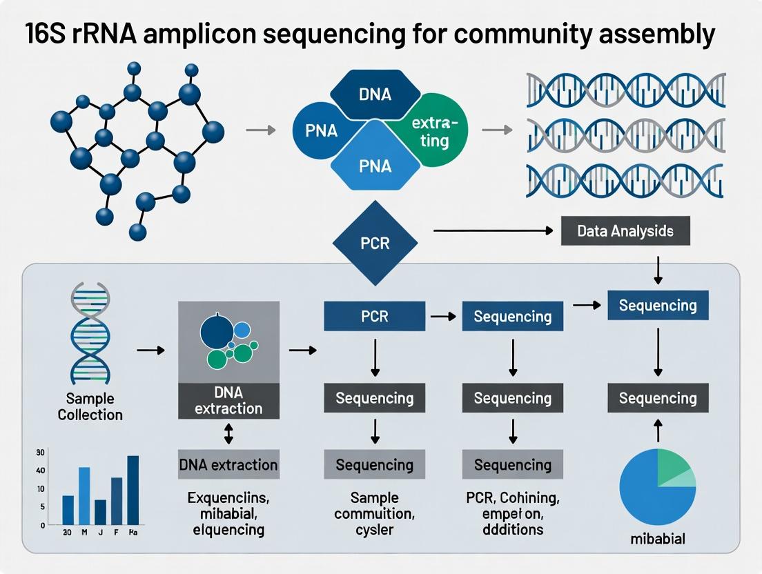
Within the context of 16S rRNA amplicon sequencing for microbial community assembly research, the 16S rRNA gene serves as the cornerstone for taxonomic identification and phylogenetic analysis. Its universal presence, conserved structure with hypervariable regions, and extensive reference databases enable researchers to profile complex microbial communities from diverse environments, from the human gut to extreme ecological niches.
Key Quantitative Data: Primer Performance and Sequencing Metrics
Table 1: Common 16S rRNA Gene Primer Pairs and Their Coverage
| Primer Pair (Name) | Target Region | Approx. Amplicon Length (bp) | Estimated Bacterial Coverage* (%) | Estimated Archaeal Coverage* (%) | Key References |
|---|---|---|---|---|---|
| 27F / 338R | V1-V2 | ~310 | 80-85 | Low | Klindworth et al., 2013 |
| 338F / 806R | V3-V4 | ~468 | 90-95 | Moderate | Caporaso et al., 2011 |
| 515F / 806R (515F-Y) | V4 | ~291 | 92-98 | High (with modifications) | Parada et al., 2016; Apprill et al., 2015 |
| 515F / 926R | V4-V5 | ~411 | 95-99 | High | Parada et al., 2016 |
| 8F / 534R | V1-V3 | ~526 | 75-80 | Very Low | Baker et al., 2003 |
Coverage estimates based on *in silico analysis against databases like SILVA or Greengenes. Performance varies with sample type and sequencing platform.
Table 2: Typical 16S Amplicon Sequencing Output and Analysis Metrics
| Metric | Illumina MiSeq v2 (2x250) | Illumina MiSeq v3 (2x300) | Illumina NovaSeq (2x250) | Notes |
|---|---|---|---|---|
| Reads per Run | 15-25 million | 20-30 million | 2-4 billion | Total output; can multiplex hundreds of samples. |
| Recommended Reads per Sample | 20,000 - 50,000 | 30,000 - 70,000 | 50,000 - 100,000 | Depends on community complexity and saturation. |
| Post-QC Read Length (merged) | ~250-420 bp | ~400-550 bp | ~250-420 bp | Affected by overlap and primer region. |
| Typical ASV/OTU Yield | 100 - 5,000+ | 100 - 5,000+ | 100 - 5,000+ | Varies drastically with ecosystem. |
| Alpha Diversity (Shannon Index) Range | 1.0 - 10.0+ | 1.0 - 10.0+ | 1.0 - 10.0+ | Soil: High (8-10); Clinical: Often lower (1-4). |
Core Experimental Protocol: 16S rRNA Gene Amplicon Library Preparation for Illumina Sequencing
Protocol: Library Preparation using Dual-Indexed Primers This protocol is adapted from the Earth Microbiome Project and widely used for community assembly studies.
I. Sample Lysis and Genomic DNA Extraction
- Method: Use a standardized kit (e.g., DNeasy PowerSoil Pro Kit) to ensure reproducibility.
- Steps:
- Aliquot 0.25g of sample (soil, stool) or pellet from 1-2mL liquid culture into a PowerBead Tube.
- Add Solution CD1. Secure tubes and homogenize using a bead-beater (45 sec, 5 m/s).
- Incubate at 65°C for 10 minutes. Centrifuge (10,000 x g, 30 sec).
- Transfer supernatant to a clean tube. Add Solution CD2, vortex, incubate on ice (5 min), centrifuge.
- Load supernatant onto a silica membrane column. Wash with buffers CB and EA.
- Elute DNA in 50-100 µL of Solution EB. Quantify using a fluorometric assay (e.g., Qubit).
II. First-Stage PCR: Target Amplification with Barcoded Primers
- Objective: Amplify the target hypervariable region (e.g., V4) while attaching sample-specific dual indices and Illumina adapter sequences.
- Reaction Mix (25 µL):
- 12.5 µL 2x High-Fidelity Master Mix (e.g., KAPA HiFi)
- 5.5 µL PCR-grade water
- 0.5 µL Forward Primer (10 µM; e.g., 515F with Illumina i5 overhang)
- 0.5 µL Reverse Primer (10 µM; e.g., 806R with Illumina i7 overhang)
- 1.0 µL Template DNA (1-10 ng)
- Thermocycling Conditions:
- 95°C for 3 min (initial denaturation)
- 25-35 cycles of:
- 95°C for 30 sec (denaturation)
- 55°C for 30 sec (annealing)
- 72°C for 30 sec (extension)
- 72°C for 5 min (final extension)
- Hold at 4°C.
- Clean-up: Purify amplicons using a magnetic bead-based clean-up kit (e.g., AMPure XP beads) at a 0.8x bead-to-sample ratio. Elute in 30 µL.
III. Library Validation and Quantification
- Assess library quality and size on a Bioanalyzer or TapeStation using a High Sensitivity DNA kit. Expect a single peak ~550 bp (for V4 with adapters).
- Quantify libraries fluorometrically. Normalize all libraries to 4 nM.
IV. Pooling and Sequencing
- Combine equal volumes of normalized libraries into a single pool.
- Denature the pool with NaOH, dilute to 8-12 pM in hybridization buffer, and load onto the Illumina cartridge. Include a 10-15% PhiX control to mitigate low-diversity issues.
- Sequence using a 2x250 bp or 2x300 bp paired-end kit.
Visualization of Workflows
Diagram 1: 16S Amplicon Sequencing Analysis Pipeline
Diagram 2: Primer Binding on the 16S rRNA Gene
The Scientist's Toolkit: Research Reagent Solutions
Table 3: Essential Materials for 16S rRNA Amplicon Workflow
| Item | Function & Rationale | Example Product |
|---|---|---|
| High-Efficiency DNA Extraction Kit | Consistent lysis of diverse cell walls (Gram+, Gram-, spores). Inhibitor removal is critical for downstream PCR. | DNeasy PowerSoil Pro Kit (Qiagen), MagMAX Microbiome Kit (Thermo) |
| High-Fidelity PCR Master Mix | Reduces PCR errors, essential for accurate Amplicon Sequence Variant (ASV) calling. | KAPA HiFi HotStart ReadyMix, Q5 High-Fidelity Master Mix (NEB) |
| Validated 16S Primer Cocktails | Primer sets with balanced coverage for Bacteria and/or Archaea, pre-fused to Illumina adapters. | 16S V4 Primer Set (515F/806R) from Integrated DNA Technologies (IDT) |
| Magnetic Bead Clean-up Reagent | For size-selective purification of PCR amplicons and library normalization. Less biased than column methods. | AMPure XP Beads (Beckman Coulter) |
| Fluorometric DNA Quantification Kit | Accurate quantification of low-concentration DNA and libraries. More accurate than absorbance (A260). | Qubit dsDNA HS Assay Kit (Thermo Fisher) |
| Library Quality Control Kit | Assesses library fragment size distribution and detects adapter dimers. | Agilent High Sensitivity DNA Kit (Agilent) |
| Sequencing Control | Improves base calling on low-diversity amplicon runs by adding nucleotide diversity. | PhiX Control v3 (Illumina) |
| Bioinformatics Pipeline Software | Containerized, reproducible analysis suite for processing raw reads to biological insights. | QIIME 2 Core Distribution, DADA2 R package |
Application Notes
The application of 16S rRNA amplicon sequencing within community assembly research frameworks has become pivotal for elucidating the microbiome's role in human pathophysiology and therapeutic outcomes. These studies move beyond correlation to investigate principles of ecological assembly—such as selection, drift, dispersal, and speciation—that govern microbiome composition in health and its disruption in disease. Insights into these assembly rules are critical for developing microbiota-targeted diagnostics and interventions.
1. Dysbiosis and Disease Association: Comparative case-control studies identify microbial taxa and community structures (e.g., reduced diversity, specific pathogen enrichment) associated with conditions like Inflammatory Bowel Disease (IBD), colorectal cancer, and metabolic syndrome. Quantitative metrics derived from sequencing data are analyzed through an ecological lens to determine if disease states exert a stronger "selection" pressure on the community.
2. Drug Metabolism and Efficacy: The gut microbiota directly modulates the pharmacokinetics and pharmacodynamics of numerous drugs, including chemotherapeutics (e.g., 5-fluorouracil), cardiac glycosides (digoxin), and immunotherapies (checkpoint inhibitors). Research focuses on identifying bacterial taxa and genes responsible for biotransformation and linking inter-individual microbiome variation to drug response heterogeneity.
3. Microbiome as a Therapeutic Target: Evaluating the impact of interventions (e.g., probiotics, prebiotics, fecal microbiota transplantation) on community reassembly. Protocols assess whether interventions can shift a dysbiotic community state toward a healthier assembly, often measuring the resilience of new states.
Table 1: Key Quantitative Metrics in Microbiota-Disease Research
| Metric | Typical Value in Health (Fecal) | Typical Shift in Disease (e.g., IBD) | Ecological Interpretation |
|---|---|---|---|
| Alpha Diversity (Shannon Index) | 3.5 - 5.5 | Often decreased (e.g., 2.0 - 3.5) | Reduced niche diversity or increased host selection. |
| Firmicutes/Bacteroidetes Ratio | Highly variable (~0.1 - 10) | Often altered, direction inconsistent | Shift in dominant community assembly processes. |
| Faecalibacterium prausnitzii Abundance | High (common core taxon) | Consistently decreased | Loss of a beneficial taxa possibly due to hostile environment. |
| Beta Diversity (Bray-Curtis) Distance | -- | Significant separation between health/disease groups (PERMANOVA p<0.05) | Distinct community state types driven by disease. |
Table 2: Microbial Impact on Drug Response
| Drug Class | Example Drug | Microbial Modifier | Effect | Consequence |
|---|---|---|---|---|
| Immunotherapy | Anti-PD-1/PD-L1 | Akkermansia muciniphila, Bifidobacterium spp. | Enhances efficacy | Higher response rates in patients with high abundance. |
| Cardiac Glycoside | Digoxin | Eggerthella lanta | Inactivates drug | Reduces therapeutic effect. |
| Chemotherapy | 5-Fluorouracil | Fusobacterium nucleatum | Potential resistance | Associated with poorer outcomes in colorectal cancer. |
| Parkinson's Therapy | Levodopa (L-dopa) | Enterococcal tyrosine decarboxylase | Decarboxylation in gut | Reduces drug bioavailability. |
Experimental Protocols
Protocol 1: 16S rRNA Amplicon Sequencing for Community Assembly Analysis
Objective: To profile microbial community composition from fecal samples and analyze data within an ecological assembly framework.
Materials:
- Fecal Sample Collection Kit: (e.g., OMNIgene•GUT kit) Stabilizes microbial DNA at ambient temperature.
- DNA Extraction Kit: (e.g., Qiagen DNeasy PowerSoil Pro Kit) Efficiently lyses tough bacterial cell walls and removes PCR inhibitors.
- PCR Reagents: High-fidelity DNA polymerase (e.g., Q5 Hot Start), primers targeting the V3-V4 hypervariable region (e.g., 341F/806R).
- Sequencing Platform: Illumina MiSeq or NovaSeq, using 2x300 bp paired-end chemistry.
- Bioinformatics Pipeline: QIIME 2 (2024.2), DADA2 for ASV inference, SILVA database v138 for taxonomy assignment, and R packages (phyloseq, picante) for analysis.
Procedure:
- Sample Collection & Stabilization: Collect fecal sample in stabilization solution, homogenize, and store at room temperature or -80°C.
- Genomic DNA Extraction: Follow kit protocol. Include bead-beating step. Quantify DNA using fluorometry (e.g., Qubit).
- Library Preparation:
- Perform first-stage PCR (25-30 cycles) with barcoded primers to amplify the 16S target region.
- Clean amplicons using magnetic beads (e.g., AMPure XP).
- Optional: Perform a second, limited-cycle PCR to add full sequencing adapters.
- Pool libraries equimolarly based on qPCR or fragment analyzer quantification.
- Sequencing: Load pooled library onto sequencer following manufacturer's instructions. Aim for >50,000 reads per sample.
- Bioinformatic Analysis:
- Demultiplex sequences and quality filter using QIIME 2.
- Denoise with DADA2 to generate Amplicon Sequence Variants (ASVs).
- Assign taxonomy using a pre-trained classifier.
- Construct a phylogenetic tree (e.g., with MAFFT/FastTree).
- Calculate diversity metrics (alpha: Shannon, Faith PD; beta: Weighted/Unweighted UniFrac, Bray-Curtis).
- Community Assembly Statistics:
- Use null model analysis (e.g.,
picante::ses.mpd) to calculate standardized effect sizes of phylogenetic diversity, inferring the relative roles of deterministic vs. stochastic assembly. - Apply PERMANOVA (e.g.,
vegan::adonis2) to partition variance in beta diversity among factors (e.g., disease state, drug treatment).
- Use null model analysis (e.g.,
Protocol 2: In Vitro Culturing for Drug-Biotransformation Assay
Objective: To validate the ability of a specific bacterial isolate to metabolize a target drug.
Materials:
- Anaerobic Workstation: (e.g., Whitley A95) for cultivating obligate anaerobes.
- Reduced Culture Medium: Pre-reduced brain heart infusion (BHI) or specific defined medium.
- Target Drug: Pharmaceutical grade.
- Analytical Instrumentation: LC-MS/MS for drug and metabolite quantification.
Procedure:
- Culture Inoculation: Grow the bacterial strain of interest to mid-log phase in appropriate anaerobic conditions.
- Drug Exposure: Aliquot bacterial culture into multiple vials. Add the target drug at a physiologically relevant concentration (e.g., 10 µM). Include controls: drug + sterile medium (chemical stability), and drug + killed bacteria (non-enzymatic binding).
- Incubation: Incubate anaerobically at 37°C for a defined period (e.g., 2, 6, 24 hours).
- Reaction Termination: At each time point, add an equal volume of ice-cold acetonitrile or methanol to precipitate proteins and stop metabolism. Centrifuge to pellet cells and debris.
- Sample Analysis: Analyze supernatant by LC-MS/MS to quantify the depletion of parent drug and appearance of known metabolites. Compare peak areas against standard curves.
- Kinetic Analysis: Calculate the rate of drug depletion/metabolite formation per unit of bacterial cell density (OD600 or cell count).
The Scientist's Toolkit: Research Reagent Solutions
| Item | Function & Rationale |
|---|---|
| OMNIgene•GUT Kit (DNA Genotek) | Stabilizes microbial composition at room temperature for up to 60 days, preventing shifts and enabling feasible sample transport. |
| Qiagen DNeasy PowerSoil Pro Kit | Optimized for soil/fecal samples; includes bead-beating for mechanical lysis and reagents to remove humic acids/PCR inhibitors. |
| ZymoBIOMICS Microbial Community Standard | Defined mock community of bacteria and fungi. Serves as a positive control and standard for evaluating extraction, sequencing, and bioinformatics pipeline accuracy. |
| PMA (Propidium Monoazide) Dye | Binds DNA of dead cells with compromised membranes. Used with PMA-seq to profile only the viable microbiome component. |
| AnaeroPack System (Mitsubishi Gas Chemical) | Creates anaerobic atmosphere in jars for culturing oxygen-sensitive gut bacteria without a full workstation. |
| Picodent Twinsil Dental Impression Material | For creating custom gaskets to seal 96-well plates for anaerobic high-throughput screening of bacterial growth/drug effects. |
Visualizations
Title: 16S rRNA Sequencing Workflow for Microbiota Applications
Title: Microbiota-Mediated Modulation of Drug Response
Title: Community State Transitions and Intervention
Within the framework of 16S rRNA amplicon sequencing for community assembly research, the fundamental step of grouping sequences into biologically meaningful units has evolved significantly. This evolution reflects a broader thesis shift from inferring community structure based on operational definitions to characterizing it based on exact biological sequences. The choice of metric—Operational Taxonomic Units (OTUs) versus Amplicon Sequence Variants (ASVs) or Exact Sequence Variants (ESVs)—is not merely technical but philosophical, impacting downstream ecological interpretations, cross-study comparisons, and translational applications in drug development and microbiome therapeutics.
Conceptual Definitions & Philosophical Underpinnings
Operational Taxonomic Unit (OTU): An OTU is a cluster of sequencing reads grouped based on a user-defined sequence similarity threshold (typically 97% for species-level). It is an operational definition, acknowledging that sequencing errors and intra-genomic variation exist, and that clustering is a practical method to estimate species diversity. The philosophy is one of approximation and noise reduction through clustering.
Amplicon/Exact Sequence Variant (ASV/ESV): An ASV (or ESV) is a unique, exact ribosomal sequence generated by error-correcting algorithms (e.g., DADA2, Deblur, UNOISE). It treats each unique sequence as a biologically relevant unit, distinguishing true biological variation from sequencing error. The philosophy is one of precision and reproducibility, aiming to identify the exact biological sequences present.
Core Philosophical Difference: OTU clustering is a phenetic approach (grouping by overall similarity), while ASV generation is a discrete approach (identifying unique entities). This impacts the perception of microbial diversity, stability of identifiers across studies, and resolution for detecting subtle shifts.
Table 1: Comparative Analysis of OTU vs. ASV Methodologies
| Feature | OTU (97% Clustering) | ASV/ESV (DADA2, Deblur) |
|---|---|---|
| Definition Basis | Similarity threshold (e.g., 97%, 99%) | Exact, error-corrected sequence |
| Primary Algorithm | Hierarchical/UPARSE, VSEARCH, CD-HIT | DADA2 (Divisive Amplicon Denoising), Deblur, UNOISE3 |
| Treatment of Errors | Clustered together, assumed to be noise | Modeled and removed statistically |
| Resolution | Species or genus-level (97% threshold) | Single-nucleotide, sub-species level |
| Reproducibility Across Studies | Low (cluster composition is dataset-dependent) | High (exact sequences are portable) |
| Perceived Richness | Generally lower (clustering reduces units) | Generally higher (retains subtle variants) |
| Computational Demand | Moderate | Higher (intensive error modeling) |
| Common File Output | OTU Table (BIOM format) | ASV Table (BIOM/TSV format) |
| Downstream Taxonomic ID | Assigned to cluster consensus/repr. seq | Assigned to each exact sequence |
Table 2: Impact on Key Alpha-Diversity Metrics (Hypothetical Data from Mock Community)
| Metric | True Composition | OTU-based (97%) | ASV-based |
|---|---|---|---|
| Number of Units | 20 strains | 18 (± 3) | 22 (± 2)* |
| Shannon Index | 2.85 | 2.70 (± 0.15) | 2.88 (± 0.10) |
| Observed Richness | 20 | 17.5 (± 1.8) | 21.1 (± 1.2)* |
| Notes: *ASV methods may slightly overestimate due to residual artifacts or genuine intra-genomic variation. |
Detailed Experimental Protocols
Protocol 4.1: Traditional OTU Picking via VSEARCH (Open-Source Pipeline)
Objective: To generate an OTU table from demultiplexed 16S rRNA paired-end reads using a 97% similarity threshold.
Materials: Demultiplexed FASTQ files, QIIME2 (2024.5+) or standalone VSEARCH, SILVA/GTDB reference database.
Procedure:
- Primer Removal & Quality Filtering: Use
cutadaptto remove primer sequences. Merge paired-end reads usingvsearch --fastq_mergepairswith quality filtering (expected error--fastq_maxee_rate 1.0). - Dereplication: Combine all sequences and dereplicate:
vsearch --derep_fulllength merged.fasta --output uniques.fasta --sizeout. - Chimera Detection (Reference-based):
vsearch --uchime_ref uniques.fasta --db reference_db.fasta --nonchimeras nonchimeras.fasta. - OTU Clustering: Cluster non-chimeric sequences at 97%:
vsearch --cluster_size nonchimeras.fasta --id 0.97 --centroids otus.fasta --relabel OTU_ --sizein --sizeout. - OTU Table Construction: Map all quality-filtered reads back to OTUs:
vsearch --usearch_global merged.fasta --db otus.fasta --id 0.97 --otutabout otu_table.tsv. - Taxonomic Assignment: Assign taxonomy to OTU representative sequences using a classifier (e.g.,
qiime feature-classifier classify-sklearn) against a reference database.
Protocol 4.2: ASV Generation via DADA2 (R Pipeline)
Objective: To infer exact Amplicon Sequence Variants from raw 16S rRNA reads.
Materials: Raw FASTQ files, R (4.3.0+), DADA2 package (1.30.0+), high-performance computing recommended.
Procedure:
- Filter & Trim: Inspect quality profiles (
plotQualityProfile). Filter reads:filterAndTrim(fwd, filt_fwd, rev, filt_rev, truncLen=c(240,200), maxN=0, maxEE=c(2,2), truncQ=2, rm.phix=TRUE, compress=TRUE). Adjust truncation length based on quality drop. - Learn Error Rates: Model the sequencing error rate:
errF <- learnErrors(filt_fwd, multithread=TRUE);errR <- learnErrors(filt_rev, multithread=TRUE). - Dereplication:
derepF <- derepFastq(filt_fwd, verbose=TRUE); similarly for reverse. - Core Sample Inference: Run the DADA algorithm:
dadaF <- dada(derepF, err=errF, multithread=TRUE);dadaR <- dada(derepR, err=errR, multithread=TRUE). - Merge Paired Reads: Merge denoised forward and reverse reads:
mergers <- mergePairs(dadaF, derepF, dadaR, derepR, verbose=TRUE). - Construct Sequence Table:
seqtab <- makeSequenceTable(mergers). - Remove Chimeras:
seqtab.nochim <- removeBimeraDenovo(seqtab, method="consensus", multithread=TRUE, verbose=TRUE). - Assign Taxonomy: Assign taxonomy via
assignTaxonomy(seqtab.nochim, "reference_db.fasta.gz", multithread=TRUE). The resultingseqtab.nochimis the ASV count table.
Visualization of Methodologies
Diagram 1: Comparative Workflow: OTU Clustering vs ASV Inference (67 chars)
The Scientist's Toolkit: Research Reagent & Computational Solutions
Table 3: Essential Reagents, Software, and Databases for 16S rRNA Amplicon Analysis
| Item Name | Type | Function & Brief Explanation |
|---|---|---|
| KAPA HiFi HotStart ReadyMix | Wet-Lab Reagent | High-fidelity polymerase for accurate amplification of the 16S target region, minimizing PCR bias. |
| Nextera XT Index Kit | Wet-Lab Reagent | Used for dual-indexing PCR to allow multiplexing of hundreds of samples on Illumina sequencers. |
| PhiX Control v3 | Wet-Lab Reagent | Internal sequencing control for Illumina runs; improves base calling accuracy on low-diversity amplicon libraries. |
| QIIME 2 (2024.5+) | Software Platform | Reproducible, extensible microbiome analysis pipeline supporting both OTU and ASV workflows. |
| DADA2 (R Package) | Software Package | Primary algorithm for modeling sequencing errors and inferring exact ASVs from amplicon data. |
| VSEARCH | Software Tool | Open-source, 64-bit alternative to USEARCH for OTU clustering, chimera detection, and read merging. |
| SILVA SSU Ref NR 99 | Reference Database | Curated database of aligned ribosomal RNA sequences for taxonomic assignment (updated regularly). |
| GTDB (R07-RS220) | Reference Database | Genome-based Taxonomy Database, provides phylogenetically consistent taxonomy for genomes/ASVs. |
| Mock Community (e.g., ZymoBIOMICS) | Control Standard | Defined microbial mixture used as a positive control to evaluate sequencing accuracy and bioinformatic pipeline performance. |
| Mag-Bind TotalPure NGS | Wet-Lab Reagent | Magnetic beads for PCR clean-up and library normalization, ensuring even representation in final pool. |
Within the framework of a thesis on 16S rRNA amplicon sequencing community assembly, primer selection is a foundational experimental design choice. The 16S rRNA gene contains nine hypervariable regions (V1-V9) interspersed with conserved sequences. No single region universally provides the highest taxonomic resolution across all bacterial phyla, making the selection of an optimal region—or combination of regions—critical for accurate microbial community profiling. This document synthesizes current data and provides protocols to guide this selection process.
Comparative Analysis of Hypervariable Regions
The following table summarizes the key attributes of each V region based on current literature, focusing on their utility for taxonomic resolution.
Table 1: Characteristics and Taxonomic Resolution of 16S rRNA Hypervariable Regions
| Region | Approx. Length (bp) | Taxonomic Resolution (General) | Key Strengths | Key Limitations |
|---|---|---|---|---|
| V1-V2 | ~340 | High for many Firmicutes, Bacteroidetes | Often provides species-level resolution for gut microbiota; well-suited for short-read platforms (e.g., MiSeq). | Poor resolution for Actinobacteria; prone to chimerism. |
| V3-V4 | ~460 | Medium-High (Broadly applicable) | Most commonly used (e.g., 341F/806R); good balance of length and information; comprehensive database coverage. | May miss discrimination for specific genera (e.g., Streptococcus). |
| V4 | ~290 | Medium (Broadly applicable) | Highly accurate and reproducible; minimal chimera formation; recommended by Earth Microbiome Project. | Shorter length limits phylogenetic information compared to longer spans. |
| V4-V5 | ~390 | Medium-High | Good resolution for environmental and diverse communities; often used in marine studies. | Slightly lower resolution for some gut taxa compared to V1-V2 or V3-V4. |
| V5-V7 / V6-V8 | ~400-500 | Varies by taxa | Useful for specific phyla like Cyanobacteria and Planctomycetes. | Not universally optimal; requires validation for target community. |
| Full-length (V1-V9) | ~1500 | Highest (Gold Standard) | Enables near-complete phylogenetic reconstruction and highest species/strain-level discrimination. | Requires long-read sequencing (PacBio, Oxford Nanopore); higher cost/per-sample. |
Table 2: Recommended Region Selection by Primary Research Goal
| Primary Research Goal | Recommended Region(s) | Rationale |
|---|---|---|
| Broad microbial profiling (e.g., human gut) | V3-V4 or V4 | Optimal balance of fidelity, coverage, and compatibility with Illumina MiSeq (2x300bp). |
| Maximizing species-level resolution in specific environments | V1-V2 or V1-V3 | For studies focusing on Firmicutes/Bacteroidetes-dominated systems (e.g., vaginal microbiome). |
| High-resolution community assembly for novel taxa | Full-length 16S (V1-V9) | Essential for discovering and phylogenetically placing novel lineages in complex environments. |
| Pathogen detection / strain tracking | Full-length or V1-V3/V3-V4 multi-region | Combines broad profiling (V3-V4) with high-discrimination power (V1-V3) for precise identification. |
Experimental Protocols
Protocol 1:In SilicoAssessment of Primer Pairs
Objective: To computationally predict the coverage and taxonomic discrimination of primer pairs for your target community.
- Obtain Reference Databases: Download curated 16S rRNA gene databases (e.g., SILVA, Greengenes, RDP).
- Define Target Sequences: Extract full-length 16S sequences representing your expected microbial community or isolate genomes of interest.
- Primer Matching: Use tools like
TestPrime(in mothur) orecoPCRto evaluate:- Coverage: The percentage of target sequences that perfectly match or have ≤1 mismatch to the primer.
- Specificity: The proportion of matches that are to the target domain (Bacteria/Archaea).
- Amplicon Length Distribution: Confirm the expected product size is uniform.
- Resolution Simulation: Use alignment and simple tree-building (e.g., FastTree) on the in silico amplicons from different V regions to compare branch lengths and clustering patterns at genus/species levels.
Protocol 2: Wet-Lab Validation via Mock Community Sequencing
Objective: To empirically evaluate the accuracy, resolution, and bias of selected primer pairs.
Materials: Defined Mock Microbial Community (e.g., ZymoBIOMICS Microbial Community Standard), selected primer pairs, high-fidelity PCR mix, magnetic bead cleanup system, sequencer.
- PCR Amplification: Amplify the mock community DNA in triplicate with each primer pair candidate. Use a minimal number of PCR cycles (e.g., 25-30) to reduce bias.
- Library Preparation & Sequencing: Purify amplicons, attach dual-index barcodes and sequencing adapters per standard Illumina protocols. Pool libraries and sequence on an appropriate platform (e.g., MiSeq for V3-V4, PacBio Sequel IIe for full-length).
- Bioinformatic Analysis:
- Process reads through a standard pipeline (DADA2, QIIME 2, mothur).
- Generate Amplicon Sequence Variants (ASVs).
- Accuracy Assessment: Map ASVs to the known mock community reference sequences. Calculate the rate of spurious ASVs, chimeras, and the sensitivity of detecting all expected taxa.
- Bias Quantification: Compare the observed read count proportions to the known genomic DNA abundance in the mock community. Calculate the log2 fold-change deviation for each member.
Visualizations
Diagram 1: Primer Selection Workflow for Community Assembly (99 chars)
Diagram 2: Primer Binding and Amplicon Span Across V Regions (99 chars)
The Scientist's Toolkit: Research Reagent Solutions
Table 3: Essential Materials for Hypervariable Region Selection Studies
| Item | Function in This Context | Example Product(s) |
|---|---|---|
| Defined Mock Community | Ground truth standard for validating primer accuracy, bias, and limit of detection. | ZymoBIOMICS Microbial Community Standard, ATCC Mock Microbial Communities. |
| High-Fidelity DNA Polymerase | Minimizes PCR errors during amplicon generation, critical for creating accurate ASVs. | Q5 Hot Start (NEB), KAPA HiFi HotStart ReadyMix. |
| Magnetic Bead Cleanup Kits | For size selection and purification of amplicons post-PCR and post-ligation to remove primer dimers and contaminants. | AMPure XP Beads (Beckman), SPRISelect (Beckman). |
| Dual-Index Barcoding Kit | Allows multiplexing of hundreds of samples with unique barcodes for Illumina sequencing. | Nextera XT Index Kit, 16S Metagenomic Sequencing Library Prep (Illumina). |
| Long-read Sequencing Kit | Essential for generating full-length (V1-V9) amplicons. | SMRTbell Express Template Prep Kit 3.0 (PacBio), Ligation Sequencing Kit (Oxford Nanopore). |
| Curated 16S Database | Essential for in silico primer testing and downstream taxonomic classification. | SILVA SSU NR, Greengenes, RDP Database. |
| Primer Design/Testing Software | For in silico evaluation of primer coverage, specificity, and amplicon length. | ecoPCR (OBITools), TestPrime (mothur), Primer-BLAST (NCBI). |
The analysis of microbial communities via 16S rRNA gene amplicon sequencing is a cornerstone of modern microbiome research, with direct implications for drug development, diagnostics, and therapeutic discovery. This Application Note delineates the foundational bioinformatics concepts—raw sequencing reads, demultiplexing, and the primary analysis ecosystems—framed within a thesis on community assembly dynamics. The accurate processing of raw data is critical for downstream ecological inference, including alpha/beta diversity metrics, differential abundance testing, and biomarker identification, which inform translational applications.
Core Concepts: Reads and Demultiplexing
Sequencing Reads: Raw output from next-generation sequencing platforms (e.g., Illumina MiSeq, NovaSeq), representing short DNA sequences from amplified target regions (e.g., V4 region of 16S rRNA). Quality is quantified per base position using Phred scores (Q).
Demultiplexing: The process of assigning each sequencing read to its sample of origin based on sample-specific barcode sequences (indexes) added during PCR preparation. This is the first computational step post-sequencing.
Table 1: Common Illumina Sequencing Output Metrics for 16S Studies
| Metric | Typical Value (MiSeq V4-V5) | Significance |
|---|---|---|
| Read Length (bp) | 250 - 300 (paired-end) | Determines gene region coverage. |
| Total Reads/Run | 15 - 25 million | Defines sampling depth per sample. |
| Q-score Threshold (Q) | ≥ 30 (Q30) | Indicates 99.9% base call accuracy. |
| Barcode Length (bp) | 8 - 12 | Uniquely identifies each sample. |
Detailed Protocol: Demultiplexing and Initial Quality Control
Protocol Title: Demultiplexing of Dual-Indexed 16S Amplicons and Generation of Raw Read Tables.
Reagents & Materials:
- Raw sequencing data (
.fastq.gzfiles) for Read 1, Read 2, and Index reads. - Sample metadata file containing barcode sequences for each sample ID.
- Computing resources (minimum 8GB RAM, 4 cores).
Procedure (using QIIME 2 tools as exemplar):
- Create a QIIME 2 Manifest File: Format a comma-separated file specifying the absolute filepaths for
forward-fastq,reverse-fastq, andbarcode-fastqfiles, and the sample identifier. - Import Data: Use
qiime tools importwith theSampleData[PairedEndSequencesWithQuality]type and theEMPPairedEndSequencesformat. - Execute Demultiplexing: Run
qiime demux emp-pairedusing the imported data. This step matches barcodes, assigns reads to samples, and discards unmatched reads. - Summarize Output: Generate and visualize a summary with
qiime demux summarizeto assess per-sample sequence counts and initial quality scores. - Generate Raw Data Table: The output is a
FeatureTable[Sequences]artifact, representing the count of raw reads per sample.
Troubleshooting: Low yield per sample may indicate barcode hopping/index switching. Apply strict quality filtering on barcode reads or use dual-index-aware demultiplexing algorithms.
Ecosystem Comparison: QIIME 2, MOTHUR, and Usearch/Vsearch
Table 2: Comparison of Major 16S rRNA Analysis Ecosystems
| Feature | QIIME 2 | MOTHUR | Usearch/Vsearch |
|---|---|---|---|
| Primary Architecture | Plugin-based, extensible platform. | Monolithic, all-in-one executable. | Suite of fast, individual commands. |
| Core Methodology | Deblur (error correction) or DADA2 (denoising). | Traditional OTU clustering (e.g., dist.seqs, cluster). |
High-speed OTU clustering (cluster_fast) and dereplication. |
| Input/Output | Artifact system (.qza/.qzv) with provenance tracking. |
Multiple file formats (.fasta, .names, .groups). |
Standard .fasta/.fastq with custom report files. |
| User Interface | Command-line (qiime) with visualizations. |
Command-line interactive or scripted. | Command-line non-interactive. |
| Strengths | Reproducibility, comprehensive tutorials, visualization. | Extensive SOPs, fine-grained control, stable algorithms. | Exceptional speed, low memory footprint. |
| Best Suited For | End-to-end reproducible analysis, large collaborative projects. | Research closely following classic 16S literature, custom pipelines. | Large datasets where computational speed is critical. |
Protocol: From Raw Reads to Amplicon Sequence Variants (ASVs) in QIIME 2
Protocol Title: DADA2 Denoising Pipeline for Generating ASVs in QIIME 2.
Procedure:
- Import Demultiplexed Reads: Start with the
SampleData[PairedEndSequencesWithQuality]artifact from Section 3. - Denoise with DADA2: Execute
qiime dada2 denoise-paired. Key parameters:--p-trunc-len-fand--p-trunc-len-r: Set based on quality plots (e.g., 220, 200).--p-trim-left-fand--p-trim-left-r: Remove primer sequences (e.g., 15, 15).--p-max-ee: Maximum expected errors per read (e.g., 2.0).--p-chimera-method:consensus.
- Outputs: The command produces:
FeatureTable[Frequency]: Count table of ASVs per sample.FeatureData[Sequence]: Representative sequences for each ASV.SampleData[DADA2Stats]: Denoising statistics per sample.
- Filter Singletons (Optional): Remove ASVs with total abundance = 1 using
qiime feature-table filter-features --p-min-frequency 2.
Workflow Diagram: 16S Amplicon Data Processing Pipeline
Diagram Title: 16S Amplicon Processing Workflow.
The Scientist's Toolkit: Key Research Reagent Solutions
Table 3: Essential Materials for 16S rRNA Amplicon Sequencing Experiments
| Item | Function & Application Notes |
|---|---|
| PCR Primers with Adapters (e.g., 515F/806R) | Amplify the target hypervariable region; contain flow cell adapter and barcode landing sites. |
| Dual Index Barcode Kits (e.g., Illumina Nextera XT) | Provide unique sample identifiers for multiplexing, reducing index hopping rates. |
| High-Fidelity DNA Polymerase (e.g., Phusion, KAPA HiFi) | Ensures accurate amplification with minimal PCR errors that confound sequence variants. |
| Magnetic Bead Cleanup Kits (e.g., AMPure XP) | Size selection and purification of amplicon libraries, removing primer dimers and contaminants. |
| Quantification Kits (e.g., Qubit dsDNA HS Assay) | Accurate pre-sequencing library quantification for precise pooling and loading. |
| PhiX Control v3 | Spiked into sequencing runs (1-5%) for low-diversity libraries to improve cluster detection and base calling. |
| Positive Control Mock Community DNA (e.g., ZymoBIOMICS) | Validates entire wet-lab and bioinformatics pipeline from extraction to analysis. |
| Negative Extraction Control (NEC) | Identifies contamination introduced during sample preparation. |
Logical Diagram: Ecosystem Selection Decision Path
Diagram Title: Selecting a 16S Analysis Ecosystem.
A Step-by-Step Pipeline: From Sample Collection to Community Analysis
Within the context of a 16S rRNA amplicon sequencing thesis investigating microbial community assembly, rigorous Phase 1 experimental design is foundational. This phase dictates the reliability, reproducibility, and interpretability of downstream sequencing data. Careful attention to cohort stratification, comprehensive control strategies, and statistical power analysis is required to mitigate biases and draw robust ecological inferences.
Cohort Selection and Stratification
Cohort selection aims to minimize confounding variation while capturing the biological signal of interest (e.g., disease state, treatment effect). Key considerations include host-intrinsic and extrinsic factors known to influence microbiota composition.
Table 1: Key Confounding Factors and Stratification Recommendations for 16S Cohort Design
| Factor | Impact on Microbiota | Recommended Stratification/Matching |
|---|---|---|
| Age | Taxonomic composition shifts dramatically over lifespan. | Cohort bands (e.g., 20-30, 40-50 years) or regression covariate. |
| BMI | Strongly associated with Firmicutes/Bacteroidetes ratio. | Match cases/controls within ±3 BMI points. |
| Diet | Major driver of short-term and long-term community structure. | Use validated FFQ and include as covariate or exclude extremes. |
| Antibiotics | Causes profound, long-lasting dysbiosis. | Exclude participants with antibiotic use within 3-6 months. |
| Geography | Influences microbial exposure and prevalent taxa. | Single-center study or multi-center stratified sampling. |
| Sample Collection | Time of day, fasting state, collection method affect data. | Standardize protocols across all participants. |
Control Strategy
Incorporating controls at each step distinguishes technical artifacts from biological signals.
Extraction Controls
- Negative Control: A "blank" extraction using no biological sample (e.g., lysis buffer only). Identifies contamination from extraction kits and laboratory environment.
- Positive Control: A mock microbial community with known, quantifiable composition (e.g., ZymoBIOMICS Microbial Community Standards). Assesses extraction efficiency, bias, and fidelity.
PCR Amplification Controls
- No-Template Control (NTC): Contains all PCR reagents except template DNA. Detects contamination in PCR master mix or primers.
- Positive PCR Control: Uses a well-characterized DNA template (e.g., from positive extraction control) to confirm PCR reagent efficacy.
ZymoBIOMICS Solutions as Integrated Controls
The ZymoBIOMICS product suite provides calibrated standards for end-to-end workflow validation.
Table 2: ZymoBIOMICS Controls for 16S Amplicon Sequencing Workflow
| Product Name | Composition | Function in Experimental Design |
|---|---|---|
| ZymoBIOMICS Microbial Community Standard (D6300) | Defined ratios of 8 bacterial and 2 fungal strains, with known genome copies. | Process Positive Control. Spiked into sample matrix or used alone to evaluate total workflow accuracy from extraction to bioanalysis. |
| ZymoBIOMICS Spike-in Control I (MOCK I) (D6320) | Even community of 10 bacteria. | Internal Control. Can be spiked into every sample pre-extraction to normalize and identify technical variation across samples. |
| ZymoBIOMICS DNA/RNA Miniprep Kit (R2002/R2003) | Kit includes a positive control. | Validates nucleic acid extraction and purification performance. |
Power and Sample Size Analysis
An a priori power analysis is essential to determine the minimum sample size required to detect a hypothesized effect. For microbial community data, this often relies on metrics like UniFrac distance or Shannon diversity.
Current Guidance (2024): Recent meta-analyses suggest microbiome effect sizes are often smaller than previously estimated. A conservative approach is recommended.
- For detecting differences in alpha diversity (e.g., Shannon index), a minimum of 15-20 samples per group is often required for moderate effects.
- For beta diversity (community composition), sample size needs are higher and depend on expected effect size (e.g., R² in PERMANOVA). Simulations using tools like
HMPorMKpowerin R are necessary.
Table 3: Example Power Analysis Output for a Two-Group Comparison (Case vs. Control)
| Target Metric | Effect Size (Assumed) | Significance Level (α) | Desired Power (1-β) | Minimum N per Group |
|---|---|---|---|---|
| Bray-Curtis Dissimilarity | R² = 0.05 (Small-Moderate) | 0.05 | 0.80 | ~45 |
| Weighted UniFrac Distance | R² = 0.10 (Moderate) | 0.05 | 0.80 | ~22 |
| Shannon Diversity | Cohen's d = 0.8 (Large) | 0.05 | 0.80 | ~20 |
Note: Effect size estimates (R², Cohen's d) should be derived from pilot data or published literature in your specific research niche.
Detailed Protocols
Protocol 1: Cohort Sample Collection and Preservation
Objective: Standardize collection of fecal samples for 16S analysis.
- Provide participants with a pre-labelled, sterile collection tube containing a stabilizing solution (e.g., DNA/RNA Shield).
- Instruct participants to collect a small aliquot (~200mg) immediately after defecation, using the provided spoon or stick.
- Ensure sample is fully immersed in stabilizer, tube is tightly sealed, and immediately refrigerated or frozen at -20°C.
- Transport to lab on ice and store at -80°C until extraction.
Protocol 2: Integrated Extraction with Controls
Objective: Extract microbial DNA incorporating negative and positive controls. Reagents: ZymoBIOMICS DNA Miniprep Kit, ZymoBIOMICS Microbial Community Standard (Positive Control), DNA/RNA Shield (Negative Control).
- Sample Lysis: Add 200μL of sample (or 200μL positive control resuspension, or 200μL Shield for negative control) to a BashingBead tube. Add 750μL lysis solution. Homogenize on a bead beater for 5 min.
- DNA Binding: Centrifuge at 10,000 x g for 1 min. Transfer 400μL supernatant to a Zymo-Spin III-F filter in a collection tube. Centrifuge at 8,000 x g for 1 min.
- Wash: Add 400μL DNA Wash Buffer to the filter. Centrifuge at 8,000 x g for 1 min. Repeat wash step.
- Elution: Transfer filter to a clean 1.5mL tube. Apply 20μL DNase/RNase-Free Water directly to the filter matrix. Centrifuge at 10,000 x g for 30 sec to elute DNA.
- Quantify DNA using a fluorometric assay (e.g., Qubit).
Protocol 3: 16S rRNA Gene Amplicon PCR with Controls
Objective: Amplify the V3-V4 hypervariable region with dual-index barcodes. Primers: 341F (5'-CCTACGGGNGGCWGCAG-3'), 806R (5'-GGACTACHVGGGTWTCTAAT-3') with Illumina overhang adapters. Reagents: 2x KAPA HiFi HotStart ReadyMix, PCR-grade water, template DNA (extracted samples, extraction positive control, extraction negative control, and a No-Template Control).
- Set up 25μL reactions: 12.5μL Master Mix, 1.25μL each forward/reverse primer (10μM), 5-20ng template DNA, water to volume.
- Thermocycling: 95°C for 3 min; 25 cycles of: 95°C for 30s, 55°C for 30s, 72°C for 30s; final extension 72°C for 5 min.
- Check PCR success and specificity via agarose gel electrophoresis (expect ~550bp band). The positive controls should show a strong band; negative controls should show no band.
The Scientist's Toolkit
Table 4: Essential Research Reagent Solutions for 16S Amplicon Study Design
| Item | Function & Rationale |
|---|---|
| DNA/RNA Shield (Zymo Research) | A sample preservation solution that instantly inactivates nucleases and stabilizes microbial community profiles at room temperature, crucial for cohort studies. |
| ZymoBIOMICS DNA Miniprep Kit | Optimized for mechanical lysis of diverse microbes and removal of PCR inhibitors from complex samples like stool and soil. |
| ZymoBIOMICS Microbial Community Standard | Defined mock community with published expected 16S profile. Serves as the primary process control to quantify technical error and batch effects. |
| KAPA HiFi HotStart ReadyMix | High-fidelity polymerase mix designed for robust amplification of complex amplicons like the 16S V3-V4 region, minimizing chimera formation. |
| Dual-Indexed PCR Primers (Nextera XT Index Kit) | Allows unique barcoding of hundreds of samples prior to pooling for multiplexed Illumina sequencing. |
| Agencourt AMPure XP Beads | For post-PCR purification to remove primer dimers and size-select the target amplicon, ensuring clean sequencing libraries. |
Visualizations
Title: Phase 1 Experimental Workflow for 16S Study
Title: Hierarchical Control Strategy for 16S Workflow
Within the context of a broader thesis investigating microbial community assembly via 16S rRNA amplicon sequencing, the integrity of the wet lab phase is paramount. This phase converts an environmental or clinical sample into a sequence-ready amplicon library. The selection between primer sets, notably the 515F-806R (targeting the V4 region) and 27F-338R (targeting the V1-V2 regions), is a critical methodological decision that influences downstream taxonomic resolution and bias. This document provides detailed Application Notes and Protocols for DNA extraction and PCR amplification, tailored for researchers, scientists, and drug development professionals.
Research Reagent Solutions Toolkit
| Reagent / Material | Function / Application |
|---|---|
| PowerSoil Pro Kit (Qiagen) | Efficiently lyses a wide range of microbial cells and removes PCR inhibitors (e.g., humic acids) from complex environmental samples. |
| Phusion High-Fidelity DNA Polymerase | Provides high fidelity and processivity for accurate amplification of the 16S rRNA gene, minimizing PCR errors. |
| Agencourt AMPure XP Beads | For post-PCR clean-up, size selection, and normalization of amplicon libraries, removing primer dimers and nonspecific products. |
| Qubit dsDNA HS Assay Kit | Fluorometric quantification of double-stranded DNA with high specificity, essential for accurate library pooling. |
| PNA Clamp Mix (for host-rich samples) | Peptide Nucleic Acid clamps block amplification of host (e.g., human) mitochondrial and chloroplast 16S rDNA, enriching for bacterial signal. |
| Dual-Indexed Primer Sets (e.g., Nextera XT) | Allows for combinatorial multiplexing of hundreds of samples in a single sequencing run with minimal index hopping risk. |
Protocol: DNA Extraction from Complex Microbial Communities
Principle: To obtain high-quality, inhibitor-free genomic DNA representative of the entire microbial community.
Detailed Protocol:
- Homogenization: Weigh 0.25 g of sample (soil, stool, biofilm) into a PowerSoil Bead Tube.
- Cell Lysis: Add provided Solution CD1 and secure on a vortex adapter. Vortex horizontally at maximum speed for 10 minutes.
- Inhibition Removal: Centrifuge at 10,000 x g for 30 sec. Transfer supernatant to a clean tube. Add 250 µL of Solution CD2, vortex for 5 sec, and incubate at 4°C for 5 min. Centrifuge at 10,000 x g for 1 min.
- DNA Binding: Transfer supernatant to a tube with 400 µL of Solution CD3 and 400 µL of ethanol. Vortex and load onto an MB Spin Column.
- Washes: Centrifuge and flow-through is discarded. Add 500 µL of Solution EA (ethanol-based), centrifuge, and discard flow-through. Add 500 µL of Solution EB (ethanol-based), centrifuge, and discard flow-through.
- Elution: Centrifuge empty column at 10,000 x g for 1 min to dry. Transfer column to a clean elution tube. Apply 50 µL of Solution C6 (10 mM Tris, pH 8.5) to the center of the membrane, incubate for 2 min, and centrifuge at 10,000 x g for 1 min to elute DNA.
- Quantification & Quality Control: Measure DNA concentration using Qubit. Assess purity via A260/A280 (expected: ~1.8) and A260/A230 (expected: >2.0) ratios. Verify integrity by running 1 µL on a 1% agarose gel (high molecular weight smear expected).
Protocol: PCR Amplification of the 16S rRNA Gene
Principle: To specifically amplify the target hypervariable region(s) of the bacterial/archaeal 16S rRNA gene with minimal bias and error.
Reaction Setup (25 µL):
| Component | Volume (µL) | Final Concentration |
|---|---|---|
| Nuclease-free Water | To 25 µL | - |
| 5X Phusion HF Buffer | 5 | 1X |
| 10 mM dNTPs | 0.5 | 200 µM each |
| 10 µM Forward Primer (e.g., 515F) | 1.25 | 0.5 µM |
| 10 µM Reverse Primer (e.g., 806R) | 1.25 | 0.5 µM |
| Template DNA (1-10 ng/µL) | 2 | ~1-10 ng total |
| Phusion DNA Polymerase (2 U/µL) | 0.25 | 1 unit/50 µL |
Cycling Conditions:
| Step | Temperature | Time | Cycles |
|---|---|---|---|
| Initial Denaturation | 98°C | 30 sec | 1 |
| Denaturation | 98°C | 10 sec | |
| Annealing | 50°C (27F-338R) or 55°C (515F-806R) | 30 sec | 25-30 |
| Extension | 72°C | 30 sec | |
| Final Extension | 72°C | 5 min | 1 |
| Hold | 4°C | ∞ |
Post-PCR Clean-up (SPRI Beads):
- Vortex AMPure XP beads and add 25 µL (1.0x ratio) to the 25 µL PCR reaction. Mix thoroughly.
- Incubate for 5 min at room temperature.
- Place on a magnetic stand for 2 min until supernatant is clear.
- Carefully remove and discard supernatant.
- With tube on magnet, wash beads twice with 200 µL of freshly prepared 80% ethanol. Discard ethanol.
- Air-dry beads for 5-7 min. Remove from magnet.
- Resuspend dried beads in 22.5 µL of 10 mM Tris-HCl (pH 8.5). Incubate for 2 min.
- Place on magnet for 2 min. Transfer 20 µL of clean eluate to a new tube.
- Quantify cleaned amplicon using Qubit dsDNA HS Assay.
Primer Selection: Comparative Data
The choice of primer pair directly influences community profiles. The most current data indicate the following performance characteristics.
Table 1: Comparison of 16S rRNA Gene Primer Pairs
| Primer Pair (Region) | Consensus Sequence (5' -> 3')* | Target Length (bp) | Key Taxonomic Biases & Notes | Optimal Use Case |
|---|---|---|---|---|
| 515F (Parada) / 806R (Apprill) (V4) | 515F: GTGYCAGCMGCCGCGGTAA806R: GGACTACNVGGGTWTCTAAT | ~292 (without adapters) | Improved coverage of Thaumarchaeota and marine clades; lower bias against Bacteroidetes. Recommended for most general profiling. | Earth Microbiome Project; diverse environmental and host-associated samples. |
| 27F (Lane) / 338R (Lane) (V1-V2) | 27F: AGAGTTTGATCMTGGCTCAG338R: GCTGCCTCCCGTAGGAGT | ~310 (without adapters) | May underrepresent Bifidobacteria and certain Proteobacteria; shorter length suits older 454 or MiSeq platforms. | Studies focusing on deeper phylogenetic resolution among early-diverging bacterial lineages. |
*Commonly used versions with degenerate bases shown. M=A/C, V=A/C/G, N=A/C/G/T, Y=C/T, W=A/T.
Workflow and Decision Pathway Visualization
Title: 16S Amplicon Sequencing Wet Lab Workflow
Title: Primer Pair Selection Decision Tree
In 16S rRNA amplicon sequencing for community assembly research, the choice between paired-end (PE) and single-read (SR) sequencing, coupled with appropriate sequencing depth, is critical. This phase directly influences the resolution of microbial community composition, the accuracy of taxonomic assignment, and the statistical power to detect differentially abundant taxa. Optimal strategies maximize data quality while ensuring cost-effectiveness for large-scale studies in drug development research, where microbiome signatures are increasingly relevant.
Comparative Analysis: Paired-End vs. Single-Read for 16S Sequencing
Table 1: Strategic Comparison of Single-Read and Paired-End Sequencing for 16S Amplicons
| Feature | Single-Read (SR) Sequencing | Paired-End (PE) Sequencing |
|---|---|---|
| Read Configuration | Sequences from one end of the fragment only. | Sequences from both ends (forward & reverse) of the fragment. |
| Typical Read Length | Up to 300 bp (common on Illumina MiSeq). | 2x250 bp or 2x300 bp (common for full-length overlap of V3-V4). |
| Effective Amplicon Length | Limited to single read length (~300 bp). | Combined length after merging (e.g., ~450-550 bp for V3-V4). |
| Primary Advantage | Lower cost per sample; simpler data processing. | Higher sequencing accuracy; ability to resolve longer amplicons. |
| Key Disadvantage | Higher error rates; limited phylogenetic resolution. | Higher cost; requires computational merging (assembly) of reads. |
| Error Correction | Limited to single-read quality filtering. | Overlapping regions allow for consensus building, significantly reducing errors. |
| Best Suited For | Short hypervariable regions (e.g., V4 ~250 bp); preliminary, low-complexity, or budget-constrained studies. | Longer regions (e.g., V3-V4, V1-V3); studies requiring higher taxonomic resolution (genus/species level). |
| Impact on Community Assembly | May under-represent diversity due to higher error noise and chimeras. | Yields higher-fidelity sequences, improving OTU/ASV clustering and alpha/beta diversity metrics. |
Determining Optimal Sequencing Depth
Table 2: Guidelines for Determining Sequencing Depth in 16S Studies
| Factor | Consideration & Quantitative Impact |
|---|---|
| Sample Complexity | Soil/gut microbiota: 50,000-100,000 reads/sample. Low-biomass sites (skin, air): 20,000-50,000 reads/sample. |
| Rarefaction Threshold | Depth should be beyond the "knee" of rarefaction curves where species richness plateaus. Typically >10,000 reads/sample. |
| Statistical Power | For differential abundance testing, >20,000 reads/sample often required to detect 2-fold changes in low-abundance taxa. |
| Saturation Analysis | Use pilot data: sequencing depth is sufficient when adding 1000 new reads yields <10 new OTUs/ASVs. |
| Cost-Benefit Trade-off | Diminishing returns beyond 100,000 reads/sample for most environments. Balance depth with increased sample replication. |
| Common Benchmarks | Human Gut Microbiome Project: 10,000-50,000 reads. Earth Microbiome Project: 50,000-100,000 reads. |
Protocol 3.1: Experimental Workflow for Pilot Study to Determine Sequencing Depth
- Sample Selection: Randomly select a subset of 10-15 samples representing the full range of expected community diversity (e.g., different treatment groups, time points).
- Library Preparation & Deep Sequencing: Prepare 16S amplicon libraries (e.g., V4 region) using a standardized protocol (see 4.1). Sequence this pilot batch at very high depth (>200,000 reads per sample) on an Illumina MiSeq or NovaSeq platform using paired-end 2x250 bp chemistry.
- Bioinformatic Processing: Process raw reads through a standard pipeline (QIIME 2, DADA2, or mothur) to generate Amplicon Sequence Variants (ASVs) or Operational Taxonomic Units (OTUs).
- Generate Rarefaction Curves: Using a tool like
qiime diversity alpha-rarefactionor the R packagevegan, plot species richness (e.g., Observed ASVs) against sequencing depth for each sample. - Analyze Saturation: Determine the depth at which the rarefaction curve for the most diverse sample approaches an asymptote. Identify the point where the gain in new ASVs per 1000 added reads falls below 1-5%.
- Set Final Depth: The optimal depth is the lowest number of reads that captures >95-97% of the asymptotic richness for the most diverse sample. Add a 10-20% buffer to account for sample-to-sample variation.
Detailed Experimental Protocols
Protocol 4.1: Standardized Protocol for 16S rRNA Gene Amplicon Library Preparation (Illumina)
- Principle: Amplify the target hypervariable region (e.g., V3-V4) with primers containing Illumina adapter overhangs.
- Reagents: KAPA HiFi HotStart ReadyMix, locus-specific primers (e.g., 341F/805R), PCR-grade water, Agencourt AMPure XP beads.
- Steps:
- Primary PCR: In a 25 µL reaction, combine 12.5 µL 2X KAPA HiFi Mix, 1 µL each of forward and reverse primer (10 µM), 5-50 ng genomic DNA, and water to volume. Cycle: 95°C 3 min; 25 cycles of [95°C 30s, 55°C 30s, 72°C 30s]; 72°C 5 min.
- PCR Clean-up: Purify amplicons using AMPure XP beads at a 0.8:1 bead-to-sample ratio. Elute in 30 µL Tris buffer.
- Index PCR (Dual Indexing): Attach unique i5 and i7 indices to each sample using the Nextera XT Index Kit. Use 5 µL of purified PCR product as template in a 50 µL reaction. Cycle: 95°C 3 min; 8 cycles of [95°C 30s, 55°C 30s, 72°C 30s]; 72°C 5 min.
- Final Library Clean-up: Clean indexed libraries with AMPure XP beads (0.8:1 ratio). Quantify using fluorometry (Qubit).
- Pooling & Normalization: Normalize all libraries to 4 nM, then pool equimolarly.
- Sequencing: Denature and dilute the pool per Illumina guidelines. Load onto a MiSeq flow cell with a 10-15% PhiX spike-in for internal control. Use a 2x250 bp or 2x300 bp paired-end run.
Protocol 4.2: Protocol for In Silico Subsampling to Validate Sufficient Depth
- Principle: Use existing deep-sequenced data to simulate the effects of lower sequencing depth.
- Tools: QIIME 2's
qiime diversity alpha-rarefactionor custom R scripts withvegan::rarefy. - Steps:
- Start with the ASV/OTU table and metadata from your pilot or full-depth study.
- Perform repeated rarefaction (e.g., 100 iterations) at progressively lower depths (e.g., 1000, 5000, 10000, 25000, 50000 reads).
- At each depth, calculate core alpha diversity metrics (Observed Features, Shannon Index) and beta diversity (e.g., Weighted UniFrac distance).
- Compare the diversity metrics and distance matrices at each subsampled depth to those from the full-depth dataset using Procrustes analysis or Mantel tests.
- Identify the depth where the correlation (e.g., Mantel r) between subsampled and full beta diversity matrices exceeds 0.95-0.98.
Visualization: Decision Workflow and Data Processing
Title: Sequencing Strategy Decision Workflow for 16S Studies
Title: 16S Amplicon Data Processing Pathways: PE vs. SR
The Scientist's Toolkit: Essential Research Reagents & Materials
Table 3: Key Reagent Solutions for 16S rRNA Amplicon Sequencing Workflow
| Item | Function & Rationale |
|---|---|
| High-Fidelity DNA Polymerase (e.g., KAPA HiFi) | Ensures low error rates during PCR amplification of the 16S gene, critical for accurate ASV calling. |
| Dual-Indexed Primers (Nextera XT Index Kit) | Allows multiplexing of hundreds of samples in a single run by attaching unique barcode combinations to each. |
| Magnetic Bead Clean-up Kits (e.g., AMPure XP) | For size-selective purification of amplicons, removing primer dimers and non-specific products. |
| Fluorometric Quantitation Kit (e.g., Qubit dsDNA HS) | Accurate quantification of library concentration, essential for equitable pooling. |
| PhiX Control v3 Library | Spiked into runs (5-20%) to provide a balanced nucleotide diversity for Illumina's base calling calibration. |
| Standardized Mock Community DNA | A defined mix of genomic DNA from known bacterial strains. Serves as a positive control to assess sequencing accuracy, bias, and limit of detection. |
| PCR Inhibitor Removal Beads (e.g., OneStep PCR Inhibitor Removal Kit) | For difficult samples (e.g., soil, feces), improves amplification efficiency by removing humic acids and other inhibitors. |
Within the framework of a thesis on 16S rRNA amplicon sequencing community assembly, Phase 4 represents the critical computational step of distinguishing true biological sequences from sequencing errors. This phase transitions from raw sequence reads to Amplicon Sequence Variants (ASVs), which are high-resolution, reproducible units for microbial ecology. DADA2, Deblur, and UNOISE3 are three prominent algorithms for this denoising task, each with distinct methodological approaches. The choice of tool directly impacts downstream ecological inferences regarding diversity, composition, and differential abundance, making protocol selection a cornerstone of robust microbiome research and its applications in drug development and therapeutic discovery.
Table 1: Core Algorithmic Comparison of Denoising Tools
| Feature | DADA2 | Deblur | UNOISE3 (USEARCH) |
|---|---|---|---|
| Core Principle | Probabilistic model of substitution errors; partitions reads based on p-values. | Positive (subtractive) error correction; iteratively removes reads identified as errors. | Clustering-based denoising via greedy 1% radius clustering and chimera removal. |
| Input Requirement | Demultiplexed FASTQ; recommended quality filtering first. | Demultiplexed FASTQ; requires stringent length trimming to a single length. | Demultiplexed FASTQ; recommended quality filtering first. |
| Error Model | Learns a sample-specific error model from the data. | Uses a pre-computed global error profile. | Implicitly corrects errors via clustering at a 1% divergence threshold. |
| Read Orientation | Processes forward & reverse reads separately, then merges. | Works on single-end reads only (requires prior merging). | Works on single-end reads (requires prior merging or use of forward reads only). |
| Output Resolution | Infers biological sequences up to single-nucleotide differences. | Infers biological sequences up to single-nucleotide differences. | Infers biological sequences; clusters at 1% (OTU-like but error-corrected). |
| Key Advantage | Models errors, handles paired ends natively, high sensitivity. | Extremely fast, low memory footprint, simple command structure. | Fast, integrated within USEARCH toolkit, effective chimera filtering. |
| Consideration | Computationally intensive; sensitive to parameter tuning. | Requires fixed-length reads; may discard more reads. | Proprietary software (free 32-bit limited); clustering step reduces some resolution. |
Table 2: Typical Performance Metrics from Benchmarking Studies (Summary)
| Metric | DADA2 | Deblur | UNOISE3 | Notes |
|---|---|---|---|---|
| Runtime (on 1 sample) | ~30-60 min | ~5-10 min | ~5-15 min | Varies significantly with read depth and hardware. Deblur is consistently fastest. |
| Memory Usage | Moderate-High | Low | Low | DADA2 requires more RAM for error model learning. |
| Reported Sensitivity | High | High | Moderate-High | DADA2 and Deblur often recover more rare variants. |
| Precision (Fewer FPs) | High | High | High | All three significantly outperform traditional OTU methods. |
| Chimera Removal | Integrated (removeBimeraDenovo) |
Post-hoc recommended (uchime2_ref) |
Integrated in algorithm | All require careful checking; DADA2's is sample-inference based. |
Detailed Experimental Protocols
Protocol 3.1: DADA2 Workflow in R
This protocol follows the standard DADA2 pipeline (Callahan et al., 2016) within an R environment.
1. Prerequisite and Installation:
2. Environment Setup and File Parsing:
3. Quality Profiling and Filtering:
4. Error Model Learning:
5. Sample Inference (Denoising):
6. Read Merging:
7. Sequence Table Construction and Chimera Removal:
8. Output:
The seqtab.nochim object is the ASV table (samples x sequences). Export using:
Protocol 3.2: Deblur Workflow via QIIME 2
This protocol utilizes the QIIME 2 framework (Bolyen et al., 2019) and the Deblur plugin.
1. Prerequisite:
- Install QIIME 2 (https://qiime2.org).
- Import demultiplexed paired-end sequences into a QIIME 2 artifact (
demux.qza).
2. Join Paired-End Reads:
3. Quality Filter and Trim to Uniform Length:
4. Run Deblur Denoising:
5. Chimera Filtering (Recommended Post-Deblur):
6. Export Data:
Protocol 3.3: UNOISE3 Workflow via USEARCH
This protocol uses the USEARCH tool (Edgar, 2016) for UNOISE3 denoising.
1. Prerequisite:
- Install USEARCH (http://www.drive5.com/usearch).
- Merge paired-end reads and perform quality filtering prior to input. (e.g., using
-fastq_mergepairsand-fastq_filterin USEARCH or VSEARCH).
2. Combine All Quality-Filtered Reads:
3. Dereplicate and Sort by Abundance:
4. Run UNOISE3 Denoising Algorithm:
5. Generate ZOTU (ASV) Table:
6. (Optional) Remove Chimeras Post-hoc:
Visualizations
Title: DADA2 Bioinformatic Processing Workflow
Title: Deblur Denoising Pipeline in QIIME2
Title: Decision Tree for Selecting a Denoising Algorithm
The Scientist's Toolkit: Research Reagent Solutions
Table 3: Essential Computational Tools & Resources for Denoising
| Item | Function / Purpose | Example / Note |
|---|---|---|
| High-Performance Computing (HPC) Access | Provides necessary CPU, RAM, and parallel processing for error model learning (DADA2) and large dataset handling. | Local cluster, cloud computing (AWS, GCP), or a robust workstation (≥16 cores, ≥64 GB RAM). |
| Bioinformatics Container | Ensures reproducibility and ease of installation by packaging software, dependencies, and environment. | Docker images (e.g., quay.io/qiime2/core), Singularity containers, or Conda environments (bioconda). |
| Quality Assessment Tool | Visualizes read quality to inform trimming parameters (truncLen, maxEE). |
FastQC, MultiQC, or the plotQualityProfile function in DADA2. |
| Reference Databases | Used for phylogenetic placement, taxonomy assignment, and optional reference-based chimera checking post-denoising. | SILVA, Greengenes, GTDB, NCBI RefSeq. Must be formatted for the specific tool (e.g., .fasta for USEARCH). |
| Sequence Alignment & Phylogeny Tool | For constructing phylogenetic trees from ASVs for downstream diversity metrics (e.g., Faith's PD). | MAFFT (alignment), FastTree or IQ-TREE (tree inference), integrated in QIIME2 or phyloseq R pipeline. |
| Metadata Management File | Tab-separated text file linking sample IDs to experimental variables (e.g., treatment, timepoint, patient ID). | Critical for all downstream statistical analyses and visualization. Must be meticulously curated. |
| Taxonomy Classifier | Assigns taxonomic labels to representative ASV sequences. | Pre-trained classifiers for QIIME2, DADA2's assignTaxonomy function (using RDP, SILVA), or VSEARCH/USEARCH -sintax. |
Within a comprehensive thesis on 16S rRNA amplicon sequencing for community assembly research, taxonomic classification represents the critical step of translating sequenced amplicon reads into biological identities. This phase directly informs downstream ecological and statistical analyses. The selection of reference database and classifier algorithm significantly impacts the resolution, accuracy, and interpretability of results. This protocol details the application of Naive Bayes classifiers in conjunction with three primary ribosomal databases: SILVA, Greengenes, and the RDP.
The choice of reference database influences taxonomic nomenclature, update frequency, coverage, and the phylogenetic depth of classification. Below is a comparative analysis.
Table 1: Comparative Analysis of 16S rRNA Reference Databases
| Feature | SILVA | Greengenes | RDP |
|---|---|---|---|
| Current Version | v138.1 (SSU Ref NR) | gg138 | RDP Release 11.9 |
| Update Frequency | Biannual | Discontinued (2013) | ~Yearly |
| Taxonomy | Bergey's-based, curated | NCBI-based, curated | RDP proprietary |
| # of Quality-checked Seqs | ~2.7 million (Ref NR) | ~1.3 million | ~3.6 million |
| Alignment | Manually curated, ARB-based | NAST-based, PyNAST | Infernal, covariance models |
| Primary Use Case | High-resolution, full-length & V-region; widely adopted in Europe. | Legacy compatibility; human microbiome (HMP). | Well-established for shorter reads (e.g., 454, Ion Torrent). |
| License | Free for academic use | Public Domain | Free for academic use |
Detailed Experimental Protocol
Protocol 5.1: Taxonomic Classification with QIIME 2 and Naive Bayes
This protocol assumes prior completion of sequence quality control, denoising (e.g., DADA2, Deblur), and chimera removal, resulting in a feature table of Amplicon Sequence Variants (ASVs) or OTUs.
Part A: Classifier Training
Research Reagent Solutions & Essential Materials:
- QIIME 2 Core Distribution (2024.5 or later): Open-source bioinformatics platform.
- Reference Database FASTA & Taxonomy Files: Downloaded from respective project websites (e.g., SILVA SSU Ref NR 99% OTUs).
- Extracted Region Sequences: In-silico amplicons matching your primers.
- High-Performance Computing (HPC) Cluster or Workstation: Minimum 16GB RAM, multi-core processor.
Procedure:
- Download and Prepare Reference Data:
Extract Primer-Specific Region:
Train the Naive Bayes Classifier:
Part B: Classification of Sequences
Procedure:
- Run Taxonomic Classification:
Visualize Results:
View the
taxonomy.qzvfile at https://view.qiime2.org.
Protocol 5.2: Evaluation and Cross-Database Comparison (Critical for Thesis Validation)
Procedure:
- Train separate classifiers for SILVA, Greengenes, and RDP databases following Protocol 5.1, Part A.
- Classify a representative subset of your ASVs (e.g.,
rep_seqs.qza) with each classifier. - Use a mock community (known composition) sequenced alongside your samples as a positive control. Classify the mock community sequences with each database/classifier combination.
- Compare classification consistency at the genus and family levels across databases for your samples and assess accuracy against the known mock community.
Visualizing the Classification Workflow
Database Comparison Workflow
The Scientist's Toolkit: Key Research Reagent Solutions
Table 2: Essential Materials for Taxonomic Classification
| Item | Function / Relevance |
|---|---|
QIIME 2 (qiime2.org) |
Primary platform for executing end-to-end microbiome analysis, including classifier training and classification. |
| DADA2 / Deblur | Denoising algorithms that produce the Amplicon Sequence Variants (ASVs) to be classified. |
| scikit-learn Library | Machine learning library within QIIME 2 that powers the Naive Bayes classifier implementation. |
| SILVA SSU Ref NR 99% OTUs | High-quality, curated, and comprehensive reference database for general microbial diversity studies. |
| Greengenes 13_8 99% OTUs | Legacy database essential for comparative studies or projects requiring compatibility with older Human Microbiome Project (HMP) data. |
| RDP 16S Reference Files | Database with robust training sets for the RDP classifier, often used with shorter read platforms. |
| Mock Community (ZymoBIOMICS, etc.) | Control standard of known microbial composition to validate and benchmark classification accuracy across databases. |
| NCBI BLAST+ Suite | Tool for manual verification of ambiguous classifications or novel sequences not well-represented in curated databases. |
Within a thesis investigating microbial community assembly via 16S rRNA amplicon sequencing, ecological diversity metrics are fundamental. They transform raw sequence counts into ecological insights, testing hypotheses about community structure under different experimental conditions (e.g., drug treatment, environmental gradient). Alpha diversity measures species richness and evenness within a sample, while beta diversity quantifies differences in community composition between samples. This phase is critical for linking microbial ecology to drug development outcomes, such as understanding how a therapeutic modulates gut microbiota.
Core Alpha & Beta Diversity Metrics: Definitions and Applications
Alpha Diversity:
- Chao1: A non-parametric estimator of total species richness, particularly sensitive to rare species. It addresses undersampling.
- Shannon Index (H'): A measure of species diversity that incorporates both richness (number of species) and evenness (abundance distribution). It is more influenced by common species.
Beta Diversity:
- Principal Coordinates Analysis (PCoA): An ordination method that plots samples in 2D/3D space based on a pairwise distance matrix (e.g., Bray-Curtis, UniFrac). It captures the greatest variance in the data along principal axes.
- Non-metric Multidimensional Scaling (NMDS): An ordination technique that attempts to represent the rank-order of pairwise dissimilarities between samples in low-dimensional space. It is robust to non-linear relationships.
Table 1: Comparison of Key Diversity Metrics
| Metric | Type | What it Measures | Sensitivity | Common Distance Metric Used |
|---|---|---|---|---|
| Chao1 | Alpha | Estimated minimum species richness. | Rare species. | N/A |
| Shannon | Alpha | Species diversity (richness & evenness). | Common species. | N/A |
| Bray-Curtis | Beta | Compositional dissimilarity. | Abundance. | Used directly in PCoA/NMDS. |
| Weighted UniFrac | Beta | Phylogenetic dissimilarity (weighted by abundance). | Abundant lineages. | Used directly in PCoA/NMDS. |
| Unweighted UniFrac | Beta | Phylogenetic dissimilarity (presence/absence). | Rare lineages. | Used directly in PCoA/NMDS. |
Experimental Protocol: From ASV Table to Diversity Analysis
Input: Amplicon Sequence Variant (ASV) or Operational Taxonomic Unit (OTU) count table with associated sample metadata and phylogenetic tree (for UniFrac).
Software Tools: QIIME 2, R (phyloseq, vegan, ape packages), mothur.
Protocol Steps:
A. Data Preparation & Normalization
- Import Data: Load the ASV table, taxonomic assignments, and sample metadata into your analysis environment (e.g.,
phyloseqobject in R). - Rarefaction (Optional but common): Subsample all samples to an even sequencing depth to mitigate bias from unequal library sizes. Note: This is debated; alternatives like DESeq2-style variance stabilization exist.
- Protocol: Use
rarefy_even_depth()inphyloseqorqiime diversity core-metrics-phylogeneticin QIIME 2.
- Protocol: Use
B. Alpha Diversity Calculation & Visualization
- Calculate Indices: Compute Chao1, Shannon, Simpson, and Observed Species indices on the normalized table.
- R Command (
phyloseq):estimate_richness(physeq, measures = c("Chao1", "Shannon"))
- R Command (
- Statistical Testing: Compare alpha diversity between sample groups (e.g., Control vs. Treated) using non-parametric tests (Kruskal-Wallis, pairwise Wilcoxon rank-sum test).
- Visualization: Generate boxplots grouped by experimental condition.
C. Beta Diversity Calculation & Ordination
- Calculate Distance Matrix:
- Bray-Curtis:
distance(physeq, method = "bray")(vegan). - UniFrac:
UniFrac(physeq, weighted=TRUE/FALSE)(phyloseq).
- Bray-Curtis:
- Perform Ordination:
- PCoA:
ordinate(physeq, method = "PCoA", distance = "bray") - NMDS:
ordinate(physeq, method = "NMDS", distance = "bray")(Note: Check stress value; <0.2 is acceptable).
- PCoA:
- Statistical Testing (PERMANOVA): Test if centroid and/or dispersion of groups are significantly different using
adonis2()in vegan (e.g.,adonis2(distance_matrix ~ Treatment, data = metadata)). - Visualization: Plot ordination results, coloring points by sample group, and optionally overlay environmental vectors or ellipses.
Title: Workflow for 16S rRNA Diversity Analysis
The Scientist's Toolkit: Research Reagent Solutions
Table 2: Essential Materials and Tools for Ecological Analysis
| Item | Function & Application |
|---|---|
| QIIME 2 Core | Primary pipeline for processing raw sequences through diversity analysis. Provides reproducibility via plugins. |
| R with phyloseq/vegan | Flexible statistical programming environment for custom analysis, advanced visualization, and statistical modeling. |
| Silva / GTDB rRNA Database | Curated reference databases for taxonomic assignment of 16S sequences, essential for phylogenetic metrics (UniFrac). |
| FastTree | Software for generating phylogenetic trees from alignments, required for calculating UniFrac distances. |
| Positive Control Mock Community | Genomic DNA from a defined mix of known species. Used to validate sequencing accuracy and bioinformatic pipeline performance. |
| Beta Diversity Distance Matrix | The computed pairwise sample dissimilarity object (Bray-Curtis, UniFrac) that is the direct input for PCoA/NMDS and PERMANOVA. |
Title: Decision Logic for Beta Diversity Distance Metric Selection
Within a 16S rRNA amplicon sequencing thesis investigating microbial community assembly, this phase transitions from descriptive alpha/beta diversity to statistical and predictive functional analysis. It aims to identify taxa that are differentially abundant between defined sample groups (e.g., treatment vs. control, different disease states) and to predict the metagenomic functional content and microbial phenotypes of the observed communities. This bridges the gap between taxonomic composition and potential ecosystem function, crucial for hypothesis generation in therapeutic development.
Differential Abundance Analysis
DESeq2 Protocol for Count Data
DESeq2 models raw ASV/OTU counts using a negative binomial distribution and is robust for studies with small sample sizes.
Detailed Protocol:
- Input Data: A raw count table (ASVs/OTUs x Samples) and a metadata table with grouping variables.
- Data Object Creation: In R, create a
DESeqDataSetobject. Incorporate experimental design formula (e.g., ~ Group). - Normalization: DESeq2 performs internal size factor estimation (median-of-ratios method) to correct for library size differences.
- Model Fitting & Statistical Testing: Estimate dispersion for each feature, fit negative binomial generalized linear models (GLMs), and perform Wald tests or likelihood ratio tests.
- Result Extraction: Apply
results()function to extract log2 fold changes, p-values, and adjusted p-values (Benjamini-Hochberg FDR). - Thresholding: Significantly differentially abundant taxa are typically identified using an FDR-adjusted p-value (padj) < 0.05 and an absolute log2 fold change > 1.
Table 1: Key Parameters & Outputs for DESeq2 Differential Abundance
| Parameter/Output | Typical Setting/Description | Interpretation in Community Assembly Context | |
|---|---|---|---|
| Size Factors | Calculated automatically. | Corrects for sequencing depth, isolating biological variation. | |
| Dispersion Estimation | Gene-wise → Mean → Fit. | Models biological variability within groups. | |
| Test Type | Wald test (standard), LRT (for multi-factor designs). | Assesses significance of the grouping variable effect. | |
| Fold Change Threshold | [log2FC] > 1 | Identifies taxa with a doubling/halving in abundance. | |
| FDR (padj) | < 0.05 | Confidence threshold for calling significant taxa. | |
| Base Mean | Average normalized count across all samples. | Indicator of a taxon's overall abundance. |
LEfSe Protocol for Multi-Class Comparisons
LEfSe (Linear Discriminant Analysis Effect Size) is designed for high-dimensional biomarker discovery and class comparisons.
Detailed Protocol:
- Input Data: A relative abundance table (features x samples) and a class/subclass hierarchy (e.g., Disease_State → Subject).
- Non-parametric Factorial Kruskal-Wallis Test: Identifies features with significant abundance differences among classes (p < 0.05, typically).
- Pairwise Wilcoxon Tests: Assesses consistency of differences between subclasses.
- LDA Effect Size Calculation: Estimates the magnitude of the effect of each differentially abundant feature (log10 LDA score threshold often set to > 2.0).
- Output: A list of biomarkers (taxa) statistically significant and consistent across groupings, ranked by effect size.
Table 2: Comparison of DESeq2 and LEfSe for Differential Abundance
| Feature | DESeq2 | LEfSe |
|---|---|---|
| Primary Input | Raw Count Table | Relative Abundance Table |
| Statistical Core | Negative Binomial GLM | Non-parametric tests (K-W, Wilcoxon) + LDA |
| Group Design | Best for simple contrasts (A vs. B). | Handles multi-class and subclass hierarchies. |
| Output Emphasis | Log2 fold change and precise p-values. | Biomarker identification and effect size (LDA score). |
| Best For | Controlled experiments with replicates. | Observational studies, cohort comparisons, biomarker discovery. |
Functional Inference & Phenotype Prediction
PICRUSt2 Protocol for Metagenome Prediction
PICRUSt2 (Phylogenetic Investigation of Communities by Reconstruction of Unobserved States) predicts Kyoto Encyclopedia of Genes and Genomes (KEGG) pathway abundances.
Detailed Protocol:
- Input: A QIIME2-compatible feature table of ASVs and their sequences.
- Placement of ASVs into Reference Tree: ASVs are placed into a reference phylogeny (e.g., GTDB) using
EPA-ngandgappa. - Hidden-State Prediction of Gene Families: Gene content (KEGG Orthologs, KOs) is predicted for each ASV using
castor, based on evolutionary modeling of reference genomes. - Metagenome Inference: Predicted KOs per ASV are multiplied by ASV abundances and summed across the community.
- Pathway Abundance Calculation: KO abundances are summed into MetaCyc or KEGG pathways using
MinPath. - Downstream Analysis: The resulting pathway abundance table can be analyzed for differential abundance (DESeq2/LEfSe) or visualized.
BugBase Protocol for Phenotype Prediction
BugBase predicts biologically interpretable microbial phenotypes (e.g., Gram staining, oxygen tolerance, pathogenicity) from 16S data.
Detailed Protocol:
- Input: An OTU/ASV table (BIOM format) and associated metadata.
- Normalization: OTU table is normalized by 16S rRNA gene copy number (from reference database).
- Phenotype Prediction: Uses a pre-compiled database mapping microbial taxa to known phenotypes.
- Abundance Calculation: Calculates the relative abundance of each phenotype present in each sample.
- Statistical Analysis: Built-in tools for comparing phenotype abundances across sample groups.
Table 3: Functional & Phenotypic Prediction Tools Comparison
| Tool | Primary Prediction | Key Database | Output for Downstream Analysis |
|---|---|---|---|
| PICRUSt2 | Metagenomic functional potential (enzyme, pathway abundance). | KEGG, MetaCyc | Table of KO or pathway abundances per sample. |
| BugBase | Microbial phenotypes (e.g., aerobic, anaerobic, Gram-positive). | Manually curated phenotype database. | Table of predicted phenotype proportions per sample. |
Visualization & Workflow Diagrams
Diagram 1: Phase 7 Analysis Workflow (98 chars)
Diagram 2: PICRUSt2 Functional Inference Logic (96 chars)
The Scientist's Toolkit: Research Reagent Solutions
Table 4: Essential Materials & Tools for Statistical & Functional Inference
| Item | Function/Description | Example/Note |
|---|---|---|
| R Statistical Environment | Open-source platform for running DESeq2 and other statistical analyses. | Version 4.0+. |
| DESeq2 R/Bioconductor Package | Performs differential abundance analysis on raw count data. | Critical for controlled experiments. |
| Galaxy or HutLab Server | Web-based platform offering LEfSe, PICRUSt2, and BugBase without command-line use. | Enhanges accessibility. |
| QIIME2 (q2-picrust2 Plugin) | Integrates PICRUSt2 into the QIIME2 pipeline for streamlined analysis. | Recommended workflow. |
| PICRUSt2 Reference Database | Collection of reference genomes and phylogenies for hidden-state prediction. | Regularly updated (e.g., version 2.5.0). |
| BugBase Phenotype Database | Curated mapping of microbial taxa to known phenotypic traits. | Internal to BugBase tool. |
| High-Performance Computing (HPC) Cluster | For computationally intensive steps like phylogenetic placement in PICRUSt2. | Often necessary for large datasets. |
| KEGG & MetaCyc Pathway Databases | Functional databases used to interpret predicted gene/pathway abundances. | Required for biological interpretation. |
Navigating Pitfalls and Maximizing Data Fidelity in 16S rRNA Studies
Within the context of 16S rRNA amplicon sequencing for community assembly research, contamination is a pervasive threat to data integrity. Contaminants can originate at any stage, from reagent manufacture to sample analysis, leading to erroneous conclusions about microbial diversity and abundance. These artifacts are particularly problematic in low-biomass samples or studies seeking to identify subtle ecological shifts. This document provides application notes and detailed protocols for identifying, quantifying, and mitigating common contamination sources to ensure robust and reproducible results.
Contaminants can be broadly categorized by their source. The following table summarizes common sources, their typical constituents, and their estimated impact on sequencing data based on current literature.
Table 1: Common Contamination Sources in 16S rRNA Amplicon Workflows
| Source Category | Specific Source | Typical Contaminant Taxa | Estimated Contribution to Total Reads (Range) | Primary Impact |
|---|---|---|---|---|
| Molecular Biology Reagents | PCR Master Mix, DNA Extraction Kits | Delftia, Pseudomonas, Burkholderia, Comamonadaceae, Sphingomonadaceae | 0.1% - 90% (highly sample-biomass dependent) | False positives, skews community composition |
| Laboratory Environment | Ambient Air, Benchtops, Equipment | Human skin flora (Staphylococcus, Corynebacterium), Environmental genera (Bacillus, Penicillium fungi) | <0.01% - 10% | Introduction of exogenous DNA |
| Human Handling | Saliva, Skin, Hair | Streptococcus, Staphylococcus, Propionibacterium | 0.01% - 5% | Sample cross-contamination |
| Cross-Contamination | Between samples, from positive controls | Varies (often high-abundance taxa from other samples) | Highly variable; can be >50% in affected samples | Compromises sample-specific signals |
| Sequencing Process | Index hopping, cross-talk between lanes | Varies (from other samples in the same run) | ~0.1% - 1% (with dual-unique indexing) | Misassignment of reads to samples |
Detailed Experimental Protocols
Protocol 1: Systematic Negative Control Strategy
Purpose: To identify and profile contamination inherent to reagents and laboratory processes.
Materials:
- Sterile, DNA-free water (e.g., certified nuclease-free)
- Full suite of DNA extraction and purification kits
- PCR reagents (polymerase, buffers, nucleotides)
- Sterile collection tubes (pre-treated with UV irradiation)
Procedure:
- Prepare Extraction Controls: Include at least three types of negative controls per extraction batch: a. Process Control: A tube containing only sterile water taken through the entire extraction protocol. b. Kit Reagent Control: Combine all liquid kit reagents (lysis buffers, wash buffers, elution buffer) in their used volumes into a single tube and co-extract. c. Environmental Control: Leave an open, sterile collection tube on the bench during the extraction process to capture ambient DNA.
- PCR Amplification: Amplify controls using the same primer set and cycling conditions as experimental samples. Use a low cycle number (e.g., 30-35 cycles).
- Sequencing: Sequence controls in the same run as experimental samples, using unique dual-indexed primers to track index hopping.
- Bioinformatic Analysis: Process control sequences through the same pipeline as samples. Generate an ASV/OTU table and identify taxa present in controls.
Protocol 2: Quantification of Contaminant Load via qPCR
Purpose: To assess the absolute level of contaminating bacterial DNA in reagents.
Materials:
- Universal 16S rRNA gene qPCR primers (e.g., 341F/518R)
- qPCR master mix (SYBR Green or probe-based)
- Standard curve generated from a known quantity of a cloned 16S gene (e.g., from E. coli)
Procedure:
- Sample Preparation: Aliquot key reagents (elution buffer, PCR water, master mix) into sterile tubes.
- qPCR Reaction: Perform reactions in triplicate for each reagent. Use a 10-fold dilution series of the standard (10^1 - 10^8 copies) to generate a standard curve.
- Analysis: Calculate the mean copy number of 16S rRNA genes per microliter of reagent. Reagents yielding >10^2 copies/µL should be considered high-risk for low-biomass studies.
Visualization of Contamination Pathways
Title: Contamination Pathways in 16S Workflow
Title: Bioinformatic Contaminant Identification Logic
The Scientist's Toolkit: Research Reagent Solutions
Table 2: Essential Materials for Contamination Mitigation
| Item | Function & Rationale |
|---|---|
| UV Sterilization Cabinet | Exposes plasticware and surfaces to UV-C light (254 nm) to fragment contaminating DNA prior to use. Critical for pre-treating tubes and pipette tips. |
| DNA Degradation Reagents (e.g., DNA-ExitusPlus, DNA-away) | Chemical solutions applied to benches and equipment to hydrolyze DNA, reducing environmental contamination. |
| PCR Workstation with UDL/HEPA Filtration | Provides a clean, UV-treated, laminar-flow air environment for setting up PCR reactions to prevent amplicon and environmental contamination. |
| Ultra-Pure, Certified DNA-Free Water | Water tested via stringent qPCR to ensure absence of amplifiable bacterial DNA. Used for all master mixes and sample elution. |
| High-Fidelity, Low-DNA Polymerase | Polymerase formulations (e.g., AmpliTaq Gold LD) that are extensively purified to minimize bacterial DNA carryover from manufacturing. |
| Duplex-Specific Nuclease (DSN) | Enzyme used in pre-PCR steps to selectively degrade contaminating double-stranded DNA from reagents while protecting single-stranded template from low-biomass samples. |
| Unique Dual-Indexed Primers | 8-base indexes on both forward and reverse primers. Dramatically reduces index hopping (crosstalk) between samples during sequencing compared to single indexing. |
| Synthetic Spike-In Controls (e.g., SEQwiki ZymoBIOMICS) | Known, non-biological DNA sequences added to samples. Used to differentiate true sample signal from contamination and to monitor PCR/sequencing efficiency. |
- Design: Always include multiple, well-distributed negative controls (extraction and PCR) in every batch.
- Dedicate: Use separate, isolated workspaces for pre-PCR and post-PCR steps. Employ dedicated equipment and lab coats for each area.
- Decontaminate: Routinely treat workspaces with UV and chemical DNA degradants. Use filter-barrier pipette tips.
- Quantify: Use qPCR to assess total bacterial load in both samples and reagent blanks to gauge contamination risk.
- Bioinformate: Systematically identify and subtract contaminants present in controls using validated computational tools (e.g., R package
decontamusing the prevalence or frequency method). - Report: Transparently document all controls, mitigation steps, and bioinformatic filtering parameters in publications.
Within 16S rRNA amplicon sequencing for microbial community analysis, primer bias remains a primary determinant of observed taxonomic composition. The selective amplification of certain taxa over others, compounded by conserved region variability across the tree of life, leads to significant coverage gaps. This Application Note addresses strategies to mitigate these biases, thereby enhancing the fidelity of community assembly research critical for ecological studies and therapeutic development.
The Challenge of Primer Bias: Quantitative Landscape
The performance of common primer pairs varies significantly across bacterial phyla. The following table summarizes the in silico coverage of frequently used primer sets against the SILVA SSU 138.1 reference database.
Table 1: In silico Coverage of Common 16S rRNA Gene Primer Pairs
| Primer Pair Name | Target Region | Approx. Amplicon Length (bp) | Percent Coverage of Bacteria (SILVA 138.1) | Notable Taxonomic Gaps or Biases |
|---|---|---|---|---|
| 27F-338R | V1-V2 | ~350 | 74.5% | Underrepresents Bifidobacterium, Lactobacillus; poor for some Actinobacteria. |
| 341F-805R | V3-V4 | ~465 | 89.2% | Standard for MiSeq; misses some Bacilli and Clostridia. |
| 515F-926R | V4-V5 | ~410 | 92.1% | Recommended for Earth Microbiome Project; improved for diverse environments. |
| 8F-1391R | Nearly Full-Length | ~1380 | >95% | Highest coverage but challenging for short-read sequencing. |
| Bact-0341F/Bact-0785R (Pro341F/Pro805R) | V3-V4 | ~465 | 95.8% | Prokaryote-specific; improved for Archaea and hard-to-amplify Bacteria. |
| MiFish-U-F/MiFish-U-R | 12S rRNA (Vertebrate) | ~170 | N/A | Example of eukaryotic-specific primer, highlighting cross-kingdom design. |
Table 2: Impact of Experimental Modifications on Bias Reduction
| Strategy | Protocol Modification | Effect on Shannon Diversity Index (Mean Increase) | Notes on Artifact Risk |
|---|---|---|---|
| Standard PCR (35 cycles) | Baseline | 0.0 (Reference) | High bias for dominant taxa. |
| Reduced PCR Cycles | 25 cycles | +0.45 | Lower yield, requires careful library prep. |
| Polymerase Blend | Mix of Taq and high-fidelity enzyme | +0.32 | Reduces chimera formation. |
| Increased Template Dilution | 10-fold lower template concentration | +0.28 | Mitigates primer dimer formation. |
| Multiplex Primer Sets | Using 2-3 primer pairs in parallel | +1.15 | Greatest improvement but increases cost/complexity. |
Protocols for Enhanced Taxonomic Capture
Protocol 3.1: Multiplexed Primer Set PCR for Broader Coverage
Objective: To simultaneously amplify the 16S rRNA gene from multiple variable regions using primer sets with complementary biases, followed by pooling for sequencing. Materials:
- DNA template (10-20 ng/µL)
- Primer Sets (e.g., Set A: 341F-805R; Set B: 515F-926R) with unique Illumina linker sequences.
- High-fidelity PCR master mix.
- Thermocycler.
- Magnetic bead-based purification kit.
Procedure:
- Separate PCRs: Set up individual 25 µL PCR reactions for each primer pair.
- 12.5 µL PCR master mix
- 2.5 µL forward primer (10 µM)
- 2.5 µL reverse primer (10 µM)
- 2.5 µL template DNA
- 5.0 µL nuclease-free water
- Cycling Conditions:
- 98°C for 30 sec (initial denaturation)
- 25 cycles of:
- 98°C for 10 sec (denaturation)
- 55°C for 15 sec (annealing)
- 72°C for 30 sec (extension)
- 72°C for 5 min (final extension)
- Amplicon Purification: Purify each reaction separately using a magnetic bead-based cleanup kit (e.g., 0.8x ratio). Elute in 20 µL.
- Quantification & Pooling: Quantify each purified product using a fluorometric method (e.g., Qubit). Pool amplicons in equimolar ratios.
- Library Construction: Proceed with standard Illumina library preparation steps (index PCR, cleanup, pooling).
Protocol 3.2: Wet-Lab Validation of Primer Coverage (Mock Community)
Objective: To empirically assess the bias of a primer set using a defined genomic mock community. Materials:
- Genomic DNA from ZymoBIOMICS Microbial Community Standard (or similar).
- Primer set(s) to be tested.
- qPCR reagents (SYBR Green).
- Sequencing platform (e.g., Illumina MiSeq).
Procedure:
- Amplification & Sequencing: Amplify the mock community DNA using the primer set from Protocol 3.1. Perform sequencing on an appropriate platform using a minimum of 50,000 reads per sample.
- Bioinformatic Processing:
- Process raw reads through DADA2 or QIIME 2 pipeline to generate amplicon sequence variants (ASVs).
- Classify ASVs against a curated reference database (e.g., GTDB, SILVA).
- Bias Calculation:
- Calculate the observed-to-expected ratio for each taxon in the mock community:
(Observed Read Count / Total Reads) / (Known Genomic 16S Copy Number Proportion). - A ratio of 1 indicates perfect representation; <1 indicates under-amplification; >1 indicates over-amplification.
- Calculate the observed-to-expected ratio for each taxon in the mock community:
- Analysis: Generate a bar plot of these ratios. Primer sets with ratios closer to 1 across all taxa exhibit lower bias.
Visualization of Strategies and Workflows
Title: Decision Workflow for Mitigating 16S Primer Bias
Title: Detailed Experimental Protocols for Broader Capture
The Scientist's Toolkit: Research Reagent Solutions
Table 3: Essential Reagents and Kits for Bias-Reduced 16S Studies
| Item | Function & Rationale | Example Product(s) |
|---|---|---|
| High-Fidelity Polymerase Blend | Reduces PCR errors and chimera formation, which are misinterpreted as novel diversity. | Q5 High-Fidelity (NEB), KAPA HiFi HotStart ReadyMix. |
| Defined Genomic Mock Community | Provides ground-truth standard for empirical validation of primer bias and protocol performance. | ZymoBIOMICS Microbial Community Standard, ATCC MSA-1003. |
| Magnetic Bead Cleanup Kits | Enable precise size selection and cleanup of PCR products, removing primer dimers that affect quantification. | AMPure XP beads (Beckman Coulter), SPRIselect (Beckman Coulter). |
| Fluorometric Quantification Kit | Accurate quantification of DNA for equitable pooling of multiplexed amplicons, critical for data balance. | Qubit dsDNA HS Assay (Thermo Fisher), Quant-iT PicoGreen. |
| Degenerate or Tailored Primer Panels | Primer mixes with degeneracy or specific modifications to broaden binding affinity across taxa. | Pro341F/Pro805R, Pan-bacterial arrays. |
| PCR Inhibitor Removal Kit | Removes humic acids, salts, etc., that can cause differential amplification and exacerbate bias. | OneStep PCR Inhibitor Removal Kit (Zymo), PowerClean Pro (Qiagen). |
| Low-Bias Library Prep Kit | Kits optimized for low-input and even amplification across diverse genomes. | Nextera XT DNA Library Prep Kit (Illumina). |
1. Introduction In 16S rRNA amplicon sequencing for community assembly research, the accuracy of the inferred microbial composition is paramount. The polymerase chain reaction (PCR) step, necessary for amplifying target hypervariable regions, introduces systematic artifacts that can distort true biological signals. This application note details the primary PCR artifacts—chimera formation, biased amplification efficiency, and the impact of cycle number—within the context of a thesis investigating soil microbiome assembly under drought stress. We provide updated protocols and data to mitigate these artifacts, ensuring higher fidelity in downstream ecological analyses.
2. Quantitative Data on PCR Artifacts Table 1: Impact of PCR Cycle Number on Artifact Formation (Mock Community Data)
| PCR Cycle Number | % Chimeric Reads (Mean ± SD) | % Relative Abundance Distortion (Max Error) | Alpha Diversity Inflation (Observed OTUs) |
|---|---|---|---|
| 25 | 0.8 ± 0.3 | 15% | +5% |
| 30 | 2.5 ± 1.1 | 35% | +18% |
| 35 | 8.9 ± 2.4 | 75% | +45% |
| 40 | 22.3 ± 5.6 | >150% | +110% |
Table 2: Comparative Performance of Polymerases for 16S Amplicon PCR
| Polymerase Blend | Chimera Formation Rate (Relative) | Amplification Efficiency (Relative) | Error Rate (subs/bp) |
|---|---|---|---|
| Standard Taq | High (1.0) | Low (1.0) | 2.4 x 10^-5 |
| High-Fidelity (w/ Proofreading) | Low (0.3) | High (1.8) | 5.5 x 10^-6 |
| Mock Community Optimized* | Very Low (0.15) | Optimal (1.5) | 3.2 x 10^-6 |
*Note: Optimized blends often combine Taq with a proofreading enzyme like Pfu.
3. Detailed Experimental Protocols
Protocol 3.1: Determination of Optimal Cycle Number (Cycling Gradient PCR) Objective: To empirically determine the minimum number of PCR cycles required for sufficient library yield while minimizing artifacts. Reagents: Microbial genomic DNA (e.g., ZymoBIOMICS Microbial Community Standard), high-fidelity polymerase mix, target-specific primers (e.g., 341F/806R for V3-V4), dNTPs, nuclease-free water. Procedure:
- Prepare a master mix for 8 reactions, containing (per reaction): 12.5 µL 2X HiFi master mix, 1 µL each primer (10 µM), 2 µL template DNA (1 ng/µL), 8.5 µL H₂O.
- Aliquot 25 µL of master mix into 8 PCR tubes.
- Run samples in a thermal cycler with the following program:
- Initial Denaturation: 95°C for 3 min.
- Cycling: Denature at 95°C for 30 sec, Anneal at 55°C for 30 sec, Extend at 72°C for 60 sec. Run 8 separate tubes for 20, 23, 25, 28, 30, 32, 35, and 40 cycles.
- Final Extension: 72°C for 5 min.
- Purify all reactions using a magnetic bead-based clean-up system.
- Quantify yield via fluorometry (e.g., Qubit). Plot yield vs. cycle number. The optimal cycle number (C_opt) is the last cycle before the yield curve deviates from exponential growth (typically 25-30 cycles for most mock communities).
Protocol 3.2: Chimera Detection and Filtration In Silico Objective: To identify and remove chimeric sequences from FASTQ files prior to OTU/ASV clustering. Software: Use the DADA2 pipeline (current version) within R, which models and removes chimeras de novo. Procedure:
- After quality filtering and error learning in DADA2, generate an error-corrected sequence table.
- Execute the core chimera removal command:
- The function compares each sequence to more abundant "parent" sequences and removes those that can be constructed from left and right segments of two parent sequences.
- Output the percentage of chimeric reads removed (typically 5-20% for 30+ cycles) and proceed with taxonomy assignment on the non-chimeric table.
4. Diagrams
Title: PCR Artifact Generation and Mitigation Workflow
Title: Logic for Determining Optimal PCR Cycle Number
5. The Scientist's Toolkit: Research Reagent Solutions Table 3: Essential Reagents for High-Fidelity 16S Amplicon PCR
| Item | Function & Rationale |
|---|---|
| High-Fidelity Polymerase Blend (e.g., Q5, KAPA HiFi) | Combines processivity with 3'→5' proofreading activity to reduce substitution errors and limit chimera formation by preventing mis-extension of incompletely annealed strands. |
| Mock Microbial Community Standards (e.g., ZymoBIOMICS D6300) | Defined mix of known bacterial genomes. Serves as a positive control to quantitatively measure amplification bias, chimera rates, and error profiles in your specific protocol. |
| Magnetic Bead Clean-up Kits (e.g., AMPure XP) | For size-selective purification of amplicons post-PCR. Removes primer dimers and non-specific products that can consume sequencing depth and complicate analysis. |
| Fluorometric Quantification Kits (e.g., Qubit dsDNA HS) | Provides accurate concentration measurements of double-stranded DNA amplicon libraries, critical for equimolar pooling prior to sequencing. |
| Dual-Indexed Barcoded Primers (e.g., Nextera XT Index Kit) | Allow unique multiplexing of hundreds of samples, minimizing index hopping cross-talk and enabling precise sample identification post-sequencing. |
| PCR Inhibitor Removal Kit (e.g., OneStep PCR Inhibitor Removal) | Critical for complex samples (soil, stool). Removes humic acids, polyphenols, and other co-extracted compounds that inhibit polymerase, causing biased amplification. |
Batch Effect Correction and Normalization Techniques (CSS, TSS, Rarefaction)
Within the broader thesis investigating 16S rRNA Amplicon Sequencing Community Assembly Research, the accurate comparison of microbial communities across samples is paramount. Technical artifacts, known as batch effects, introduced during sample collection, DNA extraction, PCR amplification, and sequencing, can confound biological signals. This necessitates robust bioinformatic normalization to mitigate these effects before downstream ecological and statistical analysis. This protocol details the application and evaluation of three primary normalization techniques: Total Sum Scaling (TSS), Cumulative Sum Scaling (CSS), and Rarefaction.
Table 1: Comparison of Normalization Methods for 16S Data
| Technique | Principle | Key Parameter | Handles Zero Inflation | Preserves Sparsity | Recommended Use Case |
|---|---|---|---|---|---|
| Total Sum Scaling (TSS) | Scales each sample's counts to a common total (e.g., 1M reads). | None (global sum). | No | Yes | Initial exploratory analysis; input for some downstream metrics (e.g., Bray-Curtis). |
| Cumulative Sum Scaling (CSS) | Scales counts using the cumulative sum of counts up to a data-derived percentile. | lts (percentile threshold, often 50%). |
Yes | Yes | Standard for differential abundance analysis (e.g., with metagenomeSeq). |
| Rarefaction (Subsampling) | Randomly subsamples each sample to an equal sequencing depth. | depth (minimum library size). |
Partially | Yes, but reduces data. | Comparing alpha diversity indices across samples with uneven sequencing effort. |
Table 2: Impact of Normalization on Simulated 16S Dataset (n=100 samples)
| Metric | Raw Counts | After TSS | After CSS | After Rarefaction |
|---|---|---|---|---|
| Median Library Size | 85,432 | 1,000,000 | NA | 50,000 |
| Std. Dev. of Library Size | 45,678 | 0 | NA | 0 |
| Observed ASVs (Mean) | 155 | NA | 155 | 122 |
| Signal-to-Noise Ratio (PC1) | 1.2 | 1.5 | 3.8 | 2.1 |
Experimental Protocols
Protocol 3.1: Pre-normalization Quality Control
- Objective: To filter out low-quality sequences and non-biological features prior to normalization.
- Input: ASV/OTU table (raw counts), Taxonomy table, Sequence metadata.
- Procedure:
- Low-Abundance Filtering: Remove ASVs with less than 10 total counts across all samples.
- Prevalence Filtering: Remove ASVs present in fewer than 5% of samples.
- Contaminant Removal: Use
decontam(R) with prevalence-based or frequency-based methods to identify and remove putative contaminants. - Non-Bacterial Sequence Removal: Filter out chloroplast, mitochondrial, and archaeal sequences if the research focuses solely on bacterial communities.
- Output: Filtered ASV table ready for normalization.
Protocol 3.2: Application of Total Sum Scaling (TSS)
- Objective: To normalize by relative abundance.
- Input: Filtered ASV table.
- Software: R with
phyloseqorvegan. Procedure:
- Calculate the total number of sequences (library size) for each sample.
- Divide the count of each ASV in a sample by that sample's library size.
- Multiply by a scaling factor (e.g., 1,000,000) to generate counts per million (CPM).
Output: Relative abundance table.
Protocol 3.3: Application of Cumulative Sum Scaling (CSS)
- Objective: To normalize using a data-driven, quantile-based approach that reduces bias from highly variable ASVs.
- Input: Filtered ASV table.
- Software: R with
metagenomeSeq. Procedure:
- Create an
MRexperimentobject from the ASV table. - Calculate the appropriate percentile for scaling (
lts) by comparing the distribution of cumulative sums across samples. - Use the
cumNorm()function to perform the scaling, which calculates scaling factors for each sample. - Extract the normalized matrix with
MRcounts(..., norm=TRUE).
- Create an
Output: CSS-normalized count matrix.
Protocol 3.4: Application of Rarefaction
- Objective: To standardize sequencing depth by random subsampling.
- Input: Filtered ASV table.
- Software: R with
phyloseqorvegan. Note: Rarefy only once for analysis. Procedure:
- Determine the minimum library size among all samples post-filtering.
- Use a random seed for reproducibility.
- Subsample (rarefy) each sample's counts without replacement to the chosen depth.
- Discard samples with a library size below the threshold prior to this step.
Output: Rarefied ASV table with equal sequencing depth per sample.
The Scientist's Toolkit: Research Reagent Solutions
Table 3: Essential Materials for 16S Sequencing & Analysis Pipeline
| Item | Function | Example/Note |
|---|---|---|
| PCR Barcoded Primers (e.g., 515F/806R) | Amplify the hypervariable V4 region of 16S gene with sample-specific indexes. | Illumina-tailed primers for dual-indexing. |
| Mock Community DNA | Positive control for sequencing run and bioinformatic pipeline validation. | ZymoBIOMICS Microbial Community Standard. |
| DNA Extraction Kit | Standardized cell lysis and DNA purification from diverse sample types. | DNeasy PowerSoil Pro Kit (Qiagen). |
| High-Fidelity Polymerase | Reduces PCR errors during library amplification. | KAPA HiFi HotStart ReadyMix. |
| AMPure XP Beads | Size selection and purification of amplified libraries. | Beckman Coulter. |
| Bioinformatic Pipeline | Process raw sequences to ASV table. | DADA2 or QIIME 2. |
| Normalization Software | Implement CSS, TSS, or rarefaction. | R packages: metagenomeSeq, phyloseq, vegan. |
Visualization of Workflows
Normalization Technique Decision Workflow
Cumulative Sum Scaling (CSS) Protocol Steps
Within a broader thesis on 16S rRNA amplicon sequencing community assembly, the analysis of host-dominated samples (e.g., tissue biopsies, blood, lung aspirates) presents a critical methodological frontier. The overwhelming abundance of host nucleic acids can obscure microbial signals, leading to false negatives, skewed diversity metrics, and erroneous conclusions about community structure. This document outlines application notes and protocols to mitigate these challenges, ensuring that resulting microbial community data is robust and biologically meaningful.
Table 1: Impact of Host Biomass on Sequencing Output
| Metric | High-Host Sample (Typical) | After Optimization (Goal) | Common Challenge |
|---|---|---|---|
| Host DNA Proportion | 95 - 99.9% | 20 - 70% | Microbial reads insufficient for analysis |
| Microbial Reads per Sample | 1,000 - 10,000 | 50,000 - 200,000 | Low statistical power for diversity |
| Observed ASV/OTU Richness | Artificially low, skewed | Closer to true richness | Loss of rare taxa, biased community assembly |
| Probability of Contamination | Highly increased (signal-to-noise <1) | Mitigated | Reagent & environmental contaminants dominate |
Detailed Protocols
Protocol 1: Pre-Sequencing Host DNA Depletion
Objective: Selectively reduce host genomic DNA prior to library preparation. Methodology:
- Sample Lysis: Use a gentle, enzymatic lysis buffer (e.g., lysozyme, mutanolysin for bacteria) to preserve microbial cell walls while solubilizing host cells.
- Nuclease Treatment: Treat the lysate with a host-selective nuclease (e.g., Benzonase). Conditions: 37°C for 30-60 min. This degrades unprotected host DNA from lysed eukaryotic cells.
- Microbial Cell Enrichment: (Optional) Perform differential centrifugation. Low-speed spins (300-500 x g) pellet host cells/debris; supernatant containing microbial cells is then pelleted at high-speed (10,000-16,000 x g).
- DNA Extraction: Use a mechanical lysis method (e.g., bead beating) on the microbial pellet/enriched lysate to ensure robust breakage of all microbial cell walls. Employ extraction kits designed for inhibitor removal. Critical Controls: Include a positive control (mock community spiked into host matrix) and a negative extraction control.
Protocol 2: Post-Extraction Host DNA Depletion with Probe Hybridization
Objective: Remove host DNA remnants after total DNA extraction. Methodology:
- DNA Shearing: Fragment total DNA to ~200-300 bp using a focused-ultrasonicator or enzymatic shearing.
- Probe Hybridization: Use biotinylated oligonucleotide probes complementary to conserved regions of the host genome (e.g., human Alu repeats, mitochondrial DNA). Conditions: Incubate at 55°C for 1 hr in hybridization buffer.
- Capture & Removal: Add streptavidin-coated magnetic beads to bind biotinylated host DNA-probe complexes. Use a magnet to separate and discard the beads.
- Cleanup: Purify the supernatant (enriched microbial DNA) using SPRI beads. Quantify via qPCR targeting the 16S rRNA gene versus a host gene (e.g., GAPDH) to assess depletion efficiency.
Protocol 3: 16S rRNA Gene Amplification & Library Prep Optimization
Objective: Maximize microbial target amplification while minimizing host co-amplification. Methodology:
- Primer Selection: Use high-fidelity, degenerate primers targeting the V1-V3 or V4 hypervariable regions, which offer lower conservation with eukaryotic rRNA genes.
- PCR Additives: Include 1-5% DMSO or 1M Betaine to reduce secondary structure and improve priming efficiency on diverse microbial templates.
- Cycle Number Optimization: Limit first-stage PCR cycles to 25-30 cycles to reduce chimera formation and amplification of contaminant sequences.
- Dual-Indexing Strategy: Use unique dual indices (Nextera-style) for each sample to control for index hopping and improve sample multiplexing accuracy.
Visualizations
Workflow for Host-Dominated 16S Analysis
Decision Logic for Host DNA Depletion
The Scientist's Toolkit: Research Reagent Solutions
Table 2: Essential Materials for Low-Biomass, Host-Dominated Studies
| Item | Function & Rationale |
|---|---|
| Host-Specific Nuclease (e.g., Benzonase) | Degrades linear host DNA post-lysis while intact microbial cells are protected by their cell walls. |
| Biotinylated Host Depletion Probes | Sequence-specific probes (e.g., for human Alu repeats) enable hybridization-based removal of host DNA post-extraction. |
| Streptavidin Magnetic Beads | Used in conjunction with biotinylated probes to physically capture and remove host DNA fragments. |
| Mechanical Lysis Beads (0.1mm) | Essential for thorough disruption of tough microbial (esp. Gram-positive) cell walls during DNA extraction. |
| Inhibitor-Removal DNA Extraction Kit | Critical for removing PCR inhibitors (e.g., heme, humic acids) common in tissue/blood samples. |
| High-Fidelity DNA Polymerase | Reduces PCR errors during 16S amplification, crucial for accurate sequence variant (ASV) calling. |
| Synthetic Mock Community | Defined mix of microbial genomes used as a positive control to quantify bias, loss, and reproducibility. |
| DNA-Free PCR Reagents & Tubes | Validated to be free of bacterial DNA contaminants that would amplify in negative controls. |
Application Notes
Within a thesis investigating microbial community assembly via 16S rRNA amplicon sequencing, parameter selection in the bioinformatic preprocessing phase is a critical determinant of downstream ecological inference. Inaccurate parameterization can skew diversity estimates, inflate error rates, and obscure true biological signals.
1. Quality Trimming: This process removes low-quality bases from read termini, where sequencing errors most commonly accumulate. Aggressive trimming conserves data fidelity but may discard excessive sequence, while lenient trimming retains more data at the risk of incorporating errors. The optimal threshold balances retained read length and overall sequence quality.
2. Error Rate Specification: Denoising algorithms (e.g., DADA2, UNOISE3) require a prior estimation of the expected error rate. Setting this too low can cause the algorithm to overfit noise, generating spurious Amplicon Sequence Variants (ASVs). Setting it too high can lead to the erroneous merging of biologically distinct sequences, reducing resolution.
3. Truncation Length: For paired-end reads, truncation defines the position at which reads are cut before merging. Reads beyond this point with low quality scores are discarded. Optimal truncation length is determined by the intersection of per-base quality profiles for both forward and reverse reads, ensuring maximal overlap for reliable merging without incorporating low-quality regions.
Quantitative Parameter Comparison Table
| Parameter | Typical Range (V4 region, Illumina MiSeq) | Impact if Too High/Too Aggressive | Impact if Too Low/Too Lenient | Recommended Determination Method |
|---|---|---|---|---|
| Quality Score (Q) Trimming Threshold | Q20 - Q30 | Loss of sequence data, reduced read length for merging. | Inclusion of sequencing errors, inflated ASV diversity. | Plot per-base quality; trim where median score drops below selected threshold. |
| Maximum Expected Error (maxEE) | 1-2 (for denoising) | Over-merging of true biological variants, loss of diversity. | Generation of error-driven false ASVs, artificial inflation of richness. | Evaluate denoising output stability across a range of maxEE values. |
| Forward/Reverse Truncation Length | F: 240-250; R: 230-250 | Loss of informative sequence, reduced overlap for merging. | Inclusion of low-quality bases, failed merges, or high merger errors. | Use quality profile plots; truncate before median quality crashes. |
| Minimum Overlap for Read Merging | 12-20 bp | Inability to merge reads from the same fragment. | Increased chance of spurious merges from non-overlapping fragments. | Set to ~12bp + length of primer variability region. |
Experimental Protocols
Protocol 1: Determining Optimal Truncation Length and Quality Trim Threshold Using FastQC and MultiQC
- Input: Demultiplexed raw FASTQ files (forward and reverse).
- Quality Assessment: Run FastQC on a representative subset of samples:
fastqc *.fastq.gz. - Aggregate Reports: Generate a summary report using MultiQC:
multiqc .. - Visual Inspection: Open the MultiQC report. Navigate to the "Per Base Sequence Quality" plot.
- Parameter Decision:
- Identify the position at which the median quality score (central red line) for the forward reads drops consistently below Q30 (or your chosen threshold, e.g., Q20). This is your
truncLen_F. - Repeat for the reverse reads to determine
truncLen_R. - Ensure
truncLen_F + truncLen_R > amplicon length + primer sequencesto guarantee sufficient overlap.
- Identify the position at which the median quality score (central red line) for the forward reads drops consistently below Q30 (or your chosen threshold, e.g., Q20). This is your
- Validation: Use the
plotQualityProfile()function in the DADA2 R package for a more targeted analysis of your specific data.
Protocol 2: Evaluating Denoising Algorithm Sensitivity to Maximum Expected Error (maxEE) Parameter
- Setup: Install DADA2 in R. Prepare a list of your sample FASTQ paths.
- Parameter Sweep: Define a vector of
maxEEvalues to test (e.g.,c(1,2,3,5,10)). - Iterative Denoising: For each
maxEEvalue, run the standard DADA2 pipeline (filterAndTrim(),learnErrors(),dada(),mergePairs(),makeSequenceTable()). - Output Metric Collection: For each run, record key outputs: Number of ASVs, Number of reads remaining post-filtering, and Number of chimeras removed.
- Analysis: Plot the number of ASVs and filtered reads against the
maxEEvalue. The optimalmaxEEis often in the "elbow" of the ASV curve, where increasing the error rate does not dramatically change the ASV count, indicating stability against random errors.
Visualization
Title: Bioinformatics Preprocessing Workflow for 16S Data
Title: Impact of Parameter Extremes on Diversity Estimates
The Scientist's Toolkit: Research Reagent Solutions
| Item | Function in 16S rRNA Amplicon Bioinformatics |
|---|---|
| DADA2 (R Package) | A core denoising algorithm that models and corrects Illumina-sequenced amplicon errors, resolving true biological sequences at the single-nucleotide level to create ASVs. |
| QIIME 2 (Pipeline) | A comprehensive, plugin-based platform that orchestrates the entire analysis workflow from raw sequences to statistical analysis, ensuring reproducibility. |
| Cutadapt | Precisely removes primer/adapter sequences from reads, which is essential for accurate downstream merging and denoising. |
| FastQC & MultiQC | Tools for initial quality control of raw sequence data and aggregation of reports across multiple samples, guiding trimming/truncation decisions. |
| USEARCH/UNOISE3 | A high-performance alternative denoising and clustering algorithm suite for deriving ASVs or OTUs from amplicon data. |
| Silva/GTDB Reference Database | Curated databases of aligned 16S rRNA sequences used for taxonomic assignment of the derived ASVs or OTUs. |
| Phred Quality Score (Q) | The logarithmic scale defining base-call accuracy (Q20 = 99% accuracy). The fundamental metric for quality filtering decisions. |
This application note, framed within a broader thesis on 16S rRNA amplicon sequencing community assembly research, details the critical methodological caveats associated with taxonomic resolution and functional inference. While 16S rRNA sequencing is a cornerstone of microbial ecology, its limitations must be rigorously understood to prevent erroneous conclusions in research and drug development contexts. This document provides current data, comparative tables, and protocols to navigate these constraints.
Quantitative Limitations in Taxonomic Resolution
The resolution of 16S rRNA amplicon sequencing is constrained by genetic similarity, amplicon region, and sequencing technology. The following table summarizes key quantitative limitations based on current literature.
Table 1: Taxonomic Resolution Limits of 16S rRNA Amplicon Sequencing (V3-V4 Region, Illumina MiSeq)
| Taxonomic Rank | Approximate % Sequence Identity in 16S Gene | Typical Resolution Capability | Key Caveats & Confounding Factors |
|---|---|---|---|
| Phylum | <80% | Highly Reliable (>99%) | Rare primer bias can lead to under-detection. |
| Class/Order | 80-85% | Highly Reliable (>98%) | Robust across most protocols. |
| Family | 85-90% | Reliable (>95%) | Some families (e.g., Enterobacteriaceae) are well-defined; others are polyphyletic. |
| Genus | 90-95% | Moderate to Good (Varies Widely) | Many genera contain species with identical/highly similar V3-V4 sequences. |
| Species | >97% | Poor to Moderate | <10% of species can be reliably distinguished. Strain-level discrimination is virtually impossible. |
| Strain | >99% | Not Possible | Requires whole-genome analysis. Functional traits (e.g., virulence, AMR) cannot be inferred. |
Note: Resolution percentages are platform and region-dependent. The V1-V3 or V4-V5 regions may offer slightly different profiles. Third-generation long-read sequencing (PacBio, Oxford Nanopore) improves but does not fully solve species-level resolution.
Protocols for Validating and Contextualizing 16S Data
Protocol 3.1:In SilicoEvaluation of Primer Specificity and Resolution
Purpose: To predict the theoretical coverage and resolution of primer pairs for your target taxa. Materials: SILVA or Greengenes reference database, TestPrime (or similar) tool, local BLAST suite. Procedure:
- Obtain the FASTA file for your chosen primer pairs (e.g., 341F/806R).
- Download the latest curated 16S rRNA reference database (e.g., SILVA SSU Ref NR 99).
- Use the
testprimetool (integrated in QIIME 2, or SILVA online) with default parameters. - Input primers and select the reference database. Run analysis.
- Output Analysis: Review the "coverage" percentage for Bacteria/Archaea. Crucially, examine the "expected amplicons" list for your taxa of interest. Note groups where multiple species/genera produce identical expected amplicon sequences—these represent inherent resolution gaps.
- Perform a local BLAST of your primer sequences against a genome database (e.g., RefSeq) to check for off-target binding.
Protocol 3.2: Spike-In Control Experiment for Sensitivity Calibration
Purpose: To empirically determine the limit of detection (LoD) and quantify bias in your specific wet-lab and bioinformatics pipeline. Materials: Genomic DNA from mock community (e.g., ZymoBIOMICS Microbial Community Standard), genomic DNA from a non-community "spike-in" strain (e.g., Salmonella enterica subsp. enterica serovar Typhimurium), your standard extraction/PCR/sequencing reagents. Procedure:
- Sample Preparation: Create a dilution series of the spike-in strain DNA (e.g., from 10% to 0.001% relative abundance) mixed with a constant amount of the mock community DNA.
- Process all samples through your standard DNA extraction, PCR amplification (with your chosen 16S primers), and sequencing protocol in parallel.
- Bioinformatics: Process raw reads through your standard pipeline (DADA2, Deblur, etc.). Use a curated reference database that includes the spike-in strain's exact 16S sequence.
- Analysis: Plot the observed vs. expected relative abundance of the spike-in strain. The point where observed abundance becomes inconsistent defines your pipeline's LoD. The slope of the linear range indicates systematic bias (e.g., primer affinity, GC bias).
Caveats in Functional Prediction from 16S Data
Functional profiling from 16S data relies on inference tools (PICRUSt2, Tax4Fun2). Their accuracy is limited by genomic diversity and the quality of reference genomes.
Table 2: Accuracy and Limitations of Functional Prediction Tools
| Tool | Core Methodology | Reported Average Accuracy* | Critical Limitations & Prerequisites |
|---|---|---|---|
| PICRUSt2 | Maps ASVs to reference tree, infers hidden-state prediction of gene families. | ~0.82 (NSTI <2) | Accuracy plummets for evolutionarily novel taxa (high NSTI score). Requires near-full-length 16S sequence. |
| Tax4Fun2 | Maps 16S profiles to functional profiles via pre-computed association matrices. | ~0.79 (for KEGG pathways) | Performance is kingdom-specific (better for Bacteria). Relies on the proportionality assumption between 16S copy number and genome content. |
| FAPROTAX | Manual curation of cultured taxa to specific functions (e.g., nitrification). | High specificity, low sensitivity | Covers only a subset of known functions. Cannot predict novel functions or functions from uncultured taxa. |
Accuracy metrics (like Pearson correlation between predicted and metagenomic abundances) are highly variable and depend on the ecosystem studied.
Protocol 3.3: Benchmarking Functional Predictions for Your Study System
Purpose: To assess the reliability of PICRUSt2/Tax4Fun2 predictions for your specific microbial community samples. Materials: A subset of your 16S rRNA amplicon sequencing samples, resources for shotgun metagenomic sequencing on the same DNA extracts. Procedure:
- Select 5-10 representative samples spanning the range of your community diversity (e.g., different treatment groups).
- Perform shotgun metagenomic sequencing on these same DNA extracts. Perform functional annotation using a standard pipeline (e.g., HUMAnN3 with MetaPhlAn for taxonomy and UniRef90/GO/KEGG for pathways).
- Process the 16S rRNA amplicon data from the same samples through PICRUSt2 and Tax4Fun2.
- Benchmarking: For each sample, compare the relative abundance of predicted KEGG pathways (at KO or Module level) from the 16S inference tools to the abundances derived from the metagenomic data. Calculate correlation coefficients (Pearson/Spearman) and error metrics (RMSE).
- Report: Generate a study-specific table of "reliably inferred" pathways (those with correlation >0.7) and "unreliable" ones. This calibrates confidence in downstream analyses.
Visualizations
Title: 16S Workflow & Key Resolution Limitation
Title: Resolution Limits: Causes, Consequences, Mitigations
The Scientist's Toolkit: Research Reagent Solutions
Table 3: Essential Materials for Validated 16S rRNA Amplicon Studies
| Item | Function & Rationale |
|---|---|
| ZymoBIOMICS Microbial Community Standard (D6300) | Defined mock community of 8 bacteria and 2 yeasts with known genome sequences. Serves as a positive control for extraction, PCR, sequencing, and bioinformatics pipeline accuracy and bias assessment. |
| ZymoBIOMICS DNase/RNase-Free Water (S6011) | Certified microbial DNA-free water. Used as a negative control throughout extraction and PCR to detect contamination. |
| BEI Resources Mock Bacterial Communities (HM-276D, etc.) | NIH-funded, defined mock communities for specific research contexts (e.g., human gut, soil). Useful for ecosystem-specific benchmarking. |
| PhiX Control v3 (Illumina) | Added during sequencing (1-5%) to improve base calling accuracy on low-diversity 16S amplicon libraries. |
| DNeasy PowerSoil Pro Kit (Qiagen 47014) | Widely adopted DNA extraction kit optimized for microbial lysis and inhibitor removal from complex samples. Provides consistent yield crucial for comparative studies. |
| KAPA HiFi HotStart ReadyMix (Roche) | High-fidelity polymerase with low bias and high processivity. Minimizes PCR artifacts and chimeras, improving ASV/OTU quality. |
| Next-generation sequencing platform | Current gold standard is paired-end 2x300 bp chemistry on Illumina MiSeq for V3-V4 amplicons (~550 bp). Enables high-quality overlapping reads for accurate ASV calling. |
| PICRUSt2 / Tax4Fun2 Software & Databases | Software packages and associated reference genome databases (e.g., GTDB, SILVA) required for functional inference. Must be kept up-to-date. |
Benchmarking 16S Sequencing: Strengths, Limitations, and Complementary Technologies
Application Notes
This document provides a direct, data-driven comparison of two foundational microbial community profiling techniques: 16S rRNA gene amplicon sequencing and shotgun metagenomic sequencing. The analysis is framed within a thesis focused on 16S rRNA amplicon sequencing community assembly research, where the choice between these methods is a critical initial decision impacting all downstream ecological inferences, hypotheses, and potential therapeutic discoveries.
Core Comparative Analysis
Table 1: High-Level Method Comparison
| Feature | 16S rRNA Amplicon Sequencing | Shotgun Metagenomics |
|---|---|---|
| Primary Target | Hypervariable regions of the bacterial/archaeal 16S rRNA gene | All genomic DNA in a sample |
| Taxonomic Scope | Primarily Bacteria and Archaea; limited resolution for fungi/viruses | All domains of life (Bacteria, Archaea, Eukarya, Viruses) |
| Taxonomic Resolution | Genus to species-level (rarely strain-level) | Species to strain-level, with phylogenetic profiling |
| Functional Insight | Indirect, via inferred metagenomes (PICRUSt2, etc.) | Direct, via gene family (e.g., KEGG, COG) and pathway annotation |
| Host DNA Interference | Minimal; primers are specific to prokaryotes | High; requires sufficient microbial biomass or host depletion |
| Experimental Workflow Complexity | Lower; standardized PCR amplification | Higher; no targeted amplification, but requires careful library prep |
| Bioinformatic Complexity | Lower; established pipelines (QIIME 2, MOTHUR) | High; demanding computational resources for assembly & annotation |
| Reference Database Dependence | High (Greengenes, SILVA, RDP) | High but broader (NCBI nr, MGnify, integrated catalogs) |
Table 2: Quantitative Cost & Depth Comparison (Per Sample Estimates)
| Parameter | 16S rRNA Amplicon Sequencing | Shotgun Metagenomics | Notes |
|---|---|---|---|
| Typical Sequencing Depth | 10,000 - 100,000 reads | 10 - 50 million reads | Depth required for robust functional analysis is 10-100x higher. |
| Sequencing Cost (USD) | $20 - $100 | $150 - $500+ | Costs vary by depth, platform (Illumina NovaSeq vs. MiSeq), and service provider. |
| DNA Input Requirement | 1 - 10 ng | 10 - 100 ng (for Illumina) | Shotgun requires high-quality, high-molecular-weight DNA. |
| Computational Storage | 10 - 50 MB per sample | 5 - 50 GB per sample | Shotgun data storage is 100-1000x larger. |
| Turnaround Time (Data Generation) | 1-3 days | 3-7 days | Depends on sequencing platform and multiplexing. |
Table 3: Suitability for Research Objectives
| Research Question | Recommended Method | Rationale |
|---|---|---|
| Broad taxonomic census of a prokaryotic community | 16S rRNA | Cost-effective for high sample number studies; established ecology metrics. |
| Strain-level tracking or phylogenomics | Shotgun Metagenomics | Provides whole-genome data for resolution below the species level. |
| Identifying functional potential & novel genes | Shotgun Metagenomics | Direct sequencing of coding regions enables functional profiling. |
| Longitudinal studies with >100s of samples | 16S rRNA | Enables extensive replication and time-series analysis within budget. |
| Studying multi-kingdom interactions | Shotgun Metagenomics | Captures bacterial, viral, archaeal, and eukaryotic DNA simultaneously. |
| Thesis research on community assembly rules | Start with 16S rRNA | Enables surveying many samples/replicates to robustly test ecological hypotheses. |
Detailed Experimental Protocols
Protocol 1: 16S rRNA Amplicon Sequencing (V4 Region) for Community Assembly Studies
Objective: To generate high-throughput sequencing data of the prokaryotic 16S rRNA V4 hypervariable region for analyzing microbial community composition, diversity, and assembly processes.
Materials:
- Genomic DNA from environmental or host-associated samples.
- PCR primers: 515F (5'-GTGYCAGCMGCCGCGGTAA-3') and 806R (5'-GGACTACNVGGGTWTCTAAT-3').
- High-fidelity DNA polymerase (e.g., Q5 Hot Start Master Mix).
- Agarose gel electrophoresis supplies.
- Kit for PCR purification and normalization (e.g., Mag-Bind Universal).
- Illumina sequencing kit (e.g., MiSeq Reagent Kit v3).
Procedure:
- PCR Amplification: Perform triplicate 25 µL reactions per sample. Use 2-10 ng template DNA, 0.2 µM each primer, and high-fidelity master mix. Cycle: 98°C 30s; 25-30 cycles of (98°C 10s, 55°C 30s, 72°C 30s); 72°C 2 min.
- Amplicon Pooling & Purification: Combine triplicate reactions. Purify pooled amplicons using a magnetic bead-based clean-up system. Elute in 30 µL nuclease-free water.
- Index PCR & Library Pooling: Attach dual indices and Illumina sequencing adapters via a second, limited-cycle (8 cycles) PCR. Quantify libraries fluorometrically, normalize equimolarly, and pool into a final sequencing library.
- Sequencing: Denature and dilute the pooled library per Illumina guidelines. Load on an Illumina MiSeq sequencer using a 2x250 bp paired-end run configuration.
Protocol 2: Shotgun Metagenomic Sequencing for Functional Profiling
Objective: To comprehensively sequence all genetic material in a sample for taxonomic and functional analysis.
Materials:
- High-quality, high-molecular-weight genomic DNA (>10 ng/µL).
- Library preparation kit (e.g., Illumina DNA Prep).
- Bead-based size selection system (e.g., SPRIselect beads).
- Fluorometric DNA quantitation kit (Qubit dsDNA HS Assay).
- qPCR-based library quantification kit (e.g., Kapa Biosystems).
- Illumina sequencing platform (NovaSeq or HiSeq).
Procedure:
- DNA Shearing: Fragment 100 ng - 1 µg of input DNA via acoustic shearing (Covaris) to a target size of 350-550 bp.
- Library Preparation: Perform end-repair, A-tailing, and adapter ligation using a commercial library prep kit. Clean up reactions using SPRIselect beads.
- Size Selection: Perform a double-sided bead-based size selection to isolate fragments in the desired insert size range (e.g., 350-550 bp).
- Library Amplification: Amplify the adapter-ligated DNA with 4-8 cycles of PCR using index-containing primers. Perform a final bead clean-up.
- Quality Control & Quantification: Assess library size distribution on a Bioanalyzer. Quantify precisely via qPCR.
- Sequencing: Pool libraries at equimolar concentrations. Sequence on an Illumina NovaSeq 6000 system using a 2x150 bp configuration to a target depth of 20-40 million paired-end reads per sample.
Visualizations
Title: Decision Workflow: Choosing Between 16S and Shotgun Sequencing
Title: Shotgun Metagenomics Bioinformatics Workflow
The Scientist's Toolkit: Research Reagent Solutions
Table 4: Essential Kits & Reagents for Microbial Community Sequencing
| Item Name | Supplier Examples | Function in Context |
|---|---|---|
| PowerSoil Pro Kit | Qiagen, MO BIO | Gold-standard for mechanical and chemical lysis of diverse, tough-to-lyse samples (soil, stool) to yield inhibitor-free DNA. |
| Nextera XT DNA Library Prep Kit | Illumina | Streamlined, low-input protocol for shotgun metagenomic library construction with integrated tagmentation. |
| Q5 Hot Start High-Fidelity Master Mix | NEB | High-fidelity polymerase for accurate amplification of 16S rRNA gene regions, minimizing PCR chimera formation. |
| SPRIselect Beads | Beckman Coulter | Magnetic beads for size selection and clean-up during library prep; critical for insert size control. |
| MiSeq Reagent Kit v3 (600-cycle) | Illumina | Standard kit for 2x300 bp 16S amplicon sequencing, providing ~25 million reads per run. |
| ZymoBIOMICS Microbial Community Standard | Zymo Research | Defined mock community of bacteria and fungi with known composition for validating 16S and shotgun protocols. |
| NEBNext Microbiome DNA Enrichment Kit | NEB | Depletes methylated host (e.g., human) DNA via enzymatic digestion to increase microbial sequence yield in host-dominated samples. |
| KAPA Library Quantification Kit | Roche | Accurate qPCR-based quantification of sequencing libraries for precise pooling and optimal cluster density. |
Within a thesis investigating microbial community assembly via 16S rRNA amplicon sequencing, a fundamental limitation is the inference of community function from taxonomic structure alone. This application note details the integration of 16S data with metatranscriptomics and metaproteomics to transition from "who is there" to "what are they doing," providing a functional validation of assembly hypotheses and revealing the active biochemical pathways in complex microbiota.
Table 1: Comparative Analysis of Multi-Omics Data Types
| Aspect | 16S rRNA Amplicon Sequencing | Metatranscriptomics | Metaproteomics |
|---|---|---|---|
| Target Molecule | Hypervariable regions of 16S rRNA gene | Total mRNA (cDNA) | Proteins/Peptides |
| Primary Output | Taxonomic profile (relative abundance) | Gene expression profile | Protein abundance & modification |
| Temporal Relevance | Potential capacity (static) | Real-time activity (hours) | Realized function (hours-days) |
| Throughput & Cost | High throughput, low cost | Moderate throughput & cost | Lower throughput, higher cost |
| Key Challenge | PCR bias, database completeness | RNA stability, host depletion | Protein extraction, database complexity |
| Typical Correlation with 16S | Self (baseline) | Moderate (r~0.3-0.7)* | Weak to Moderate (r~0.2-0.6)* |
*Reported Pearson/Spearman correlation coefficients between taxon abundance and transcript/protein levels vary widely by community type and method.
Detailed Experimental Protocols
Protocol 1: Coordinated Sample Preparation for Multi-Omics
Principle: Split a single, homogenized sample aliquot for parallel nucleic acid and protein extraction to ensure data comparability.
Materials: Sample (e.g., stool, biofilm), PBS, Lysis buffer (e.g., with SDS), Proteinase K, Phenol:Chloroform:IAA, TRIzol reagent, Protease inhibitors.
Procedure:
- Homogenization: Weigh and resuspend sample in PBS. Vortex and centrifuge briefly. Split into two aliquots (A: Nucleic Acids, B: Protein).
- Aliquot A (DNA/RNA Co-extraction): a. Add to TRIzol and lyse with bead-beating. b. Phase separate with chloroform. c. RNA Recovery: Precipitate RNA from aqueous phase with isopropanol. d. DNA Recovery: Precipitate DNA from interphase/organic phase with ethanol. e. DNase-treat RNA for metatranscriptomics. Purify DNA for 16S sequencing.
- Aliquot B (Protein Extraction for Metaproteomics): a. Lyse cells in SDS-based buffer with bead-beating and heat. b. Centrifuge to pellet debris. c. Precipitate proteins in cold acetone overnight. d. Resuspend pellet in digestion-compatible buffer (e.g., TEAB). e. Quantify via BCA assay.
Protocol 2: Bioinformatics Correlation Pipeline
Principle: Map metatranscriptomic and metaproteomic reads to a unified database derived from 16S-based genome inference.
Materials: Software: QIIME 2, DADA2, MetaPhlAn, HUMAnN, MaxQuant, Prophane, custom R/Python scripts.
Procedure:
- 16S Processing: Denoise with DADA2. Assign ASVs/OTUs. Infer metagenome-phenotype using PICRUSt2 or generate a genome database via METASPADES from available genomes of representative taxa.
- Metatranscriptomics: Trim adapters (Trimmomatic). Map reads (Bowtie2/Salmon) to a non-redundant genomic database. Quantify transcripts per gene family (e.g., KEGG Orthology). Normalize as TPM.
- Metaproteomics: Process raw MS files (MaxQuant). Search spectra against the same protein database used for transcripts. Filter at 1% FDR. Normalize by iBAQ or label-free intensity.
- Integration: a. Taxonomic Binning: Aggregate transcript/protein counts by taxa using lowest common ancestor assignment. b. Correlation Analysis: Calculate Spearman correlations between 16S relative abundance, transcript TPM, and protein iBAQ for each taxon and/or pathway across samples. c. Visualization: Generate heatmaps, correlation networks, and ternary plots.
Visualizations
Title: Multi-Omics Integration Workflow from a Single Sample
Title: Bioinformatics Pipeline for Multi-Omic Correlation
The Scientist's Toolkit: Research Reagent Solutions
Table 2: Essential Materials for Integrated Multi-Omics Studies
| Item | Function & Rationale |
|---|---|
| TRIzol or TRI Reagent | Allows simultaneous, sequential extraction of RNA, DNA, and protein from a single sample aliquot, preserving molecular integrity and enabling matched multi-omics. |
| ZymoBIOMICS Spike-in Controls | Defined microbial cells or RNA sequences added pre-extraction to monitor and correct for technical bias across extraction and sequencing protocols. |
| RNeasy PowerMicrobiome Kit (Qiagen) | Optimized for co-extraction of high-quality microbial RNA and DNA from challenging, high-inhibitor samples (e.g., soil, stool). |
| SDS-based Lysis Buffers | Effective for broad-spectrum protein extraction from diverse microbial cell walls, compatible with downstream detergent removal for MS. |
| MS-Compatible Protease Inhibitors | Prevent protein degradation during extraction without interfering with tryptic digestion or mass spectrometry analysis. |
| Nextera XT DNA Library Prep Kit | Widely used for preparing 16S amplicon (V3-V4) and metatranscriptomic libraries, ensuring protocol consistency. |
| MaxQuant Software | Standard for LFQ metaproteomic data analysis, enabling search against large, custom protein databases and iBAQ normalization. |
| MetaPhlAn & HUMAnN pipelines | Use clade-specific marker genes to profile taxonomy and functional potential directly from sequencing reads, aiding cross-omic mapping. |
Validation with Culture-Based Methods and qPCR for Absolute Quantification
Within 16S rRNA amplicon sequencing community assembly research, relative abundance data provides a distorted view of microbial community dynamics, as an increase in one taxon's relative proportion can result from the absolute increase of that taxon or the decrease of others. Absolute quantification bridges this gap, transforming compositional data into countable cell numbers or genome copies per unit volume/mass. This application note details the validation of sequencing data through the orthogonal techniques of culture-based enumeration and quantitative PCR (qPCR), establishing a robust framework for absolute microbial quantification in complex samples.
Core Methodologies for Validation
Culture-Based Enumeration
Culture methods provide viable cell counts, offering a functional validation of sequencing data for cultivable taxa.
Protocol: Serial Dilution and Plate Counting for Aerobic Heterotrophs
- Sample Homogenization: Suspend 1g of sample (e.g., soil, stool) in 9 mL of sterile phosphate-buffered saline (PBS) or 0.85% saline. Vortex vigorously for 2 minutes.
- Serial Decimal Dilutions: Prepare a logarithmic dilution series (10⁻¹ to 10⁻⁸) in sterile diluent.
- Plating: Spread plate 100 µL of appropriate dilutions (in triplicate) onto non-selective (e.g., Reasoner's 2A Agar [R2A] for environmental samples) and selective media.
- Incubation: Incubate plates at appropriate temperature and atmosphere (e.g., 30°C, aerobic for 48-72h).
- Enumeration & Calculation: Count colonies with 30-300 colonies. Calculate Colony Forming Units (CFU) per gram:
CFU/g = (number of colonies) × (dilution factor) × (10* [to correct for 0.1 mL plating])
Quantitative PCR (qPCR) for Absolute Gene Copy Number
qPCR quantifies total (viable and non-viable) copies of a target gene, typically the 16S rRNA gene, providing a phylogenetic anchor for absolute scaling.
Protocol: Universal 16S rRNA Gene qPCR for Bacterial Load
- DNA Extraction & Standard Curve Preparation: Extract total genomic DNA from samples using a kit with bead-beating. Prepare a standard curve using a plasmid containing a cloned 16S rRNA gene insert from a known organism (e.g., E. coli). Serially dilute the plasmid from 10⁸ to 10¹ copies/µL.
- qPCR Reaction Setup (20 µL):
- 10 µL of 2X SYBR Green Master Mix
- 0.8 µL each of forward and reverse primer (10 µM) (e.g., 338F: ACTCCTACGGGAGGCAGCAG, 518R: ATTACCGCGGCTGCTGG)
- 2 µL of template DNA (sample or standard)
- 6.4 µL of PCR-grade water
- qPCR Run Parameters:
- Stage 1: 95°C for 5 min (initial denaturation)
- Stage 2: 40 cycles of [95°C for 15 sec, 60°C for 30 sec, 72°C for 30 sec (data acquisition)]
- Melting curve analysis: 65°C to 95°C, increment 0.5°C.
- Data Analysis: Plot the Cq values of the standards against the log10 of their known copy number. Use the generated linear regression equation to calculate the 16S rRNA gene copy number in unknown samples. Correct for 16S rRNA gene copy number variation across taxa using databases like rrnDB.
Data Integration and Comparative Analysis
Table 1: Comparative Output of Validation Methods for a Fecal Sample
| Target / Metric | qPCR (16S rRNA copies/g) | Culture (CFU/g) | Notes & Conversion Factor |
|---|---|---|---|
| Total Bacterial Load | 4.2 x 10¹¹ ± 0.3 x 10¹¹ | 8.5 x 10⁹ ± 1.1 x 10⁹ | Ratio ~50:1 (Gene Copies:CFU). Accounts for non-viable cells, multi-copy 16S genes, and culturability bias. |
| Escherichia coli | 3.1 x 10⁹ ± 0.4 x 10⁹ | 2.8 x 10⁹ ± 0.5 x 10⁹ | Good agreement for readily cultivable genus. Validates taxon-specific primer/probe set. |
| Bifidobacterium spp. | 2.8 x 10¹⁰ ± 0.6 x 10¹⁰ | 1.5 x 10⁹ ± 0.3 x 10⁹ | ~19:1 ratio highlights lower recovery on culture media despite optimized anaerobic conditions. |
| Method LOD | ~10² copies/reaction | ~10¹ CFU/g | qPCR is more sensitive for direct detection from DNA. |
Table 2: Scaling 16S Amplicon Relative Abundance to Absolute Abundance
| Taxon (from 16S data) | Relative Abundance (%) | Total 16S Gene Copies/g (from qPCR) | Calculated Absolute Abundance (Copies/g) | Culture Check (CFU/g) |
|---|---|---|---|---|
| Firmicutes | 65.2 | 4.2 x 10¹¹ | 2.74 x 10¹¹ | 6.1 x 10⁹ |
| Bacteroidetes | 28.5 | 4.2 x 10¹¹ | 1.20 x 10¹¹ | 1.8 x 10⁹ |
| Akkermansia muciniphila | 1.3 | 4.2 x 10¹¹ | 5.46 x 10⁹ | 4.9 x 10⁸ (on mucin media) |
Experimental Workflow Diagram
The Scientist's Toolkit: Key Research Reagent Solutions
Table 3: Essential Materials for Validation Experiments
| Item | Function & Application | Example Product / Note |
|---|---|---|
| Bead-Beating DNA Kit | Mechanical lysis of robust cell walls (e.g., Gram-positives) in complex matrices for unbiased DNA extraction. | MP Biomedicals FastDNA Spin Kit for Soil; Qiagen DNeasy PowerLyzer PowerSoil Kit. |
| Universal 16S qPCR Primer Set | Amplifies a conserved region of the bacterial 16S rRNA gene for total bacterial load quantification. | 338F/518R (for SYBR Green) or TaqMan assays targeting V3-V4 regions. |
| Cloned Plasmid Standard | Contains a known copy number of the target gene for generating the qPCR standard curve. Must be purified and quantified. | pCR2.1-TOPO vector with a cloned 16S insert from E. coli; use linearized plasmid. |
| Selective & Non-Selective Media | Enumerates specific taxa (selective) or total cultivable bacteria (non-selective). Culture conditions must be optimized. | R2A Agar (environmental); Brain Heart Infusion Agar (fecal); MRS Agar for Lactobacillus. |
| Anaerobe System | Creates an oxygen-free environment for cultivating obligate anaerobic members of the microbiome. | Anaerobic jars with gas-generating pouches (e.g., AnaeroGen) or chamber. |
| Digital PCR (dPCR) Master Mix | Optional orthogonal method for absolute quantification without a standard curve; offers high precision for low-abundance targets. | Bio-Rad ddPCR Supermix for Probes; suitable for partitioning-based absolute count. |
Validation Pathway Logic
This analysis is framed within a doctoral thesis investigating microbial community assembly dynamics in the human gut in response to dietary interventions, using 16S rRNA amplicon sequencing. The choice of bioinformatics pipeline directly influences downstream ecological inferences (e.g., alpha/beta diversity, differential abundance), making a comparative assessment of the leading tools—QIIME 2 (version 2024.5), Mothur (version 1.48.0), and DADA2 standalone (version 1.28.0)—critical for robust, reproducible research.
Table 1: Foundational Algorithm & Output Comparison
| Feature | QIIME 2 | Mothur | DADA2 (Standalone) |
|---|---|---|---|
| Core Denoising/Clustering | DADA2, Deblur, or open-reference clustering via VSEARCH. | Mothur's own implementation of distribution-based clustering and chimera removal. | Amplicon Sequence Variants (ASVs) via Divisive Amplicon Denoising Algorithm. |
| Output Unit | ASVs (via DADA2/Deblur) or OTUs. | Typically Operational Taxonomic Units (OTUs). | Amplicon Sequence Variants (ASVs). |
| Error Model | Learns sample-specific error rates (via DADA2 plugin). | Uses pseudo-single linkage pre-clustering and average neighbor clustering. | Sample-specific error model learned from data. |
| Chimera Removal | Integrated (e.g., via DADA2, VSEARCH). | chimera.vsearch, remove.seqs. |
Integrated (removeBimeraDenovo). |
| Primary Strength | Reproducible, extensible ecosystem with interactive visualizations. | Highly customizable, single-software suite adhering to SOP. | High-resolution ASVs, simple R workflow, precise error correction. |
| Primary Limitation | Steeper learning curve due to framework concept. | Can be slower for very large datasets; less ASV-centric. | Primarily a denoiser; needs companion tools for full taxonomy/phylo. |
| Typical Run Time (for 10M reads)* | ~90 mins (DADA2 plugin). | ~120 mins (standard SOP). | ~75 mins (denoising only). |
| Key Citation | Bolyen et al., 2019. | Schloss et al., 2009. | Callahan et al., 2016. |
*Benchmarked on a 24-core server with 128GB RAM for a V3-V4 16S dataset.
Table 2: Taxonomic Classification & Database Support
| Tool | Default Classifier | Common 16S Databases | Flexibility |
|---|---|---|---|
| QIIME 2 | feature-classifier plugin (e.g., Naive Bayes). |
SILVA, Greengenes, GTDB via pre-trained classifiers. | High; plugins for k-mer, blast, etc. |
| Mothur | Wang algorithm with Bayesian classifier. | SILVA, RDP, Greengenes formatted for Mothur. | Moderate; uses provided formatted databases. |
| DADA2 | assignTaxonomy (RDP Naive Bayesian). |
SILVA, GTDB, RDP (requires specific formatting). | High within R; user can supply any training set. |
Detailed Experimental Protocols
Protocol A: Core 16S rRNA Amplicon Processing Workflow Objective: Generate a feature table (ASVs/OTUs) and taxonomy assignments from raw paired-end FASTQ files.
A.1 QIIME 2 Protocol (using DADA2 plugin)
- Import Data:
qiime tools import --type 'SampleData[PairedEndSequencesWithQuality]' --input-path manifest.csv --output-path demux.qza - Denoise with DADA2:
qiime dada2 denoise-paired --i-demultiplexed-seqs demux.qza --p-trunc-len-f 220 --p-trunc-len-r 200 --p-trim-left-f 10 --p-trim-left-r 10 --p-max-ee-f 2.0 --p-max-ee-r 2.0 --o-representative-sequences rep-seqs.qza --o-table table.qza --o-denoising-stats stats.qza - Assign Taxonomy:
qiime feature-classifier classify-sklearn --i-classifier silva-138-99-515-806-nb-classifier.qza --i-reads rep-seqs.qza --o-classification taxonomy.qza - Generate Tree (for diversity):
qiime phylogeny align-to-tree-mafft-fasttree --i-sequences rep-seqs.qza --o-alignment aligned-rep-seqs.qza --o-masked-alignment masked-aligned-rep-seqs.qza --o-tree unrooted-tree.qza --o-rooted-tree rooted-tree.qza
A.2 Mothur Protocol (based on SOP)
- Make Commands File & Process:
mothur "#make.contigs(file=stability.files, processors=12)" - Quality Control:
screen.seqs(fasta=current, group=current, maxambig=0, maxlength=275) - Dereplicate & Pre-cluster:
unique.seqs(fasta=current); pre.cluster(fasta=current, group=current, diffs=2) - Chimera Removal:
chimera.vsearch(fasta=current, count=current); remove.seqs(fasta=current, accnos=current) - Classify Sequences:
classify.seqs(fasta=current, count=current, reference=silva.nr_v138.align, taxonomy=silva.nr_v138.tax, cutoff=80) - Cluster into OTUs:
cluster.split(fasta=current, count=current, taxonomy=current, splitmethod=classify, taxlevel=4, cutoff=0.03)
A.3 DADA2 Standalone Protocol (in R)
Protocol B: Downstream Beta Diversity Analysis (Common to All) Objective: Compare microbial community composition between treatment and control groups.
- Normalize/Rarefy: Subsampling to even depth (e.g., using
qiime diversity core-metrics-phylogenetic,mothur sub.sample, orvegan::rrarefy). - Calculate Distance Matrix: Generate Bray-Curtis and Weighted/Unweighted UniFrac distance matrices.
- Ordination: Perform Principal Coordinates Analysis (PCoA).
- Statistical Testing: Run PERMANOVA (e.g.,
qiime diversity beta-group-significance,mothur permanova, orvegan::adonis2) to test for group differences.
Visualizations: Workflow & Decision Logic
Title: 16S Pipeline General Workflow & Tool Decision Logic
Title: Downstream Beta Diversity Analysis Protocol
The Scientist's Toolkit: Essential Research Reagents & Materials
Table 3: Key Reagent Solutions for 16S rRNA Amplicon Sequencing
| Item | Function/Application in Thesis Context | Example Product/Kit |
|---|---|---|
| PCR Polymerase for 16S | Amplifies hypervariable regions from complex community DNA with high fidelity and low bias. | KAPA HiFi HotStart ReadyMix or Q5 High-Fidelity DNA Polymerase. |
| Dual-Indexed Barcoded Primers | Allows multiplexing of hundreds of samples in a single sequencing run. | Nextera XT Index Kit v2 or custom Golay-coded primers. |
| Magnetic Bead Clean-up Kit | For PCR product purification and size selection prior to library pooling. | AMPure XP Beads. |
| Library Quantification Kit | Accurate fluorometric quantification of final library for equitable pooling. | Qubit dsDNA HS Assay Kit. |
| Sequencing Reagents | For generating paired-end reads on the chosen platform. | Illumina MiSeq Reagent Kit v3 (600-cycle). |
| Positive Control (Mock Community) | Validates the entire wet-lab and bioinformatics pipeline. | ZymoBIOMICS Microbial Community Standard. |
| Negative Extraction Control | Identifies contaminating bacterial DNA introduced during sample processing. | Molecular grade water processed alongside samples. |
| DNA/RNA Shield | Preserves microbial community integrity in fecal samples during collection/storage. | Zymo Research DNA/RNA Shield. |
Long-Read (PacBio, Nanopore) vs. Short-Read (Illumina) Sequencing for Full-Length 16S Analysis
Within the broader thesis on 16S rRNA amplicon sequencing community assembly research, the choice of sequencing technology is foundational. This application note details the technical and practical considerations for employing long-read (PacBio, Oxford Nanopore) versus short-read (Illumina) platforms for full-length 16S rRNA gene analysis. Full-length sequencing (≈1,500 bp) offers superior taxonomic resolution to species and strain levels, crucial for hypothesis-driven research in microbial ecology and drug development.
Table 1: Platform Comparison for Full-Length 16S Sequencing
| Feature | Illumina (Short-Read) | PacBio (HiFi) | Oxford Nanopore |
|---|---|---|---|
| Read Length | Up to 2x300 bp (paired-end) | 10-25 kb, yielding HiFi reads (Q20-30) | 10s of kb, real-time |
| 16S Approach | Hypervariable region(s) (e.g., V4) | Circular Consensus Sequencing (CCS) of full gene | Direct sequencing of full gene |
| Accuracy per Read | Very high (>Q30) | Very high (>Q30 with CCS) | Moderate (Q20-30 with latest kits) |
| Run Time | 1-3 days | 0.5-4 days | 1-48 hours (configurable) |
| Cost per Sample | $10 - $30 | $50 - $150 | $50 - $100 |
| Primary Advantage | Low cost, high throughput, precision | High accuracy long reads | Ultra-long reads, real-time, portability |
| Key Limitation | Inferior resolution; chimera from assembly | Higher input DNA requirement | Higher raw error rate requires correction |
Table 2: Bioinformatics and Data Output Comparison
| Parameter | Illumina 16S (V4) | PacBio Full-Length 16S | Nanopore Full-Length 16S |
|---|---|---|---|
| Typical ASV/OTU Resolution | Genus, sometimes species | Species, often strain | Species, strain (with error correction) |
| Chimera Formation Risk | Moderate (during PCR) | Low (CCS mitigates) | Low (minimal PCR if used) |
| Required Coverage for Saturation | 10k-50k reads/sample | 1k-5k reads/sample | 5k-10k reads/sample |
| Data Analysis Complexity | Low (established pipelines) | Moderate (e.g., DADA2, QIIME2 plugins) | High (specialized tools for error profiling) |
Detailed Protocols
Protocol 1: Full-Length 16S Amplification for Long-Read Sequencing
This protocol is optimized for generating a single, high-fidelity amplicon from the 27F to 1492R region.
- Primers: Use primers 27F (5'-AGRGTTYGATYMTGGCTCAG-3') and 1492R (5'-RGYTACCTTGTTACGACTT-3') with overhang adapters for the respective platform (e.g., PacBio SMRTbell or Nanopore rapid barcoding adapters).
- PCR Reaction: Assemble 50 µL reaction: 2X KAPA HiFi HotStart ReadyMix (25 µL), 10 µM each primer (2.5 µL each), 10-100 ng genomic DNA (5 µL), nuclease-free water (to 50 µL).
- Cycling Conditions: 95°C for 3 min; 30 cycles of 98°C for 20 s, 55°C for 15 s, 72°C for 90 s; final extension 72°C for 2 min.
- Purification: Clean amplicons using a magnetic bead-based clean-up system (e.g., AMPure PB for PacBio). Quantify via fluorometry.
Protocol 2: PacBio HiFi Library Preparation & Sequencing
- SMRTbell Library Construction: Repair and end-prep amplicons using the SMRTbell Prep Kit 3.0. Ligate platform-specific hairpin adapters to create circular templates.
- Size Selection: Perform a double size selection with AMPure PB beads to remove primer dimers and large contaminants.
- Primer Annealing & Binding: Anneal sequencing primer to the SMRTbell template. Bind polymerase to the primer-template complex using Sequel II Binding Kit.
- Sequencing: Load the complex onto a SMRT Cell 8M. Run on a Sequel IIe system with a 10-hour movie time, generating HiFi reads via CCS.
Protocol 3: Oxford Nanopore Rapid Barcoding & Sequencing
- Native Barcoding: Use the PCR Barcoding Expansion Kit (EXP-PBC096). Re-amplify purified full-length 16S amplicons (from Protocol 1) with barcoded primers in a 10-cycle PCR.
- Pooling & Clean-up: Pool equimolar amounts of barcoded samples. Purify the pool with AMPure XP beads.
- Adapter Ligation: Use the Ligation Sequencing Kit (SQK-LSK114). Perform end-prep and ligation of sequencing adapters to the pooled, barcoded amplicons.
- Sequencing: Load the library onto a primed R10.4.1 flow cell. Sequence on a GridION or PromethION for 24-48 hours, basecalling in real-time with Dorado (e.g.,
dorado basecaller super-acc).
Protocol 4: Illumina V4 Region Library Preparation
- Amplification: Amplify the V4 region using primers 515F/806R with Illumina overhangs. Use a 35-cycle PCR with a high-fidelity polymerase.
- Index PCR: Perform a limited-cycle (8 cycles) PCR to attach dual indices and full sequencing adapters.
- Purification & Normalization: Clean indexed libraries with AMPure XP beads. Quantify and normalize pools by molarity.
- Sequencing: Denature and dilute the pool. Load onto an Illumina MiSeq or iSeq for 2x250 or 2x300 bp paired-end sequencing.
Workflow & Analysis Diagrams
Title: Full-Length 16S Sequencing & Analysis Workflow
Title: Platform Trade-offs: Resolution vs. Cost
The Scientist's Toolkit: Research Reagent Solutions
Table 3: Essential Materials for Full-Length 16S Studies
| Item | Function | Example Product |
|---|---|---|
| High-Fidelity DNA Polymerase | Minimizes PCR errors during initial amplification of the 1.5 kb 16S fragment. | KAPA HiFi HotStart, Q5 High-Fidelity |
| Magnetic Bead Clean-up Kits | Size selection and purification of amplicons and final libraries. | AMPure PB (PacBio), AMPure XP (Illumina/Nanopore) |
| Platform-Specific Library Prep Kit | Prepares DNA for sequencing on the chosen instrument. | PacBio SMRTbell Prep Kit 3.0; ONT Ligation Sequencing Kit (SQK-LSK114); Illumina DNA Prep |
| Quantification System | Accurate molar quantification of libraries is critical for loading balance. | Qubit Fluorometer, Agilent Bioanalyzer/Fragment Analyzer |
| Positive Control (Mock Community) | Validates the entire workflow, from PCR to taxonomy. | ZymoBIOMICS Microbial Community Standard |
| Bioinformatics Pipeline | Processes raw data into analyzed results. | QIIME 2 with DADA2/deblur; PacBio SMRT Link; ONT Dorado/QIIME 2; Mothur |
| Reference Database | For accurate taxonomic classification of full-length reads. | SILVA, GTDB, EzBioCloud 16S database |
Within 16S rRNA amplicon sequencing for community assembly research, reproducibility is a central challenge. Variability can arise from sample collection, DNA extraction, primer selection, PCR amplification, sequencing platform, and bioinformatics pipelines. The Minimum Information about any (x) Sequence (MIxS) standards, developed by the Genomic Standards Consortium (GSC), and the use of Positive Control Communities (mock microbial communities) are two pillars supporting reproducible and comparable science. This Application Note details protocols and frameworks for integrating these tools into a robust 16S rRNA workflow.
MIxS Standards: Application and Implementation
MIxS provides a checklist of mandatory and environmental packages to contextualize sequence data. For 16S amplicon studies, the MIMARKS (Minimum Information about a MARKer gene Sequence) survey package is critical.
Table 1: Core MIxS/MIMARKS Checklist for 16S Amplicon Studies
| Field Name | Requirement | Example Entry for Soil Microbiome Study | Purpose for Reproducibility |
|---|---|---|---|
| investigation type | Mandatory | eukaryotebacterialarchaeal | Declares target domain. |
| project name | Mandatory | SoilAntibioticResistance_2023 | Links to overarching project. |
| lat_lon | Mandatory | 45.5 N 73.6 W | Precise geographic context. |
| collection_date | Mandatory | 2023-05-15 | Temporal context. |
| envbroadscale | Mandatory | soil ecosystem (ENVO:01001115) | Standardized ontology term. |
| envlocalscale | Mandatory | agricultural field (ENVO:00000116) | Standardized ontology term. |
| env_medium | Mandatory | soil (ENVO:00001998) | Standardized ontology term. |
| seq_meth | Mandatory | Illumina MiSeq | Sequencing technology. |
| pcr_primers | Mandatory | F:5'-AGAGTTTGATCMTGGCTCAG-3'; R:5'-GWATTACCGCGGCKGCTG-3' | Exact primer sequences. |
| target_gene | Mandatory | 16S rRNA | Target gene. |
| pcr_cond | Mandatory | Initial denaturation: 95°C 3min; [35 cycles: 95°C 30s, 55°C 30s, 72°C 60s]; Final extension: 72°C 5min] | PCR conditions. |
| lib_layout | Mandatory | Paired-end | Library layout. |
| sop | Recommended | DOI:10.17504/protocols.io.bakticwe | Links to detailed protocols. |
Protocol 1.1: Submitting Data with MIxS Compliance
- Sample Collection: Record all contextual data (geographic, temporal, environmental) at point of collection using standardized ontologies (e.g., ENVO).
- Laboratory Processing: Document every step (extraction kit, PCR kit, cycle count, purification beads) in a Structured Protocol. Assign a unique identifier to each sample at extraction.
- Data Generation: Record sequencing platform, kit version, and run ID from the core facility.
- Checklist Completion: Use the MIxS-compliant spreadsheet template from the GSC website. Fill all mandatory fields for each sample.
- Submission: Submit the completed checklist, raw sequence files (FASTQ), and any processed data to a public repository like the European Nucleotide Archive (ENA) or NCBI SRA. The checklist is uploaded as part of the study metadata.
Positive Control Communities: Protocols for Use
A defined mock community (e.g., from ZymoBIOMICS, BEI Resources, ATCC) with known, quantifiable strains is used to track technical error and calibrate bioinformatic pipelines.
Table 2: Example Commercial Mock Communities for 16S Research
| Product Name (Supplier) | Composition | Genomic Material | Primary Application |
|---|---|---|---|
| ZymoBIOMICS Microbial Community Standard (Zymo Research) | 8 bacterial + 2 fungal strains | Intact, lyophilized cells | Evaluating extraction efficiency, PCR bias, and full pipeline accuracy. |
| 20 Strain Staggered Mock Community (BEI Resources) | 20 bacteria, staggered abundance (10^2 – 10^9 copies/µL) | Genomic DNA mix | Quantifying limit of detection, assessing quantitative bias in sequencing. |
| ATCC Mock Microbiome Standards (ATCC) | Diverse mixes (oral, gut, soil) | Either genomic DNA or live cultures | Benchmarking pipeline performance for specific habitat types. |
Protocol 2.1: Integrating Mock Communities in Every Sequencing Run
- Design: Include at least one extraction blank (lysis buffer only) and one mock community sample per extraction batch of 20-30 samples.
- Processing: Process the mock community identically to environmental samples—same extraction kit, same PCR master mix, same cycling conditions, same sequencing lane.
- Analysis: Process the mock community data through the same bioinformatics pipeline (e.g., DADA2, QIIME 2, mothur).
- QC Metrics Calculation:
- Expected vs. Observed Composition: Compare the relative abundance of taxa in the results to the known input. Calculate Bray-Curtis dissimilarity between expected and observed.
- Limit of Detection: Verify that low-abundance strains in staggered communities are detected.
- Contamination Check: Ensure extraction blanks have minimal reads (<0.1% of sample library sizes).
Protocol 2.2: Bioinformatics Calibration Using Mock Data
- Use the known sequences of the mock community strains to create a custom, truth-set reference database.
- Run the mock community FASTQ files through your pipeline. Adjust parameters (e.g., truncation length, error rate learning, chimera removal aggressiveness) to minimize the divergence of the output from the truth set.
- The optimized parameters from Step 2 should then be locked and applied to all samples in the same batch for consistent processing.
Integrated Workflow Diagram
Title: Integrated 16S Workflow with MIxS and Mock Controls
The Scientist's Toolkit: Essential Research Reagents & Materials
Table 3: Key Reagent Solutions for Reproducible 16S Research
| Item & Example Source | Function in Workflow | Critical for Reproducibility |
|---|---|---|
| Stable Mock Community (ZymoBIOMICS, BEI) | Positive process control. Provides ground truth for benchmarking wet-lab and computational steps. | Allows cross-study comparison, quantifies technical bias, validates pipeline performance per run. |
| MOBIO PowerSoil DNA Isolation Kit (Qiagen) | Standardized, widely used kit for challenging environmental samples. | Reduces extraction bias variability between labs. SOPs are established and comparable. |
| KAPA HiFi HotStart ReadyMix (Roche) | High-fidelity PCR polymerase master mix. | Minimizes PCR error rates and reduces bias in amplicon generation, improving sequence accuracy. |
| Illumina 16S Metagenomic Sequencing Library Prep Guide | Standardized protocol for indexing and preparing amplicons for MiSeq/NovaSeq. | Ensures library compatibility and optimal loading for sequencing, reducing run-to-run variability. |
| NucleoMag NGS Clean-up and Size Select Beads (Macherey-Nagel) | For post-PCR purification and size selection. | Consistent size selection and purification is crucial for even library fragment lengths and sequencing quality. |
| Quant-iT PicoGreen dsDNA Assay Kit (Thermo Fisher) | Fluorometric quantification of DNA libraries. | Accurate, sensitive quantification ensures balanced pooling of samples, preventing read depth bias. |
| MIxS Checklist Template (Genomic Standards Consortium) | Standardized metadata spreadsheet. | Ensures all required contextual data is captured and shared in a universally understood format. |
| QIIME 2 or DADA2 (Open-source pipelines) | Standardized bioinformatics workflows for processing raw reads to ASVs/OTUs. | Code-based, version-controlled pipelines ensure identical processing, enabling true computational reproducibility. |
Application Notes
While 16S rRNA gene amplicon sequencing is a cornerstone of microbial community analysis, it has significant limitations in resolving strain-level variation and elucidating functional potential. The following notes outline advanced approaches that address these gaps within the context of 16S-based community assembly research.
Key Limitations of 16S rRNA Gene Sequencing:
- Limited Taxonomic Resolution: The conserved nature of the 16S gene prevents reliable differentiation below the species or genus level, obscuring strain diversity critical for understanding pathogenicity, virulence, and metabolic capabilities.
- Lack of Functional Insight: The 16S gene provides a phylogenetic marker but does not directly inform on the functional genes present in the community.
- PCR and Primer Bias: Amplification artifacts can distort abundance estimates and limit detection of certain taxa.
Advanced Solutions for Strain and Functional Analysis: To move beyond these limitations, integrated multi-omic strategies are required. These methods leverage the community context provided by 16S surveys but add layers of resolution and functional data.
Table 1: Comparison of Methods for Capturing Strain Diversity and Function
| Method | Primary Goal | Resolution | Key Metric/Output | Approximate Cost per Sample* | Throughput |
|---|---|---|---|---|---|
| Shotgun Metagenomics | Profile all genes in a community | Species to Strain | Mapped Reads per Gene, MGEs | $300 - $1000 | Moderate-High |
| Metatranscriptomics | Identify active gene expression | Species to Strain | Transcripts per Million (TPM) | $500 - $1500 | Moderate |
| Long-Read Sequencing | Resolve complete genomes & plasmids | Strain to Haplotype | Read Length (N50), Assembly Completeness | $200 - $1000 | Low-Moderate |
| High-Resolution 16S Regions (V1-V3, ITS) | Improve taxonomic resolution within 16S framework | Species | ASV Sequences, Shannon Index | $50 - $150 | High |
| Functional Gene Arrays (GeoChip) | Target specific functional genes | Gene Variant | Hybridization Signal Intensity | $100 - $300 | High |
*Cost estimates are broad approximations for reagent and sequencing costs as of 2023-2024 and can vary significantly by platform, depth, and service provider.
Table 2: Quantitative Outcomes from a Comparative Study of 16S vs. Shotgun Metagenomics
| Parameter | 16S rRNA Amplicon (V4) | Shotgun Metagenomics | Notes |
|---|---|---|---|
| Taxonomic Units Detected (Genus-level) | 120 ± 15 | 185 ± 22 | Shotgun reveals ~54% more genera. |
| Strain-Level Variants Identified | 0 (Not Applicable) | 450 ± 75 | Based on single nucleotide variant (SNV) analysis. |
| Functional Annotations (KEGG Orthologs) | Inferred (PICRUSt2) | Directly Observed | Inferred functions show ~70% correlation with observed. |
| Antibiotic Resistance Genes (ARGs) | Not Detected | 22 ± 5 ARG Types | Direct detection of mecA, blaTEM genes, etc. |
| Average Sequencing Depth per Sample | 50,000 reads | 20 million reads | Depth required for adequate functional coverage. |
Experimental Protocols
Protocol 1: Integrated 16S and Shotgun Metagenomics Workflow for Community Assembly
Objective: To characterize both the taxonomic composition (via 16S) and the functional gene repertoire (via shotgun) of the same microbial community sample, enabling direct correlation.
Materials:
- Purified genomic DNA (min. 1 ng/µL for shotgun, 0.1 ng/µL for 16S).
- Dual-indexed primers for 16S V4 region (e.g., 515F/806R).
- Shotgun library prep kit (e.g., Illumina DNA Prep).
- Qubit fluorometer, Bioanalyzer/TapeStation.
- Illumina MiSeq (16S) and NovaSeq (shotgun) platforms or equivalent.
Procedure:
- DNA Extraction & QC: Extract total community DNA using a bead-beating protocol (e.g., DNeasy PowerSoil Pro Kit). Quantify using Qubit and assess integrity via Bioanalyzer.
- Aliquot DNA: Split the DNA into two aliquots: one for 16S library prep (1-10 ng) and one for shotgun library prep (50-100 ng).
- 16S rRNA Gene Library Preparation:
- Amplify the V4 region in triplicate 25 µL reactions using indexed primers.
- Pool replicates, clean with AMPure XP beads, and quantify.
- Pool equimolar amounts of all samples into a final library.
- Shotgun Metagenomic Library Preparation:
- Follow manufacturer's protocol for enzymatic fragmentation, end-repair, adapter ligation, and PCR amplification (8-12 cycles).
- Clean and quantify the final library.
- Sequencing:
- Sequence the 16S library on a MiSeq with 2x250 bp chemistry (minimum 50,000 reads/sample).
- Sequence the shotgun library on a HiSeq 4000 or NovaSeq to a target depth of 20-40 million paired-end 150 bp reads per sample.
- Bioinformatic Analysis:
- 16S Data: Process with DADA2 or QIIME2 to generate amplicon sequence variants (ASVs) and taxonomic assignments (Greengenes/Silva).
- Shotgun Data: Quality-trim reads (Trimmomatic), remove host reads (Kraken2/Bowtie2), and assemble co-assembly or individual assemblies (MEGAHIT/SPAdes). Annotate genes via Prokka and functionally categorize using EggNOG-mapper or HUMAnN3.
Protocol 2: Strain-Resolved Analysis via Hybrid Long- and Short-Read Sequencing
Objective: To reconstruct high-quality metagenome-assembled genomes (MAGs), including plasmids and phage regions, to resolve strain-level differences.
Materials:
- High molecular weight (HMW) gDNA (>20 kb).
- Oxford Nanopore Technology (ONT) ligation sequencing kit (SQK-LSK114).
- Illumina DNA Prep kit.
- Magnetic bead-based clean-up beads (e.g., AMPure XP, SPRI).
- ONT MinION or PromethION flow cell, Illumina sequencer.
Procedure:
- Library Preparation (ONT):
- Repair and end-prep HMW DNA.
- Ligate ONT adapters.
- Load onto a primed flow cell and sequence for 48-72 hrs.
- Library Preparation (Illumina):
- Prepare a standard Illumina shotgun library from the same DNA extract (as in Protocol 1).
- Sequencing & Basecalling (ONT):
- Perform live basecalling using Guppy (super-acuracy model) to generate FASTQ files.
- Hybrid Assembly:
- Quality Filter: Trim Illumina reads (Trimmomatic) and filter ONT reads for length (>1 kb) and quality (Q>10).
- Assembly: Perform hybrid assembly using Unicycler or OPERA-MS. This uses short reads for accuracy and long reads for scaffold continuity.
- Binning: Use MetaBAT2 or MaxBin2 on the assembled contigs to generate draft MAGs.
- Refinement & Check: Refine bins using CheckM and DAS Tool. Assess completeness and contamination.
- Strain-Level Analysis:
- Map all short reads back to MAGs using Bowtie2.
- Call single-nucleotide variants (SNVs) using Breseq or LoFreq to identify strain populations within a MAG.
The Scientist's Toolkit: Research Reagent Solutions
| Item | Function & Rationale |
|---|---|
| DNeasy PowerSoil Pro Kit (Qiagen) | Gold-standard for mechanical lysis of diverse, tough microbial cells (e.g., Gram-positives, spores) and inhibitor removal for consistent DNA yield from complex samples like soil and stool. |
| KAPA HiFi HotStart ReadyMix (Roche) | High-fidelity polymerase essential for accurate amplification in 16S library prep and shotgun PCR, minimizing amplification bias and errors in downstream sequence data. |
| Illumina DNA Prep with Enrichment (Illumina) | Streamlined, bead-based library construction for shotgun metagenomics, offering robust performance from low (1 ng) input amounts and integrated tagmentation. |
| SQK-LSK114 Ligation Sequencing Kit (ONT) | Standard kit for preparing HMW DNA for nanopore sequencing, enabling the generation of ultra-long reads critical for resolving repetitive regions and mobile genetic elements. |
| NEBNext Microbiome DNA Enrichment Kit (NEB) | Probe-based kit to selectively deplete host (e.g., human) DNA from samples, dramatically increasing microbial sequencing depth in host-associated studies. |
| ZymoBIOMICS Microbial Community Standard | Defined mock community of bacteria and fungi with known abundances, used as a positive control to validate DNA extraction, library prep, sequencing, and bioinformatic pipeline accuracy. |
| AMPure XP & SPRIselect Beads (Beckman Coulter) | Magnetic bead-based size selection and clean-up for NGS libraries, crucial for removing primers, adapter dimers, and selecting optimal insert sizes. |
| Qubit dsDNA HS Assay Kit (Thermo Fisher) | Fluorometric quantification specific for double-stranded DNA, more accurate than absorbance (Nanodrop) for measuring low-concentration NGS library samples. |
Conclusion
16S rRNA amplicon sequencing remains an indispensable, cost-effective tool for profiling complex microbial communities, providing a foundational map of taxonomic composition and diversity. A successful study hinges on meticulous experimental design, informed primer selection, rigorous bioinformatics processing, and a critical understanding of the technique's inherent limitations, particularly regarding functional inference. As the field progresses, integration with shotgun metagenomics, metabolomics, and culturomics is essential to move beyond correlation toward mechanistic understanding. For biomedical and clinical research, especially in drug development, robust 16S pipelines can identify microbial biomarkers of disease, predict therapeutic responses, and guide the development of novel microbiome-targeted interventions, ultimately paving the way for more personalized medicine approaches.