Unlocking the Abyss: eDNA Sampling Revolutionizes Deep-Sea Microbial Discovery for Biomedicine
This article provides a comprehensive guide to environmental DNA (eDNA) sampling for deep-sea microbial communities, tailored for researchers, scientists, and drug development professionals.
Unlocking the Abyss: eDNA Sampling Revolutionizes Deep-Sea Microbial Discovery for Biomedicine
Abstract
This article provides a comprehensive guide to environmental DNA (eDNA) sampling for deep-sea microbial communities, tailored for researchers, scientists, and drug development professionals. We explore the foundational principles of deep-sea microbial ecology revealed by eDNA, detail cutting-edge methodological workflows from sample collection to bioinformatic analysis, address critical troubleshooting and optimization challenges for pristine data acquisition, and validate eDNA approaches against traditional culturing methods. The synthesis offers a roadmap for leveraging this powerful tool in the hunt for novel bioactive compounds and enzymes from Earth's most extreme environments.
The Hidden World: How eDNA Uncovers Deep-Sea Microbial Diversity and Ecological Roles
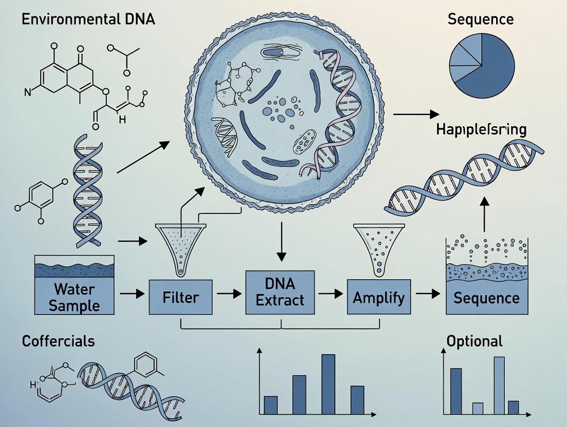
Environmental DNA (eDNA) refers to genetic material obtained directly from environmental samples such as water, sediment, soil, or air, without prior isolation of target organisms. In deep-sea microbial research, eDNA sampling bypasses the need for culturing, providing access to the vast majority (~99%) of microbial diversity that is unculturable with current methods. This application note details protocols for the collection, processing, and analysis of deep-sea eDNA, framed within a thesis on discovering novel microbial lineages and biosynthetic gene clusters for drug development.
Core eDNA Sampling Protocols
Deep-Sea Water Column Sampling Protocol
Objective: To collect microbial biomass from the deep-sea water column while preserving genetic integrity. Materials: Niskin bottles (or similar sterile, closing water samplers) mounted on a CTD rosette, peristaltic pump with tubing, in-line filters (0.22µm pore size, polyethersulfone or mixed cellulose ester), sterile filter housings, sterile gloves, RNAlater or Longmire’s buffer, liquid nitrogen or -80°C freezer for flash freezing. Procedure:
- Deployment: Lower CTD rosette with attached Niskin bottles to target depth(s). Use CTD data (conductivity, temperature, depth) to trigger bottle closure at precise layers.
- Filtration: On deck, aseptically transfer water from Niskin bottles into a sterile reservoir. Using a peristaltic pump, pass a measured volume (typically 1-10 L for deep-sea, depending on biomass) through a 0.22µm filter to capture microbial cells.
- Preservation: Immediately upon filtration completion, aseptically remove the filter using sterile forceps. Cut the filter into pieces and place in a cryovial containing preservation buffer (e.g., 1ml RNAlater). Flash-freeze in liquid nitrogen and store at -80°C until extraction.
- Controls: Process field blanks (sterile water filtered on-site) and equipment blanks to monitor contamination.
Deep-Sea Sediment Core Sampling Protocol
Objective: To collect subsurface sediment layers harboring distinct microbial communities. Materials: Multi-corer or box corer, sterile core sleeves or liners, sterile spatulas and scalpels, sub-sampling syringes (with ends cut off), 50ml sterile conical tubes, DNA/RNA shield or similar preservation solution, dry ice or -80°C freezer. Procedure:
- Core Retrieval: Deploy coring device. Upon retrieval, carefully extrude the core liner. Visibly demarcate layers based on color and texture.
- Sub-sampling: In a dedicated, clean area, use sterile cut-off syringes to sub-sample sediment from the center of each stratified layer (e.g., 0-2cm, 2-5cm, 5-10cm). Transfer ~5g of sediment into pre-labeled 50ml tubes.
- Preservation: Immediately add 10-15ml of DNA/RNA preservation solution to each tube, mix thoroughly, and flash-freeze on dry ice. Store at -80°C.
- Controls: Collect and preserve sterile sediment or blank substrate processed alongside samples.
Laboratory Processing & Metagenomic Analysis
eDNA Extraction and QC Protocol
Objective: To co-extract high-molecular-weight DNA and RNA from deep-sea filters/sediment. Materials:
- DNeasy PowerSoil Pro Kit / RNeasy PowerSoil Total RNA Kit (Qiagen) – For simultaneous lysis of tough microbial cells.
- Bead-beater (e.g., FastPrep-24) – For mechanical disruption.
- Fluorometer (Qubit) – For DNA/RNA quantification.
- Gel electrophoresis / Bioanalyzer (Agilent) – For integrity assessment.
- Inhibitor Removal Resins (e.g., OneStep PCR Inhibitor Removal Kit) – Critical for sediment samples.
Procedure:
- Lysis: For filters, cut a section. For sediment, use 0.25-0.5g. Place in bead-beating tube with provided solution. Homogenize at high speed for 45s.
- Nucleic Acid Purification: Follow kit protocol, incorporating an optional inhibitor removal spin column step for sediment extracts.
- QC: Quantify DNA/RNA with Qubit using dsDNA HS and RNA HS assays. Assess integrity via 1% agarose gel (DNA should be >10kb; RNA should show clear 16S/23S rRNA bands) or Bioanalyzer.
- Storage: Store DNA/RNA at -80°C. For metagenomics, often proceed with DNA or perform cDNA synthesis from RNA for metatranscriptomics.
Metagenomic Library Prep & Sequencing
Objective: To prepare sequencing libraries for taxonomic and functional profiling. Protocol (Illumina NovaSeq):
- Fragmentation & Size Selection: Using 100ng-1ug of input DNA, fragment via sonication (Covaris) to ~350bp. Perform double-sided size selection with SPRI beads.
- Library Construction: Use library prep kit (e.g., Illumina DNA Prep) for end-repair, A-tailing, and adapter ligation. Perform limited-cycle PCR (4-8 cycles) with dual-indexed primers.
- QC & Pooling: Quantify final libraries by qPCR (Kapa Library Quant Kit). Pool equimolar amounts.
- Sequencing: Sequence on an Illumina NovaSeq 6000 using a 2x150bp S4 flow cell, targeting 20-100 million reads per sample for deep coverage. For assembly, prefer longer reads (PacBio HiFi or Oxford Nanopore) where possible.
Table 1: Typical eDNA Yields from Deep-Sea Samples
| Sample Type | Depth/ Layer | Filtration Volume / Sediment Mass | Avg. DNA Yield (ng) | Avg. Read Depth (Illumina) Recommended | Key Contaminants to Monitor |
|---|---|---|---|---|---|
| Water Column | Mesopelagic (200-1000m) | 5 L | 50 - 200 ng | 20-40 M reads | Human DNA, surface microbes |
| Water Column | Bathypelagic (>1000m) | 10 L | 10 - 100 ng | 40-60 M reads | Polymerase inhibitors (humics) |
| Surface Sediment | 0-5 cm | 0.5 g | 500 - 5000 ng | 60-100 M reads | Extracellular "relic" DNA |
| Sub-Surface Sediment | 20-30 cm | 0.5 g | 50 - 500 ng | 80-120 M reads | Heavy metals, humic acids |
Table 2: Key Bioinformatics Tools for Deep-Sea Microbial eDNA Analysis
| Analysis Stage | Tool/Pipeline | Primary Function | Key Output for Drug Discovery |
|---|---|---|---|
| Quality Control | FastQC, MultiQC | Assess read quality, adapter content | High-quality read sets |
| Assembly & Binning | MEGAHIT, metaSPAdes, MaxBin2 | Assemble reads, bin into genomes | Metagenome-Assembled Genomes (MAGs) |
| Taxonomy | GTDB-Tk, Kaiju | Classify reads/MAGs against genome database | Novel microbial lineages |
| Function | eggNOG-mapper, antiSMASH | Annotate genes, find biosynthetic clusters | Novel Biosynthetic Gene Clusters (BGCs) |
| Comparative Analysis | Anvi'o, PhyloPhlAn | Visualize and compare community structure | Community shifts with depth/niche |
The Scientist's Toolkit: Research Reagent Solutions
| Item | Function in Deep-Sea eDNA Research |
|---|---|
| RNAlater / DNA/RNA Shield | Preserves nucleic acid integrity immediately upon sample collection, critical for labile RNA and preventing degradation during transport. |
| 0.22µm Sterivex-GP Pressure Filter | In-line, closed filtration unit that minimizes contamination risk during water filtration. |
| PowerSoil DNA/RNA Isolation Kits | Includes inhibitors removal steps essential for humic acid-rich deep-sea sediments. |
| OneStep PCR Inhibitor Removal Kit | Additional clean-up for recalcitrant samples to ensure downstream PCR/sequencing success. |
| Kapa HiFi HotStart ReadyMix | High-fidelity polymerase for accurate amplification of low-biomass eDNA and library construction. |
| ZymoBIOMICS Microbial Community Standard | Mock community used as a positive control and for benchmarking bioinformatics pipelines. |
| NEBNext Microbiome DNA Enrichment Kit | Depletes host (e.g., eukaryotic) DNA, enriching for microbial sequences in mixed samples. |
Visualizations
Title: Deep-Sea eDNA Analysis Workflow
Title: From eDNA to Drug Discovery Pathway
Application Notes: eDNA for Deep-Sea Microbial Exploration
Environmental DNA (eDNA) sampling represents a transformative, non-invasive approach for cataloging microbial diversity in the deep sea, a realm characterized by extreme hydrostatic pressure, unique geochemistry, and largely unexplored biological niches. Traditional culturing techniques recover less than 1% of deep-sea microbes. eDNA methods bypass the need for cultivation, enabling direct genetic analysis of microbial communities from water, sediment, and biofilm samples. This is critical for drug discovery, as deep-sea microbes are a prolific source of novel biosynthetic gene clusters (BGCs) for antibiotics, anti-cancer agents, and industrial enzymes.
Key Challenges & Solutions:
- Contamination & Degradation: Extreme care is required to prevent sample contamination with surface microbes and to preserve fragile eDNA. Protocols must include stringent sterile controls and instant preservation.
- PCR Biases in Extreme Chemistries: The high concentrations of salts (e.g., around hydrothermal vents) or heavy metals can inhibit polymerase enzymes. Specific buffer formulations and purification steps are essential.
- Incomplete Reference Databases: Many sequences have no match in existing databases. De novo assembly and metagenomic binning are required to reconstruct genomes from novel phyla.
Quantitative Context of the Deep-Sea Biome:
Table 1: Characteristics of Major Deep-Sea Microbial Habitats
| Habitat | Approximate Depth Range | Key Abiotic Stressors | Typical Microbial Biomass (cells/mL sediment or water) | Dominant Metabolic Strategies |
|---|---|---|---|---|
| Abyssal Plain | 3,000 - 6,000 m | High pressure (30-60 MPa), ~2-4°C, low organic input | 10^3 - 10^5 (water); 10^8 - 10^9 (sediment) | Heterotrophy, methanogenesis, sulfate reduction |
| Hydrothermal Vent | 1,000 - 4,000 m | High temperature gradients (4-400°C), toxic metals, low pH | 10^5 - 10^7 (plume water) | Chemolithoautotrophy (H2, H2S, CH4 oxidation) |
| Cold Seep | 200 - 3,000 m | Anoxia, high methane & sulfide flux, high pressure | 10^9 - 10^10 (mats & sediments) | Anaerobic methanotrophy, sulfate reduction |
| Hadal Trench | >6,000 m | Extreme pressure (>60 MPa), seismic activity, funneled organic matter | 10^4 - 10^6 (water); 10^7 - 10^10 (sediment) | Heterotrophy, fermentation, potential piezophily |
Table 2: eDNA Yield and Quality from Standardized Sampling Methods
| Sample Type | Volume Processed | Typical eDNA Yield (ng) | Recommended Preservation Method | Primary Sequencing Target |
|---|---|---|---|---|
| Deep Water Column | 2-10 L | 5 - 80 | In-line filtration onto 0.22µm filter, flash-freeze in LN2 | 16S rRNA, 18S rRNA, metagenomics |
| Surface Sediment Core | 1-5 g (sub-core) | 100 - 2,000 | Immediate subsampling into DNA/RNA shield buffer | 16S rRNA, metagenomics, metatranscriptomics |
| Biofilm/Mat (Vent/Seep) | 1 cm^2 patch | 500 - 5,000 | Excision, immersion in preservation buffer | Metagenomics, single-cell genomics |
| Particulate Organic Matter | 1-5 L filtered | 10 - 200 | Filter stored in ATL buffer at -80°C | Eukaryotic markers, functional genes |
Experimental Protocols
Protocol 1: Sterile, Pressure-Retentive Deep-Water eDNA Sampling
Purpose: To collect microbial biomass from a targeted depth while minimizing contamination and preserving pressure-sensitive taxa. Materials: Niskin bottles (sterilized with 10% bleach rinse and UV), a CTD rosette, peristaltic pump, in-line Sterivex-GP (0.22 µm) filter units, luer-lock syringes, liquid nitrogen (LN2) Dewar. Procedure:
- Deployment: Deploy sterile Niskin bottles on a CTD rosette to desired depth. Trigger closure.
- On-deck Filtration: Within 30 minutes of recovery, attach Sterivex filter to Niskin spigot via sterile tubing. Filter water (2-10L) using a peristaltic pump or gravity.
- Preservation: Immediately flush the filter with 1.5 mL of DNA/RNA Shield buffer. Cap ends. Flash-freeze the entire filter unit in LN2. Store at -80°C until extraction.
- Extraction: Thaw unit on ice. Use a syringe to push through lysis buffer (e.g., PowerWater Sterivex Kit protocol). Complete extraction using a kit optimized for inhibitor removal.
Protocol 2: Metagenomic Library Prep from eDNA for Biosynthetic Gene Cluster Mining
Purpose: To prepare sequencing-ready libraries from deep-sea eDNA, prioritizing high molecular weight DNA for assembly. Materials: High-purity eDNA (>20 ng/µL, fragment size >5 kb), NEBNext Ultra II FS DNA Library Prep Kit, size selection beads (SPRIselect), Qubit fluorometer, Bioanalyzer/TapeStation. Procedure:
- DNA Repair & End-Prep: Combine 50-100 ng eDNA with NEBNext Ultra II FS reagents. Incubate at 20°C for 15 min, then 65°C for 15 min.
- Adapter Ligation: Add NEBNext adaptor for Illumina (diluted 1:10). Incubate at 20°C for 15 min. Purify with 1X SPRIselect beads.
- Size Selection: Perform double-sided SPRI bead cleanup (e.g., 0.5X followed by 0.8X ratio) to retain fragments >300 bp.
- PCR Enrichment: Amplify with index primers for 8-10 cycles. Purify final library with 0.9X SPRI beads. Quantify via Qubit and profile via Bioanalyzer.
Visualizations
Deep Sea eDNA Research Workflow
Microbial Piezophile Adaptation Pathways
The Scientist's Toolkit
Table 3: Key Research Reagent Solutions for Deep-Sea Microbial eDNA Work
| Item | Function in Research | Key Considerations for Deep-Sea Applications |
|---|---|---|
| DNA/RNA Shield (e.g., from Zymo) | Instant chemical preservation of nucleic acids at point of collection. Inactivates nucleases. | Critical for preserving samples during long ascent from depth. Compatible with filter cartridges. |
| PowerWater Sterivex DNA Isolation Kit (Qiagen) | DNA extraction directly from Sterivex filter units, removing PCR inhibitors like humics. | Optimized for low-biomass water samples. Includes bead-beating for cell lysis of tough microbes. |
| NEBNext Microbiome DNA Enrichment Kit | Depletes host/methylated DNA to increase microbial sequence coverage. | Useful for samples from subseafloor rocks or animal symbionts where host contamination is high. |
| Pfu Turbo DNA Polymerase | High-fidelity PCR for amplifying genes from low-quality/quantity eDNA. | More error-resistant than Taq, crucial for accurate sequencing of novel taxa from challenging samples. |
| SPRIselect Beads (Beckman Coulter) | Size-selective purification of DNA fragments for library prep. | Enables selection of longer fragments from partially degraded eDNA, improving assembly. |
| GTDB-Tk Database & Toolkit | Taxonomic classification of genomes from novel microbial lineages. | More accurate than SILVA/NCBI for unknown deep-sea archaea and bacterial phyla. |
| antiSMASH Software | Identifies Biosynthetic Gene Clusters (BGCs) in metagenomic assemblies. | Primary tool for drug discovery pipelines to find genes for novel natural products. |
Application Notes: eDNA Insights into Deep-Sea Microbial Ecology
Environmental DNA (eDNA) sampling provides a non-invasive, comprehensive method to catalog deep-sea microbial diversity and infer functional potential. This approach is critical for addressing the three key ecological questions without the need for culturing, which is often impossible for extremophiles.
Note 1: Carbon Cycling in the Deep Biosphere. eDNA from sediment cores and seawater columns reveals the genetic machinery for anaerobic carbon processing. Metagenomic analysis identifies genes for extracellular enzyme production (e.g., carbohydrate-active enzymes, CAZymes), methanogenesis, and anaerobic methane oxidation (ANME), linking taxonomy to carbon transformation pathways in oxygen-depleted zones.
Note 2: Uncovering Symbioses. eDNA extracted from host-associated environments (e.g., tube worm roots, sponge tissues) or from specific micro-niches allows for the reconstruction of symbiotic metabolic networks. Co-occurrence networks derived from amplicon sequencing data can predict interactions, while metagenome-assembled genomes (MAGs) reveal complementary metabolic pathways (e.g., sulfide oxidation, carbon fixation) between hosts and symbionts.
Note 3: Adaptation to Extreme Conditions. Shotgun metagenomics and metatranscriptomics of eDNA from hydrothermal vents, cold seeps, and hadal zones identify genes associated with extreme adaptation. This includes chaperones for thermostability, osmolytes for high pressure, and efflux pumps for heavy metal resistance. Expression data pinpoints active survival strategies in situ.
Protocols
Protocol 1: Deep-Sea Water and Sediment eDNA Sampling and Preservation
Objective: To collect and preserve microbial eDNA from deep-sea water and sediment for downstream molecular analysis of community structure and function.
Materials:
- Niskin bottles (rosette-mounted) or in-situ filtration pumps
- Sterile sediment corers (multi-corer or box corer)
- Sterile filtration units (0.22µm pore size, polyethersulfone filters)
- DNA/RNA Shield preservative or RNAlater
- Liquid nitrogen or -80°C freezer for storage
- Sterile gloves, spatulas, and cutting sleeves for sediment sub-sampling
Procedure:
- Water Collection: Deploy a CTD-rosette with Niskin bottles to target depths. Trigger closure remotely. Alternatively, use an in-situ pump to filter large volumes of water (100-1000L) directly onto 0.22µm filters.
- Water eDNA Preservation: On shipboard, aseptically transfer filters to cryovials containing DNA/RNA Shield. Immediately flash-freeze in liquid nitrogen. Store at -80°C.
- Sediment Collection: Deploy a multi-corer to obtain undisturbed sediment cores. In a clean lab space, use sterile tools to sub-core (e.g., cut-off syringe) the center of the core to avoid contamination.
- Sediment eDNA Preservation: Subsample sediment slices (e.g., 0-1cm, 1-5cm) into cryovials. Completely submerge sample in DNA/RNA Shield or RNAlater. Homogenize briefly. Flash-freeze and store at -80°C.
- Controls: Collect field blanks (preservative exposed to air during sampling) and filtration blanks (sterile water processed through system).
Protocol 2: Metagenomic Library Preparation and Sequencing for Functional Inference
Objective: To prepare sequencing libraries from deep-sea eDNA for shotgun metagenomic analysis to assess functional gene content and reconstruct genomes.
Materials:
- DNeasy PowerSoil Pro Kit (QIAGEN) or similar for sediment
- DNeasy Blood & Tissue Kit (QIAGEN) for filters
- Qubit fluorometer and dsDNA HS Assay Kit
- NEBNext Ultra II FS DNA Library Prep Kit
- Size selection beads (e.g., SPRIselect)
- Illumina-compatible dual-index adapters
- Bioanalyzer or TapeStation
Procedure:
- eDNA Extraction: Follow kit protocols with modifications: increase bead-beating time to 10 minutes for sediment; incubate filters in lysis buffer with proteinase K for 1 hour at 56°C.
- DNA Quantification & QC: Measure concentration with Qubit. Assess fragment size distribution using Bioanalyzer (Agilent).
- Library Preparation: Using 10-100ng of input DNA, perform end-repair, dA-tailing, and adapter ligation per NEBNext kit instructions.
- Size Selection & PCR Enrichment: Perform double-sided SPRI bead cleanup to select fragments ~350-550bp. Amplify libraries with 8-10 PCR cycles.
- Library QC & Pooling: Quantify final libraries by Qubit. Check size profile. Pool equimolar amounts of uniquely indexed libraries.
- Sequencing: Sequence on an Illumina NovaSeq platform using a 2x150bp configuration to achieve a minimum of 20-40 million reads per sample for MAG reconstruction.
Protocol 3: Quantitative Analysis of Carbon Cycle Genes via qPCR
Objective: To quantify the abundance of key functional genes involved in microbial carbon cycling (e.g., mcrA for methanogenesis, dsrB for sulfate reduction) from deep-sea eDNA.
Materials:
- Extracted eDNA (from Protocol 1)
- Primers for target functional genes (see Table 1)
- PowerUp SYBR Green Master Mix
- Standard DNA (cloned gene fragment, gBlocks)
- Real-Time PCR system (e.g., Applied Biosystems QuantStudio)
Procedure:
- Primer Design: Use well-curated, degenerate primers from literature for environmental samples.
- Standard Curve Preparation: Serially dilute (10-fold) standard DNA from 10^7 to 10^1 gene copies/µL.
- qPCR Reaction Setup: In triplicate, mix 10µL SYBR Green Master Mix, forward and reverse primers (0.5µM final), 2µL template DNA (or standard), and nuclease-free water to 20µL.
- Thermocycling: 95°C for 2 min; 40 cycles of: 95°C for 15 sec, primer-specific annealing temperature (Ta, see Table 1) for 30 sec, 72°C for 30 sec. Include melt curve analysis.
- Data Analysis: Use system software to determine copy number/µL in each sample based on the standard curve. Normalize to mass of sediment or volume of water filtered.
Data Tables
Table 1: Key Functional Gene Targets for qPCR in Deep-Sea Carbon Cycling Studies
| Target Process | Gene | Primer Sequence (5'->3') | Annealing Temp (Ta) | Amplicon Size |
|---|---|---|---|---|
| Methanogenesis | mcrA | F: GGTGGTGTMGGATTCACACARTAYGCWACAGCR: TTCATTGCRTAGTTWGGRTAGTT | 55°C | ~470 bp |
| Sulfate Reduction | dsrB | F: CAACATCGTYCAYACCCAGGGR: GTGTAGCAGTTACCGCA | 55°C | ~350 bp |
| Anaerobic Methane Oxidation (ANME) | mcrA (ANME-1) | F: TTCGGTGGATCDCARAGRGCR: GBARGTCGWAWCCGTAGAATCC | 58°C | ~500 bp |
| Bacterial Carbon Fixation (Reductive TCA) | aclB | F: GCITGYGGIATGTAYGGIAARGGR: ARRTGRTGRTGYTCRCAIACCCA | 60°C | ~450 bp |
Table 2: Typical Sequencing Metrics for Deep-Sea eDNA Metagenomic Studies
| Sample Type | Recommended Sequencing Depth | Typical DNA Yield | Expected MAGs (Quality >Medium) | Dominant Phyla (Examples) |
|---|---|---|---|---|
| Hadal Sediment | 40-60 million read pairs | 0.5 - 5 µg/g sediment | 50-150 | Proteobacteria, Chloroflexi, Bacteroidota, Archaea (Thermoproteota) |
| Hydrothermal Vent Plume | 20-30 million read pairs | 2-20 ng/L filtered water | 10-30 | Campylobacterota (Sulfurimonas), Proteobacteria, Marine Group II Archaea |
| Cold Seep Sediment | 30-50 million read pairs | 1 - 10 µg/g sediment | 70-200 | ANME Archaea, Sulfate-Reducing Bacteria (Desulfobacterota), Halobacteria |
Diagrams
Diagram 1: Deep-Sea Microbial eDNA Analysis Workflow
Diagram 2: Key Microbial Carbon Processing Pathways
The Scientist's Toolkit
Table 3: Essential Research Reagent Solutions for Deep-Sea Microbial eDNA Studies
| Reagent/Material | Function & Rationale |
|---|---|
| DNA/RNA Shield Preservation Buffer | Immediately lyses cells and inactivates nucleases upon contact, preserving the in-situ molecular profile of the sample during long shipboard and transport periods. Critical for metatranscriptomic studies. |
| Polyethersulfone (PES) Filters (0.22µm) | Low protein binding, high flow-rate filters for collecting microbial biomass from large volumes of deep-sea water. Compatible with downstream enzymatic lysis and DNA extraction. |
| PowerSoil Pro DNA Isolation Kit | Optimized for difficult environmental matrices like deep-sea sediment. Contains inhibitors removal technology to yield PCR-ready DNA from humic acid-rich samples. |
| NEBNext Ultra II FS DNA Library Prep Kit | Designed for fragmented, low-input DNA common in environmental samples. Includes a fragmentation step that can be skipped for already-sheared eDNA, improving library yield. |
| SPRIselect Beads | Magnetic beads for precise size selection and cleanup during library prep. Essential for removing short fragments and primer dimers to optimize sequencing performance. |
| Degenerate PCR Primers | Primer sets with wobble bases (IUPAC codes) to account for genetic diversity in uncultured microbial communities, allowing amplification of target genes (e.g., mcrA, 16S rRNA) from diverse taxa. |
| Quantitative PCR Standards (gBlocks) | Synthetic double-stranded DNA fragments containing the exact target amplicon sequence. Used to generate absolute standard curves for qPCR, enabling copy number quantification of functional genes in eDNA. |
Environmental DNA (eDNA) sampling represents a paradigm shift in accessing the deep-sea microbial metabolome for drug discovery. By extracting and sequencing genetic material directly from extreme marine environments—hydrothermal vents, cold seeps, abyssal plains, and hadal zones—researchers bypass the <1% culturability barrier. This meta-genomic and meta-transcriptomic approach enables the in silico mining of biosynthetic gene clusters (BGCs) responsible for novel antimicrobial, antitumor, and anti-inflammatory compounds. This Application Note details protocols for deep-sea eDNA sampling, heterologous expression of target BGCs, and high-throughput screening, framed within a thesis on systematic bioprospecting.
Table 1: Comparative Yield of Bioactive Compounds from Different Deep-Sea Biomes via eDNA Metagenomics
| Deep-Sea Biome | Avg. eDNA Yield (ng/L seawater/sediment) | BGCs per Gb of Sequence Data | Hit Rate (% Clones with Bioactivity) | Notable Compound Classes Discovered (Examples) |
|---|---|---|---|---|
| Hydrothermal Vents | 5-50 ng/L (water); 500-5000 ng/g (sediment) | 120-180 | 0.5 - 1.2% | Thiopeptides, Polyketides, Lanthipeptides |
| Cold Seeps | 10-30 ng/L; 300-2000 ng/g | 90-140 | 0.3 - 0.8% | Non-Ribosomal Peptides (NRPs), Lipopeptides |
| Abyssal Plains | 1-10 ng/L; 100-500 ng/g | 40-80 | 0.1 - 0.4% | Alkaloids, Terpenoids |
| Hadal Trenches (>6000m) | 0.5-5 ng/L; 50-300 ng/g | 60-110 | 0.4 - 1.0% | Novel β-Lactams, Macrolides |
Table 2: Performance Metrics of Heterologous Expression Hosts for Deep-Sea Microbial BGCs
| Expression Host | Successful Expression Rate (Deep-Sea BGCs) | Avg. Titer (mg/L) | Key Advantages for Deep-Sea Genes |
|---|---|---|---|
| Streptomyces coelicolor | ~35% | 2-15 | Native for actinobacterial BGCs; robust secondary metabolism. |
| Pseudodalteromonas haloplanktis | ~25% | 1-10 | Cold-adapted; suited for psychrophilic enzyme function. |
| E. coli (specialized strains) | ~15% | 0.5-5 | High-throughput genetics; extensive toolkit available. |
| Saccharomyces cerevisiae | ~10% | 0.1-2 | Eukaryotic PTMs possible; handles high GC content moderately. |
Detailed Experimental Protocols
Protocol 3.1: Deep-Sea eDNA Sampling and Preservation (Niskin Bottle & Sediment Corer)
Objective: To collect microbial biomass from deep-water column and sediment while preserving genetic integrity.
Materials: Sterilized Niskin bottles (e.g., GO-FLO), box corer or multicorer, sterile gloves, RNAlater or DNA/RNA Shield, pressure-retaining chambers (for piezophiles), sterile syringes, filtration apparatus (0.22µm filters), liquid nitrogen.
Procedure:
- Deployment: Deploy sterilized Niskin bottles on a CTD rosette to target depths. For sediments, deploy a box corer.
- Sample Collection: Trigger bottle closure at target depth. Retrieve and immediately transfer water to sterile containers. For sediment, sub-core the center with a sterile tube to avoid contamination.
- Filtration & Preservation: Within 30 minutes of retrieval, filter 1-10L of water through a 0.22µm polyethersulfone filter under gentle vacuum. Immediately place the filter in a tube containing 2 mL of DNA/RNA Shield. For sediment, aliquot 1g into a similar preservative.
- Storage: Flash-freeze preserved samples in liquid nitrogen and store at -80°C until extraction.
- Controls: Collect field blanks (sterile water processed identically) to monitor contamination.
Protocol 3.2: Metagenomic Library Construction & Bioinformatic Screening for BGCs
Objective: To prepare large-insert fosmid or cosmid libraries from eDNA and identify putative BGCs.
Materials: High-purity eDNA extraction kit (e.g., for soil/microbes), CopyControl Fosmid Library Production Kit, electrocompetent E. coli EPI300, LB agar with chloramphenicol, sequencing reagents, bioinformatics workstation (antiSMASH, PRISM, BLAST).
Procedure:
- eDNA Extraction: Perform extraction on preserved filter/sediment using a protocol optimized for humic acid removal. Assess integrity via pulsed-field gel electrophoresis.
- Size Selection & Ligation: Size-fractionate eDNA (30-50 kb) by gel electrophoresis. Ligate into the fosmid vector following kit instructions.
- Packaging & Transformation: Package ligated DNA using phage packaging extracts. Infect/transform EPI300 cells and plate on selective media. Aim for >1 x 10^5 clones per library.
- Pooling & Sequencing: Pool colonies, extract fosmid DNA, and perform Illumina HiSeq sequencing (150bp paired-end).
- In Silico BGC Mining: Assemble reads (MEGAHIT). Run antiSMASH on contigs >10 kb to identify BGCs (PKS, NRPS, RiPPs, etc.). Prioritize BGCs with low homology to known clusters.
Protocol 3.3: Heterologous Expression and Activity Screening in a Model Actinomycete
Objective: To clone and express a prioritized deep-sea BGC in Streptomyces coelicolor and screen for bioactivity.
Materials: Gateway-compatible vectors (e.g., pCAP-based), E. coli ET12567/pUZ8002 for conjugation, S. coelicolor M1152 or M1146, ISP4 agar, MS agar, antibiotics, ethyl acetate, methanol, 96-well assay plates, pathogen lawns (S. aureus, C. albicans), cancer cell lines.
Procedure:
- BGC Capture: Amplify the target BGC (e.g., using Transformation-Associated Recombination - TAR - in yeast) and clone into a Streptomyces integrative expression vector.
- Conjugal Transfer: Transform the construct into E. coli ET12567/pUZ8002. Mate with S. coelicolor spores on ISP4 agar with 10mM MgCl2. Select exconjugants with apramycin/nalidixic acid.
- Fermentation: Inoculate 5 mL of TS broth with an exconjugant colony. Incubate at 30°C, 250 rpm for 2 days. Use 1 mL to inoculate 50 mL of production medium (e.g., R5 or SFM). Ferment for 5-7 days.
- Metabolite Extraction: Centrifuge culture. Extract supernatant with equal volume ethyl acetate (x3). Extract cell pellet with methanol. Combine, dry in vacuo, and resuspend in DMSO.
- Bioactivity Screening:
- Antimicrobial: Use agar diffusion or microbroth dilution against ESKAPE pathogens.
- Cytotoxic: Treat human cancer cell lines (e.g., HeLa, MCF-7) for 72h and assess viability via MTT assay.
Visualization Diagrams
Title: Deep-Sea eDNA to Drug Lead Workflow
Title: Stress-Induced Biosynthesis in Deep-Sea Microbes
The Scientist's Toolkit: Key Research Reagent Solutions
Table 3: Essential Reagents and Materials for Deep-Sea Microbial eDNA Drug Discovery
| Item / Solution | Function & Rationale |
|---|---|
| DNA/RNA Shield (e.g., Zymo Research) | Immediate chemical preservation of eDNA/RNA at point of collection; inhibits nucleases and microbial growth, critical for low-biomass samples. |
| CopyControl Fosmid Library Kit (Lucigen) | Optimal for cloning large, fragile eDNA fragments (30-50 kb); inducible copy number increases yield for sequencing and expression. |
| antiSMASH 7.0 (Bioinformatics Suite) | Core algorithm for automated genomic identification and analysis of BGCs from metagenomic assemblies. |
| pCAP01/pCAP02 Vectors (or similar) | Streptomyces integration vectors designed for conjugal transfer and stable expression of large, exogenous BGCs. |
| E. coli ET12567/pUZ8002 | Non-methylating E. coli strain carrying conjugation machinery, essential for efficient DNA transfer into Streptomyces. |
| Streptomyces coelicolor M1152/M1146 | Engineered heterologous hosts with minimized native antibiotic production and enhanced precursor supply for expressing cloned BGCs. |
| ISP4 & R5 Agar/Media | Standard sporulation and specialized production media for Streptomyces, promoting secondary metabolite synthesis. |
| Chromabond HR-X SPE Columns | Solid-phase extraction for fractionating complex crude extracts prior to bioassay, enabling activity-guided purification. |
From the Hadal Zone to the Lab: A Step-by-Step Guide to Deep-Sea eDNA Workflow
1. Introduction and Thesis Context Within a broader thesis on Environmental DNA (eDNA) sampling for deep-sea microbial research, the pre-sampling strategy is the critical foundation. The choice between the water column and sediment as a target zone fundamentally dictates the microbial communities recovered, the ecological insights gained, and the downstream potential for biodiscovery, including drug development. This document provides detailed application notes and protocols for defining research objectives and executing this primary strategic decision.
2. Defining Research Objectives: Key Questions The research objective must be precisely articulated to guide target zone selection. Key questions are summarized in Table 1.
Table 1: Research Objectives and Corresponding Target Zone Considerations.
| Research Objective | Primary Target Zone | Rationale |
|---|---|---|
| Biodiscovery / Bioprospecting for novel enzymes or bioactive compounds (e.g., antimicrobials). | Sediment (primarily); Deep Chlorophyll Maximum (DCM) or Chemoclines (secondarily). | Sediments are biodiversity hotspots with intense microbial competition and specialized metabolism, driving chemical diversity. Particle-attached communities in water column niches also show high functional specialization. |
| Biogeochemical Cycling (e.g., carbon sequestration, nitrogen cycling). | Both, but with distinct targets. | Requires stratified water column sampling for pelagic processes (e.g., nitrification) and sediment-water interface/core sampling for benthic processes (e.g., denitrification, sulfate reduction). |
| Baseline Biodiversity & Biomonitoring (e.g., impact assessment). | Water Column (for broad spatial signal); Sediment (for temporal integration). | Water column eDNA provides a integrated, recent snapshot over a larger area. Sediments archive eDNA over longer timescales, acting as a historical record. |
| Study of Microbial Loop & Food Web Dynamics. | Water Column (size-fractionated sampling). | Requires separation of free-living vs. particle-attached communities to understand ecological roles and interactions. |
| Extremophile Studies (high pressure, anaerobic metabolisms). | Sediment Cores & Hydrothermal Vent/Seep Fluids. | Anoxic, high-pressure conditions are best captured in deep sediment layers and vent fluid plumes, hosting unique adapted communities. |
3. Comparative Analysis: Water Column vs. Sediment eDNA
Table 2: Comparative Properties of Water Column and Sediment as eDNA Sources for Deep-Sea Microbes.
| Property | Water Column | Sediment |
|---|---|---|
| eDNA Source | Microbial cells, extracellular DNA (dissolved and particle-bound). | Intracellular DNA from live/decaying cells, strongly adsorbed extracellular DNA. |
| Spatial Signal | Integrative over transport paths (horizontal & vertical). | Highly localized, reflecting immediate microenvironment. |
| Temporal Signal | Transient (days to weeks), reflects recent community. | Accumulated/preserved (months to millennia), provides temporal archive. |
| eDNA Persistence | Lower (hours to days), degraded by UV, nucleases, dilution. | Higher (weeks to years), stabilized by adsorption to minerals/organic matter. |
| Sampling Method | Niskin/CTD Rosette, in-situ pumps, plankton nets. | Box corer, multi-corer, gravity corer, push cores. |
| Processing Complexity | Moderate (filtration, concentration). | High (homogenization, separation from inhibitors like humics). |
| Inhibitor Load | Generally lower (salt, occasional organics). | Generally high (humic acids, fulvic acids, metals). |
| Primary Bias | Filtration pore-size choice, pump shear stress. | Extraction efficiency from complex matrix, inhibitor removal. |
| Relevant for Drug Discovery | Planktonic & particle-attached specialists, photosynthetic microbes (DCM). | Benthic competitors and symbionts, anaerobic metabolisms, high chemical diversity. |
4. Detailed Experimental Protocols
Protocol 4.1: Stratified Water Column eDNA Sampling (CTD Rosette with Niskin Bottles) Objective: To collect microbial eDNA from discrete depths in the deep-sea water column. Materials: CTD rosette equipped with Niskin bottles (e.g., 12 x 10L), depth/conductivity/temperature sensors, peristaltic pump, sterile filter units (0.22µm pore-size, typically polyethersulfone or mixed cellulose ester), backup filters, sterile forceps, gloves, cryovials, liquid nitrogen or -80°C freezer, preservative (e.g., Longmire's buffer, RNALater for meta-omics). Procedure:
- Pre-cruise: Decontaminate Niskin bottles and tubing with 10% bleach rinse, followed by copious sterile water rinses. Pre-load and bag sterile filter units.
- Deployment: Lower the CTD rosette to the target maximum depth. Trigger bottle closure at pre-determined depths (e.g., surface, deep chlorophyll maximum, oxygen minimum zone, near-bottom nepheloid layer).
- Filtration: On deck, attach outlet of Niskin bottle to a peristaltic pump connected to the filter unit. Filter water (typically 2-10L per depth) onto the 0.22µm membrane under low pressure (< 5 psi).
- Preservation: Using sterile forceps, aseptically fold the filter and place it into a cryovial. Immediately immerse in liquid nitrogen or place at -80°C. Alternatively, add a DNA/RNA preservative buffer.
- Controls: Collect field blanks (filter sterile water through the same system on deck) and equipment blanks (rinse water from Niskin bottle) at regular intervals.
Protocol 4.2: Deep-Sea Sediment Core eDNA Sampling (Multi-corer) Objective: To collect undisturbed sediment cores for stratified microbial eDNA analysis. Materials: Multi-corer (preferred for intact sediment-water interface), core slicer or extruder, sterile cutoff syringes or core sub-sampler, sterile spatulas and scalpels, centrifuge tubes (50mL), gloves, cryovials, liquid nitrogen or -80°C freezer, preservative. Procedure:
- Deployment & Retrieval: Deploy multi-corer to obtain multiple, parallel, undisturbed cores (e.g., 4-12 cores, ~30-60cm length). Visually inspect for an intact sediment surface and clear overlying water.
- Sub-sampling: In a dedicated clean area, carefully extrude the core vertically. Using a sterile slicer or syringe with the end cut off, sub-sample sediment at desired depth intervals (e.g., 0-1cm, 1-5cm, 5-10cm, etc.) into pre-weighed 50mL tubes.
- Homogenization & Aliquoting: For each interval, homogenize the sediment gently but thoroughly using a sterile spatula. Transfer multiple aliquots (e.g., 0.5g-10g) into cryovials for different analyses (eDNA, metagenomics, metabolomics).
- Preservation: Immediately flash-freeze aliquots in liquid nitrogen, then store at -80°C. For some studies, preservation buffer is added to one aliquot.
- Controls: Collect and process a sterile substrate (e.g., baked silica sand) alongside core processing as an extraction blank.
5. Visualizations
Title: Decision Flow: Research Objective to Target Zone
Title: Comparative Field Sampling Workflows
6. The Scientist's Toolkit: Essential Research Reagents & Materials
Table 3: Key Reagent Solutions and Materials for Deep-Sea eDNA Pre-Sampling.
| Item | Function/Application | Key Considerations |
|---|---|---|
| Sterile Filter Units (0.22µm) | Capture microbial biomass from water. | Polyethersulfone (low binding) or mixed cellulose ester. Pre-sterilized, single-use. |
| Longmire's Buffer (100mM Tris, 100mM EDTA, 10mM NaCl, 0.5% SDS) | In-situ preservation of eDNA on filters, inhibits nucleases. | Compatible with many downstream extraction kits. Filter can be stored in buffer at room temp for short periods. |
| RNALater or DNA/RNA Shield | Preserves nucleic acid integrity for meta-transcriptomics. | Critical for RNA-based activity studies. Requires cold storage after initial stabilization. |
| Sodium Polyphosphate Solution | Dispersion agent for sediment sub-samples; helps desorb DNA from particles. | Added during initial lysis step to improve extraction yield from sediments. |
| Inhibitor Removal Buffers (e.g., PTB) | Pre-treatment for sediment extracts to remove humic acids. | Often part of commercial kit protocols (e.g., DNeasy PowerSoil Pro). |
| Sterile, DNA-free Water | Preparation of blanks and equipment rinsing. | Essential for contamination control throughout the workflow. |
| Ethanol (70% & 95%) | Surface decontamination of equipment (70%) and field use (95%). | Primary field decontaminant for corers, tools, and work surfaces. |
Within the broader thesis on Environmental DNA (eDNA) sampling for deep-sea microbes research, contamination control is the foundational challenge. Deep-sea microbial communities are often low-biomass and can be easily overwhelmed by exogenous contaminants introduced during sampling. This article details the application notes and protocols for three core sterile sampling technologies—Niskin bottles, corers, and in-situ filtration systems—that are critical for generating high-integrity eDNA data for subsequent meta-omics analysis and biodiscovery pipelines in drug development.
Application Notes & Comparative Analysis
Table 1: Comparative Specifications of Sterile Sampling Technologies for Deep-Sea eDNA
| Feature | Niskin Bottles (e.g., Sterile, Teflon-coated) | Corers (Multi- & Gravity) | In-situ Filtration Systems (e.g., McLane, Challenger Oceanic) |
|---|---|---|---|
| Primary Target | Pelagic water column microbial biomass | Benthic sediment & sediment-water interface microbes | Particulate & microbial biomass from large water volumes |
| Max. Operating Depth | ~6,500 m (standard oceanic) | ~8,000 m (gravity corer with heavy weights) | ~6,000 m (commercial systems) |
| Typical Sample Volume | 1.7 - 30 L per bottle | 0.5 - 10 m sediment core length | 50 - 1,000 L filtered in-situ |
| Key Contamination Risks | Interior bottle biofilm, messenger release mechanism, CTD rosette/hydrowire, shipboard subsampling | Liner interior, core cutter, winch/wire, core processing on deck, porewater extrusion | Filter capsule integrity, pump lubricants/ seals, sample line biofilms, deck processing |
| Sterilization Methods | Acid washing (10% HCl), Milli-Q rinse, UV irradiation, autoclaving (for certain parts). Sealed ends pre-trigger. | Autoclaving core liners, ethanol flaming of cutters, UV treatment of processing tools. | Pre-sterilized, disposable filter capsules (0.22 µm), ethanol rinses of intakes. |
| Primary eDNA Preservative | Immediate filtration onto filters, preservation in ATL buffer or Longmire's, or flash freezing in LN2. | Subsections stored at -80°C, or immersion in preservation buffer (e.g., RNAlater). | Filters preserved in nucleic acid stabilization buffer (e.g., Qiagen buffer) within the capsule. |
| Best For | Depth-resolved pelagic microbial community structure, water mass-associated taxa. | Vertical stratification of benthic microbes, biogeochemical gradients, ancient eDNA. | Rare microbial taxa, viral assemblages, large-volume requirements for metagenomics. |
Detailed Experimental Protocols
Protocol 3.1: Sterile Niskin Bottle Cast for Pelagic Microbial eDNA
Objective: To collect depth-specific microbial biomass from the deep-sea water column while minimizing exogenous contamination. Materials: Sterilized Teflon-coated Niskin bottles (e.g., General Oceanics Go-Flo) on a rosette, CTD, positive pressure laminar flow hood, peristaltic pump, Sterivex (0.22 µm) or Supor filter units, sterile tubing, preservation buffer (e.g., Longmire's buffer (100 mM Tris, 100 mM EDTA, 10 mM NaCl, 0.5% SDS)), liquid nitrogen, clean gloves.
Procedure:
- Pre-Cruise Sterilization: Disassemble Niskin bottles. Soak all internal components in 10% HCl (v/v) for 24 hrs. Rinse exhaustively with sterile Milli-Q water. Assemble under a laminar flow hood. Seal ends with sterile closures. Protect with sterile bags.
- Deployment: Mount bottles on a clean CTD rosette. Avoid deck runoff. Use a “clean” hydrocast protocol, positioning the rosette upwind of ship exhaust. Use non-toxic, metal-free messengers.
- Tripping & Retrieval: Trip bottles at desired depths. Upon retrieval, immediately move the rosette to a clean container.
- Subsampling (in laminar flow hood): Connect sterile tubing from the Niskin spigot to a peristaltic pump. Connect the outlet tubing to a Sterivex filter cartridge.
- Filtration: Filter 2-30 L of seawater (depending on biomass) at a low, steady pressure (< 5 psi) to avoid cell lysing. Record volume filtered.
- Preservation: Immediately after filtration, fill the Sterivex cartridge with 1.6 mL of Longmire's preservation buffer. Cap the ends, seal in a sterile bag, and flash freeze in liquid nitrogen. Store at -80°C.
- Blanks: Process a “field blank” using sterile water passed through the same tubing/filter setup on deck.
Protocol 3.2: Gravity Corer Operation for Sterile Sediment eDNA
Objective: To obtain intact, depth-stratified sediment cores with minimal disturbance for benthic microbial community analysis. Materials: Autoclaved acrylic core liners, ethanol-flamed core cutter and core catcher, gravity corer rig, sterile gloves, UV-irradiated spatulas and sub-sampling tools, anaerobic chamber (if required), cryovials, RNAlater or equivalent.
Procedure:
- Liner Preparation: Autoclave acrylic core liners (121°C, 20 mins). Store in sterile bags until deployment.
- Corer Assembly: Flame the core cutter and core catcher with 95% ethanol and allow to cool. Assemble the corer on a clean deck area, avoiding contamination.
- Deployment & Retrieval: Lower the corer at a controlled speed (~1 m/s). Allow free-fall for the final 3-5 m. Retrieve carefully to prevent core loss.
- On-Deck Processing (Expedited): Immediately upon retrieval, extrude the core vertically. Remove the core catcher and cut off the potentially contaminated top 1-2 cm of sediment (water interface).
- Sterile Subsampling: In a laminar flow hood or using a bench-top sterile workstation, use a UV-sterilized spatula to remove the outer layer (~1 cm) of sediment from the core section of interest (radial contamination control).
- Collection: Extract the inner, pristine sediment using a sterile cutoff syringe or mini-corer. Transfer 1-5 g aliquots into pre-weighed cryovials.
- Preservation: For DNA/RNA co-preservation, immediately add 2x volume of RNAlater to the sediment, mix gently, incubate overnight at 4°C, then decant excess liquid and store at -80°C. For metabarcoding only, flash freeze directly in LN2.
- Blanks: Collect “procedure blanks” by exposing sterile sediment or a swab to the air during processing.
Protocol 3.3:In-situFiltration with a McLane WTS-LV
Objective: To filter large volumes of deep-sea water in-situ to capture low-abundance microbial targets and avoid shipboard pressure/temperature changes. Materials: McLane WTS-LV or similar in-situ pump, pre-sterilized 0.22 µm filter capsules (e.g., 142 mm diameter), depth/flow rate controller, preservation buffer (e.g., DNA/RNA Shield), sterile wrench set, liquid nitrogen Dewars.
Procedure:
- System Preparation: In a clean lab, load pre-sterilized, disposable filter capsules into the pump head using sterile gloves. Prime the sample path with sterile, particle-free water.
- Programming: Set the controller to filter a target volume (e.g., 500 L) at a specific depth, maintaining a low flow rate (~1-2 L/min) to maximize capture efficiency.
- Deployment & Recovery: Securely mount the pump on the hydrowire. Deploy to target depth. Upon recovery, immediately transfer the pump to a clean area.
- Filter Retrieval: Using sterile tools, open the pump head and aseptically remove the filter capsule.
- Preservation: Without opening the capsule, inject 50 mL of DNA/RNA Shield or similar stabilization buffer directly into the capsule outlet port to fully immerse the filter. Seal ports, place in a sterile bag, and flash freeze in LN2.
- System Decontamination: Flush the entire pump and intake system with 10% bleach followed by sterile water between deployments to prevent cross-contamination.
Visualizations
Title: Sterile eDNA Sampling Workflow for Deep-Sea Microbes
Title: Contamination Sources & Control Points in eDNA Sampling
The Scientist's Toolkit: Research Reagent Solutions
Table 2: Essential Materials for Sterile Deep-Sea eDNA Sampling
| Item | Function & Rationale | Example Product/Brand |
|---|---|---|
| Sterivex GP Pressure Filter (0.22 µm) | Closed-system filtration cartridge for water samples; prevents post-filtration contamination. Attaches directly to tubing from Niskin. | Millipore Sigma Sterivex-GP |
| DNA/RNA Shield | A proprietary, non-toxic buffer that immediately stabilizes nucleic acids at ambient temperature, critical for in-situ preservation and long cruises. | Zymo Research DNA/RNA Shield |
| RNAlater Stabilization Solution | Aqueous, tissue storage reagent that permeates samples to stabilize and protect cellular RNA and DNA. Used for sediment/slurry preservation. | Thermo Fisher Scientific RNAlater |
| PowerWater Sterivex DNA Isolation Kit | Optimized kit for extracting high-quality genomic DNA from filters (Sterivex or flat membranes) with stringent inhibitor removal for downstream PCR. | Qiagen DNeasy PowerWater Sterivex Kit |
| DNeasy PowerSoil Pro Kit | Industry-standard kit for challenging environmental samples like deep-sea sediment; effective for microbial lysis and humic acid removal. | Qiagen DNeasy PowerSoil Pro Kit |
| Tris-EDTA (TE) Buffer, pH 8.0 | For final elution and storage of purified DNA; maintains pH and chelates ions to protect DNA from degradation. | Thermo Fisher Scientific UltraPure TE Buffer |
| Nuclease-Free Water | Certified free of nucleases for reagent preparation and dilutions where contamination must be avoided. | Invitrogen Nuclease-Free Water |
| Ethanol, Absolute (200 proof), Molecular Biology Grade | For surface decontamination, tool flaming, and precipitating nucleic acids during extraction. | Sigma-Aldrich Ethanol, Molecular Biology Grade |
The integrity of environmental DNA (eDNA) from deep-sea microbial communities is paramount for accurate taxonomic profiling, functional gene analysis, and bioprospecting for novel bioactive compounds. The transition from in-situ conditions to the laboratory, especially during long voyages, poses significant risks of nucleic acid degradation, shifts in community structure, and loss of metabolic signatures. This document outlines critical preservation strategies to arrest biological activity immediately upon sample collection, ensuring that downstream molecular analyses reflect the true in-situ state of deep-sea microbiomes.
Comparative Analysis of Preservation Methods
Table 1: Quantitative Comparison of Preservation Method Efficacy for Deep-Sea Microbial eDNA
| Method | Core Reagent | Optimal Temp | Max Hold Time (Pre-extraction) | Key Advantages | Key Limitations | Best for Analysis of |
|---|---|---|---|---|---|---|
| Flash-Freezing (LN₂/ Dry Ice) | Liquid Nitrogen (-196°C) | -80°C | 12-24 months | Halts all metabolic & enzymatic activity instantly; Gold standard for meta-omics. | Logistics & safety on ships; Risk of thaw during transit. | Whole metagenomes, metatranscriptomes, proteins. |
| Chemical Fixation (Ethanol) | 95-100% Ethanol | Room Temp or 4°C | 6-12 months | Simple, safe, non-hazardous for transport; Good for community structure. | Inhibits downstream PCR if not evaporated; Poor for RNA. | 16S/18S rRNA gene amplicons, metagenomics (with cleanup). |
| Chemical Fixation (RNAlater) | Ammonium sulfate salts | 4°C (long-term -80°C) | 1 month at 25°C, 1 yr at 4°C | Penetrates tissues, stabilizes RNA & DNA; Good for mixed samples. | High salt can interfere with extraction; Requires desalting. | Metatranscriptomics, dual DNA/RNA preservation. |
| Desiccation (Silica Gel) | Silica beads | Room Temp, Dry | Indefinite | Ultra-stable, no cold chain needed; Lightweight. | Harsh; may fragment DNA; Not suitable for wet biomass. | Long-term archiving of filter-based samples. |
Detailed Field Protocols
Protocol 1: In-Situ Filtration and Flash-Freezing for Metagenomics
Objective: To capture microbial biomass from large-volume deep-sea water samples and preserve instantly for total nucleic acid extraction.
- Equipment Setup: Assemble a peristaltic pump, sterile tubing, and 0.22µm Sterivex filter units or 47mm polyethersulfone (PES) membrane filters in a laminar hood pre-voyage.
- Filtration: Connect filter unit to pump. Filter 1-100L of seawater (volume depends on biomass). Record volume and pressure.
- Preservation:
- For Sterivex: Immediately inject 1.8mL of RNAlater into the unit. Cap, seal with parafilm, and submerge in liquid nitrogen for >2 minutes. Transfer to -80°C freezer.
- For Membrane Filters: Using flame-sterilized forceps, fold filter, place in a cryovial, and submerge directly in liquid nitrogen for >2 minutes. Store at -80°C.
- Long-Term Storage on Voyage: Maintain samples in vapor-phase liquid nitrogen dewars or at -80°C. Monitor freezer temperatures daily. Avoid freeze-thaw cycles.
Protocol 2: Ethanol Fixation of Sediment Core Subsamples
Objective: To preserve sediment-associated microbial eDNA for community profiling in a stable, non-hazardous manner.
- Subsampling: Under a sterile bench (shipboard), use a cut-off syringe or corer to take a 1-5g sub-sample from the center of a sediment core.
- Fixation: Immediately place sub-sample into a 15mL screw-cap tube containing 5x volume of pre-chilled 95% Ethanol. Vortex vigorously for 30 seconds.
- Storage: Store tubes at 4°C or room temperature in the dark for the voyage duration. For ultra-long-term (>1 year), store at -20°C.
- Pre-Extraction Processing: Pellet sediment (5000 x g, 10 min). Decant ethanol and air-dry pellet briefly to evaporate residual ethanol, which can inhibit polymerases.
Protocol 3: Post-Filtration Preservation with RNAlater for Dual DNA/RNA
Objective: To stabilize both DNA and RNA from planktonic microbes for integrated meta-omics.
- Filtration: Filter water sample onto a 0.22µm PES membrane filter as in Protocol 1.
- Immersion: Use forceps to place filter into a 2mL cryovial completely filled with RNAlater solution. Ensure the filter is fully submerged.
- Initial Incubation: Store the vial at 4°C for 24 hours to allow full penetration of the preservative.
- Long-Term Storage: After 24h, remove excess RNAlater (leaving filter moist) and store at -80°C for the duration of the voyage.
Visualizations
Diagram 1: Decision Workflow for Field Preservation Method
Diagram 2: Voyage Storage Protocol for Fixed Samples
The Scientist's Toolkit: Research Reagent Solutions
Table 2: Essential Materials for Deep-Sea eDNA Preservation
| Item | Function & Rationale |
|---|---|
| Sterivex-GP Filter Unit (0.22µm) | Closed, sterile filtration device for processing large volumes without contamination risk; allows direct in-situ fixation. |
| Polyethersulfone (PES) Membrane Filters | Low protein binding, high flow-rate filters ideal for capturing microbial biomass without inhibiting downstream enzymatic steps. |
| Liquid Nitrogen Dry Shipper | Lightweight, vacuum-insulated dewar for safe, vapor-phase transport of frozen samples without a continuous power supply. |
| RNAlater Stabilization Solution | Aqueous, non-toxic salt solution that rapidly permeates tissues to stabilize and protect cellular RNA and DNA. |
| Anhydrous Ethanol (95-100%, Molecular Grade) | Denatures enzymes instantly; effective and economical DNA preservative for bulk samples like sediment. |
| DNA/RNA Shield (Commercial Buffer) | Ready-to-use, non-hazardous buffer that inactivates nucleases and protects nucleic acids at ambient temperatures for weeks. |
| Silica Gel Desiccant | Provides rapid dehydration of filters, halting microbial activity for room-temperature storage and archiving. |
| TraceClean Cryovials (with O-rings) | Leak-proof, non-binding surface vials for long-term storage, resistant to liquid nitrogen and -80°C temperatures. |
This document outlines standardized protocols for processing environmental DNA (eDNA) samples collected from deep-sea microbial habitats, including hydrothermal vents, abyssal plains, and cold seeps. The integrity of downstream analyses—from taxonomic profiling (16S/18S rRNA gene amplicon sequencing) to functional potential assessment (shotgun metagenomics)—is critically dependent on optimized laboratory procedures that account for low biomass, high inhibitor content, and potential contamination. These protocols are designed for researchers and drug development professionals seeking to explore the unique biosynthetic potential of deep-sea microbiomes.
eDNA Extraction from Deep-Sea Filters
Objective: To obtain high-molecular-weight, inhibitor-free genomic DNA from diverse deep-sea sample types (filter membranes, sediment slurries).
Protocol (Modified from the DNeasy PowerWater Kit for Inhibitor-Rich Samples):
- Sample Lysis: Aseptically cut a 0.22µm polyethersulfone (PES) filter membrane (or use 0.5g sediment) and place in a PowerBead Tube. Add 60µL of Solution SL (containing guanidine thiocyanate and SDS) and vortex horizontally for 10 minutes at maximum speed.
- Inhibitor Removal: Incubate at 65°C for 10 minutes. Centrifuge at 13,000 x g for 1 minute. Transfer supernatant to a clean tube.
- Binding and Washes: Add 650µL of Solution IR to the supernatant, mix, and load onto an MB Spin Column. Centrifuge at 13,000 x g for 1 minute. Wash with 650µL of Solution PW (ethanol-based) twice.
- Elution: Dry the column by centrifugation (2 min). Elute DNA in 50-100µL of nuclease-free water preheated to 60°C. Incubate for 5 minutes before final centrifugation.
Quality Assessment:
- Quantification: Use Qubit dsDNA HS Assay (fluorometric) for accurate concentration measurement, as spectrophotometric methods (Nanodrop) are skewed by contaminants.
- Integrity: Run ~100ng DNA on a 0.8% agarose gel or use a Fragment Analyzer/TapeStation to confirm high molecular weight (>10kb for metagenomics).
Table 1: Performance Comparison of Common eDNA Extraction Kits for Deep-Sea Samples
| Kit Name | Principle | Avg. Yield from 1L Seawater Filter (ng) | Inhibitor Removal Efficiency (qPCR CT shift)* | Suitability for Metagenomics |
|---|---|---|---|---|
| DNeasy PowerWater (Qiagen) | Bead-beating, silica membrane | 50 - 500 | High (0-2 cycle shift) | Excellent |
| NucleoSpin Soil (Macherey-Nagel) | Bead-beating, silica membrane | 100 - 1000 | High (0-2 cycle shift) | Excellent |
| Phenol-Chloroform-Isoamyl Alcohol | Organic separation, ethanol ppt | 200 - 2000 | Moderate (2-5 cycle shift) | Good (if clean) |
| FastDNA SPIN Kit for Soil (MP) | Bead-beating, silica membrane | 150 - 800 | High (0-3 cycle shift) | Good |
*Compared to a no-template control; lower shift indicates better inhibitor removal.
Target Amplification: 16S/18S rRNA Gene Amplicon Sequencing
Objective: To amplify hypervariable regions (e.g., V4-V5 for 16S, V9 for 18S) for taxonomic profiling of prokaryotic and eukaryotic microbial communities.
Protocol (Dual-Indexing Amplification for Illumina Platforms):
- Primer Selection: Use fusion primers (e.g., 515F/926R for 16S, 1389F/1510R for 18S) containing Illumina adapter sequences, indices, and linker regions.
- First-Stage PCR (Target Amplification):
- Reaction Mix (25µL): 12.5µL 2x KAPA HiFi HotStart ReadyMix, 1µL each of forward and reverse primer (10µM), 1-10ng template DNA, nuclease-free water to volume.
- Thermocycling: 95°C for 3 min; 25-30 cycles of: 95°C for 30s, 55°C for 30s, 72°C for 30s; final extension at 72°C for 5 min.
- Purification: Clean amplicons using magnetic beads (e.g., AMPure XP) at a 0.8x ratio. Elute in 25µL.
- Indexing PCR (Add Full Adapters & Indices):
- Reaction Mix (50µL): 25µL 2x KAPA HiFi HotStart ReadyMix, 5µL each of unique i5 and i7 index primers, 5µL purified first-stage product.
- Thermocycling: 95°C for 3 min; 8 cycles of: 95°C for 30s, 55°C for 30s, 72°C for 30s; final extension at 72°C for 5 min.
- Final Purification & Pooling: Clean with magnetic beads (0.8x ratio). Quantify, normalize equimolarly, and pool for sequencing (e.g., Illumina MiSeq, 2x300bp).
Diagram 1: Dual-index amplicon library prep workflow.
Table 2: Common Primer Pairs for Deep-Sea Microbial eDNA Surveys
| Target Gene | Hypervariable Region | Primer Pair (5' -> 3') | Expected Amplicon Length (bp) | Key Taxa Covered |
|---|---|---|---|---|
| 16S rRNA (Prokaryotes) | V4-V5 | 515F (GTGYCAGCMGCCGCGGTAA), 926R (CCGYCAATTYMTTTRAGTTT) | ~410 | Bacteria & Archaea |
| 18S rRNA (Eukaryotes) | V9 | 1389F (TTGTACACACCGCCC), 1510R (CCTTCYGCAGGTTCACCTAC) | ~120 | Microbial eukaryotes, fungi |
| 16S rRNA (Archaea) | V4-V5 | Arch519F (CAGCMGCCGCGGTAA), Arch915R (GTGCTCCCCCGCCAATTCCT) | ~396 | Archaea-specific |
Shotgun Metagenomic Library Preparation
Objective: To prepare a non-targeted, fragmented, and adapter-ligated library representing the total genomic content of the sample.
Protocol (Illumina DNA Prep with Fragmentation by Sonication):
- DNA Shearing: Using a focused-ultrasonicator (e.g., Covaris M220), shear 100ng of high-integrity DNA to a target size of 350bp. Settings: 140W Peak Power, 10% Duty Factor, 200 cycles/burst for 65 seconds.
- End Repair & A-Tailing: Use bead-linked transposomes (Illumina) or enzymatic master mix to simultaneously fragment and tag DNA with partial adapters ("tagmentation"). Incubate at 55°C for 15 min. Stop with EDTA-containing buffer.
- Adapter Ligation: Add unique dual index adapters (i5 and i7) via a PCR reaction that also amplifies the library.
- Library Amplification: Perform limited-cycle PCR (8-12 cycles) to enrich for properly ligated fragments.
- Size Selection & Cleanup: Perform double-sided magnetic bead cleanup (e.g., 0.55x followed by 0.2x ratio) to select fragments ~350-700bp. Quantify by qPCR (KAPA Library Quant Kit) for accurate sequencing loading.
Diagram 2: Shotgun metagenomic library prep workflow.
Table 3: Quantitative Metrics for Deep-Sea eDNA Libraries
| Library Type | Optimal Input DNA (ng) | Final Library Concentration (nM) | Average Fragment Size (bp) | Recommended Sequencing Depth |
|---|---|---|---|---|
| 16S/18S Amplicon | 1-10 | 4-10 | ~550 (with adapters) | 50,000-100,000 reads/sample |
| Shotgun Metagenomic | 50-100 | 8-15 | ~550 (insert ~350) | 40-100 million read pairs/sample |
The Scientist's Toolkit: Research Reagent Solutions
| Item | Function & Rationale |
|---|---|
| Polyethersulfone (PES) Filter Membranes (0.22µm) | For seawater eDNA collection; low protein binding reduces biomass loss. |
| PowerBead Tubes (Garnet/Zirconia beads) | Homogenizes tough microbial cells (e.g., Gram-positives, spores) during lysis. |
| Inhibitor Removal Technology (IRT) Buffers | Contains guanidine salts and detergents to co-precipitate humic acids and polysaccharides. |
| Magnetic Beads (SPRI, e.g., AMPure XP) | For size-selective purification and cleanup of DNA fragments; critical for library prep. |
| High-Fidelity DNA Polymerase (e.g., KAPA HiFi) | Essential for accurate amplification with low error rates in both amplicon and metagenomic workflows. |
| Unique Dual Index (UDI) Primer Sets | Enables multiplexing of hundreds of samples while minimizing index hopping errors on Illumina platforms. |
| Covaris microTUBEs & AFA Beads | For reproducible, enzymatic-free mechanical shearing of DNA to desired fragment size. |
| Library Quantification Kit (qPCR-based) | Accurately measures concentration of amplifiable library fragments, ensuring balanced sequencing. |
Within the broader thesis on Environmental DNA (eDNA) sampling for deep-sea microbes research, robust bioinformatic pipelines are critical. These pipelines transform raw, complex sequence data from extreme environments into biologically interpretable information, enabling the discovery of novel microbial taxa and functional genes with potential biotechnological and pharmaceutical applications.
Core Pipeline Architecture and Quantitative Benchmarks
The standard workflow integrates quality control, assembly, taxonomic assignment, and functional annotation. Performance metrics for key tools, based on current benchmarking studies, are summarized below.
Table 1: Quantitative Performance Metrics of Key Bioinformatics Tools (2023-2024)
| Tool Category | Tool Name | Key Metric | Typical Value (Deep-sea metagenomes) | Notes for eDNA Research |
|---|---|---|---|---|
| Quality Control & Trimming | Fastp | Read Retention Post-Processing | 92-97% | Crucial for removing adapter contamination from low-input eDNA libraries. |
| Trimmomatic | Read Retention Post-Processing | 90-95% | Robust for variable quality scores common in long-sequence datasets. | |
| Metagenome Assembly | MEGAHIT (k-mer based) | N50 (bp) | 1,500 - 7,000 | Memory-efficient, suitable for high-complexity samples. |
| metaSPAdes (graph based) | N50 (bp) | 2,500 - 12,000 | Often higher contiguity but greater computational demand. | |
| Binning | MaxBin 2 | Completeness/Contamination (CheckM2) | 75% / 5% (avg.) | Effective for prokaryotic genome recovery. |
| MetaBAT 2 | Completeness/Contamination (CheckM2) | 78% / 4% (avg.) | Often used in consortia for best results. | |
| Taxonomic Classification | Kaiju (read-based) | Classification Rate | 70-85% of reads | Ultra-fast, sensitive for short reads. |
| GTDB-Tk (genome-based) | Genome Placement Rate | >95% of MAGS | Uses latest GTDB taxonomy (R214). | |
| Functional Annotation | PROKKA | Genes Annotated per Mbp | 800-1,200 | Rapid whole-genome annotation. |
| eggNOG-mapper | Functional Categories (COGs) Assigned | 65-75% of proteins | Provides robust orthology assignments. | |
| AntiSMASH | Biosynthetic Gene Clusters (BGCs) per 100 Mbp | 10-25 | Key for drug discovery potential. |
Title: End-to-End Bioinformatics Pipeline for eDNA
Detailed Application Notes and Protocols
Protocol 3.1: Quality Control and Adapter Trimming for Deep-Sea eDNA Reads
Objective: To remove low-quality sequences, sequencing adapters, and host (e.g., eukaryotic) contamination from raw Illumina paired-end reads derived from deep-sea microbial eDNA samples. Materials: High-performance computing cluster, raw FASTQ files. Procedure:
- Tool: Fastp (v0.23.4). Use for its integrated adapter trimming, quality filtering, and polyG tail correction (common in NovaSeq data).
- Command:
- Quality Assessment: Use FastQC (v0.12.1) on the cleaned files to verify improved per-base sequence quality and absence of adapter sequences.
- Host Read Removal (Optional): If sample processing involved potential host contamination, align reads to a reference host genome (e.g., Bathymodiolus mussel for vent samples) using Bowtie2 and retain unmapped pairs.
Protocol 3.2: Co-assembly and Binning for Metagenome-Assembled Genome (MAG) Recovery
Objective: To reconstruct individual microbial genomes from complex, low-biomass deep-sea metagenomic data. Materials: Quality-controlled reads from multiple related samples (e.g., from a single hydrothermal vent plume transect). Procedure:
- Co-assembly: Combine reads from multiple samples to increase coverage of low-abundance community members.
- Read Mapping: Map individual sample reads back to the co-assembly contigs to generate depth-of-coverage profiles.
- Binning: Use an ensemble approach for robust results.
Protocol 3.3: Taxonomic Classification of MAGs using the GTDB Framework
Objective: To assign accurate taxonomy to recovered MAGs using the latest phylogenetic framework, essential for characterizing novel deep-sea lineages. Materials: High-quality MAGs (completion >70%, contamination <5% as estimated by CheckM2). Procedure:
- Quality Check: Assess MAG quality with CheckM2.
- GTDB-Tk Classification: Use the Genome Taxonomy Database Toolkit (v2.3.2) with the latest reference data (R214).
- Interpretation: The
gtdbtk_output/gtdbtk.bac120.summary.tsvfile provides classification. Pay special attention to MAGs classified as "unclassified" at family or genus level, indicating potential novelty.
Protocol 3.4: Functional Annotation and Biosynthetic Gene Cluster (BGC) Mining
Objective: To annotate protein-coding genes and identify specialized metabolite BGCs for drug discovery leads. Materials: A curated, high-quality MAG in FASTA format. Procedure:
- Genome Annotation: Use PROKKA for rapid, comprehensive annotation.
- Orthology Assignment: Map predicted proteins to Clusters of Orthologous Groups (COGs) and KEGG pathways using eggNOG-mapper.
- BGC Prediction: Screen for natural product potential with AntiSMASH.
Title: Functional Annotation Workflow from a MAG
The Scientist's Toolkit: Research Reagent Solutions
Table 2: Essential Reagents and Materials for eDNA-based Deep-Sea Microbial Research
| Item | Function in Research Pipeline | Key Considerations for Deep-Sea Studies |
|---|---|---|
| Sterivex-GP Pressure Filter (0.22 µm) | In-situ filtration of microbial biomass from large volumes of seawater. | Polyethersulfone membrane minimizes eDNA adsorption; housing withstands deep-sea pressure if used on submersible. |
| RNAlater or DNA/RNA Shield | Chemical preservation of nucleic acids at point of collection. | Critical for stabilizing labile eDNA during long ascent/deployment; must be pre-loaded into filter capsules. |
| DNeasy PowerWater Kit | Co-extraction of genomic DNA and RNA from filter samples. | Optimized for low-biomass, high-inhibitor environmental samples; includes mechanical bead-beating for cell lysis. |
| NEBNext Ultra II FS DNA Library Prep Kit | Preparation of Illumina sequencing libraries from low-input eDNA. | "FS" (Fragmentase) protocol ideal for fragmented eDNA; includes adapters and PCR enzymes compatible with inhibitor carryover. |
| KAPA HiFi HotStart ReadyMix | High-fidelity PCR amplification of library constructs. | Essential for minimizing chimeras and errors in amplicon-based studies (e.g., 16S/18S rRNA gene surveys). |
| ZymoBIOMICS Microbial Community Standard | Mock community with known composition for pipeline validation. | Used as a positive control to assess biases in DNA extraction, amplification, and bioinformatic classification. |
| MetaPolyzyme | Enzyme cocktail for enhanced lysis of diverse cell walls. | Added to extraction protocol to improve recovery of fungi, gram-positive bacteria, and archaea from tough matrices. |
Navigating the Deep: Solving Common eDNA Contamination, Degradation, and Bias Challenges
Application Notes: A Systematic Framework for Contaminant Control in Deep-Sea Microbial eDNA Studies
Contamination in deep-sea microbial eDNA workflows is a multi-vector problem, introducing false positives, skewing diversity estimates, and compromising the integrity of downstream drug discovery pipelines. Effective mitigation requires a source-to-sequencer strategy.
Table 1: Quantitative Profile of Common Contamination Vectors in Marine eDNA Studies
| Contamination Source | Typical Bioburden (Cells/Particles per event) | Primary Impact | Critical Control Point |
|---|---|---|---|
| Shipboard Aerosols | 10² - 10⁵ CFU/m³ air | Sample Collection | Sterile positive-pressure sampling enclosures |
| Sampling Gear/Rosette | 10³ - 10⁷ microbial cells/cm² (if not cleaned) | Sample Collection, Filtration | Peroxide/bleach sterilization, UV treatment |
| Filtration Apparatus | 10¹ - 10³ background DNA per filter | Sample Processing | Use of pre-sterilized, DNA-free filters & housings |
| Molecular Grade Reagents | 10¹ - 10² bacterial genome copies/µL | DNA Extraction/PCR | Use of UV-irradiated reagents, droplet-digital PCR |
| Laboratory Personnel | 10⁶ - 10⁷ skin cells/hour | All Lab Stages | PPE (gloves, masks, dedicated lab coats), physical barriers |
| PCR Amplicon Carryover | N/A (catastrophic) | Library Prep & Amplification | Separate pre- and post-PCR labs, uracil-DNA glycosylase |
Table 2: Efficacy of Common Decontamination Protocols
| Decontamination Method | Target (Surface/Reagent) | Protocol Efficiency (%) | Key Limitation |
|---|---|---|---|
| 10% Sodium Hypochlorite (30 min) | Non-corrosive equipment | >99.9 | Inactivated by organics, corrosive |
| 0.22 µm Filtration | Liquids (buffers, water) | >99.9999 | Does not remove extracellular DNA |
| UV Irradiation (254 nm, 30 min) | Surfaces, open liquids | >99.9 for naked DNA | Shadowing effects, poor penetration |
| Hydrogen Peroxide Vapor | Enclosed spaces (hoods) | >99.99 | Requires specialized equipment |
| Enzymatic Removal (e.g., dsDNase) | Reagents (water, buffers) | ~99.9 | Incomplete against high biomass |
Experimental Protocols
Protocol 1: Sterile In-Situ Filtration for Deep-Sea eDNA Sampling
Objective: To collect particulate matter and microbial biomass from Niskin bottles onto sterile filters while minimizing shipboard contamination. Materials: Sterile, DNA-free filtration manifold, peristaltic pump, 0.22 µm polyethersulfone membrane filters (gamma-irradiated), sterile forceps, sterile seawater rinse (0.02 µm filtered), liquid nitrogen or RNAlater, UV-sterilized foil. Procedure:
- Pre-deployment: Assemble filtration manifold in shipboard lab. UV-irradiate (254 nm) all components for 30 minutes in a laminar flow hood.
- At-Sea Processing: Inside a dedicated positive-pressure clean air enclosure, connect a sterile silicone tube from the Niskin bottle spigot to the pump inlet.
- Rinse & Filter: Pass 500 mL of sterile-filtered seawater through the apparatus as a system rinse. Discard rinse.
- Sample Filtration: Filter 2-10 L of seawater sample per filter at a pressure not exceeding 10 psi. Record volume.
- Preservation: Using sterile forceps, aseptically fold filter, place in cryovial, and immediately flash-freeze in liquid nitrogen. Store at -80°C.
- Blanks: Process a field control blank (sterile water) and an equipment blank (system rinse) identically for every sampling batch.
Protocol 2: Rigorous DNA Extraction with Procedural Controls
Objective: To extract high-quality eDNA while monitoring and minimizing contamination from reagents and handling. Materials: DNeasy PowerWater Sterivex Kit (or equivalent), UV-treated pipette tips with aerosol barriers, dedicated extraction hood (post-PCR area), tube rotator, thermomixer, Qubit fluorometer. Procedure:
- Setup: Clean hood surface with 10% bleach, followed by 70% ethanol. UV-irradiate interior for 20 minutes. All work is performed with full PPE.
- Lysis: Add filter (or filter segment) to bead tube with solution PW1. Incubate at 65°C for 10 minutes with vortexing every 2 minutes.
- Binding & Wash: Follow kit protocol. For critical deep-sea low-biomass samples, elute in a reduced volume (e.g., 50 µL) of pre-PCR-grade elution buffer.
- Critical Controls: Include in every extraction batch:
- Negative Extraction Control: A sterile, unused filter processed identically.
- Positive Control: A mock community of known, non-environmental organisms (e.g., Aliivibrio fischeri) to assess extraction efficiency.
- Quantification: Use fluorescence-based assays (e.g., Qubit dsDNA HS Assay). Avoid qPCR for total DNA quantitation to prevent amplicon contamination.
Protocol 3: Contamination-Aware 16S rRNA Gene Amplicon Sequencing
Objective: To prepare sequencing libraries while identifying and filtering out contaminant sequences bioinformatically. Materials: PCR reagents (polymerase, dNTPs) treated with dsDNase, barcoded primers (e.g., 515F/806R) targeting V4 region, SPRI beads, library quantification kit. Procedure:
- PCR Setup in Clean Hood: Set up first-round PCR in a dedicated pre-PCR hood. Use minimal template volume (e.g., 2 µL). Include in each run:
- No-Template Control (NTC): Water instead of eDNA.
- Negative Extraction Control: DNA from Protocol 2's negative control.
- Positive Extraction Control: DNA from Protocol 2's positive control.
- Limited-Cycle Amplification: Use a high-fidelity polymerase. Limit cycles to 25-30 to reduce chimera formation and reagent-derived contamination bias.
- Library Clean-up & Quantification: Purify amplicons with SPRI beads. Quantify using a fluorometric method suitable for low DNA concentrations.
- Bioinformatic Decontamination: Post-sequencing, apply pipeline:
- Sequence Filtering: (DADA2, QIIME2).
- Contaminant Identification: Use the
decontampackage (R) in "prevalence" mode, leveraging the negative controls to identify contaminant ASVs/OTUs. - Subtraction: Remove identified contaminants from all samples prior to analysis.
Visualizations
Title: Contamination Introduction Points in eDNA Workflow
Title: Integrated Contamination Control Pipeline
The Scientist's Toolkit: Key Research Reagent Solutions
Table 3: Essential Materials for Contamination-Aware Deep-Sea eDNA Research
| Item | Function & Rationale |
|---|---|
| Gamma-Irradiated Sterivex Filters | Pre-sterilized, closed filtration units that minimize handling contamination during shipboard processing. |
| DNA/RNA-Free Water (0.02 µm filtered) | Ultra-pure water used for making blanks, rinsing equipment, and preparing reagents to eliminate background signal. |
| dsDNase-Treated Polymerase & dNTPs | Enzymatic treatment of PCR master mix components degrades contaminating double-stranded DNA from reagents. |
| Uracil-DNA Glycosylase (UDG) | Enzyme used in PCR to carryover amplicon contamination by degrading uracil-containing prior amplicons. |
| SPRI (Solid Phase Reversible Immobilization) Beads | Enable PCR clean-up and size selection without column contamination risk or ethanol carryover. |
| Synthetic Mock Community (e.g., ZymoBIOMICS) | Defined mix of microbial genomes used as a positive control to assess extraction and PCR bias, not as a spike-in for samples. |
| UV-Crosslinkable Sterilization Cabinet | Enclosure for UV irradiation (254 nm) of consumables (tubes, tips, tools) to fragment contaminating nucleic acids. |
| PCR Workstation with HEPA Filtration | Dedicated, positive-airflow hood for pre-PCR setup to protect reactions from environmental contaminants. |
Application Notes
Environmental DNA (eDNA) sampling in the deep sea presents a unique challenge: preserving fragile microbial DNA signatures from degradation between sample collection and laboratory analysis. The high-pressure, low-temperature (HPLT) environment itself can induce physical and enzymatic damage to DNA, while standard decompression during retrieval can accelerate this degradation through shear forces and pressure-driven cell lysis. Effective preservation is the critical first step for downstream applications in biodiversity assessment, functional gene analysis, and the discovery of novel bioactive compounds for drug development.
Current research indicates that the primary degradation vectors in HPLT eDNA samples are:
- Pressure Shock: Rapid decompression can cause physical shearing of DNA and osmotic shock leading to uncontrolled microbial cell lysis, releasing degradative enzymes.
- Enzymatic Activity: Despite low temperatures, residual nuclease activity from lysed cells or extracellular nucleases can persist.
- Chemical Degradation: Oxidative damage and depurination occur over time, even in cold storage.
The following protocols and data summaries are designed to maximize high-integrity DNA yield from deep-sea microbial samples for sensitive molecular techniques, including long-read metagenomic sequencing.
Data Presentation
Table 1: Comparison of DNA Preservation Methods for Deep-Sea Microbial eDNA Samples
| Method | Core Mechanism | Avg. DNA Fragment Size (kbp) Post-Preservation | Yield (ng DNA per L filtered) | Suitability for Metagenomics | On-Subsea Implementation Viability |
|---|---|---|---|---|---|
| Flash-Freezing (-80°C) Post-Retrieval | Halts enzymatic activity via rapid freezing. | 5 - 15 | 50 - 200 | Moderate (fragmentation likely) | Low (requires surface/deck lab) |
| Chemical Fixation (Ethanol) | Dehydrates and precipitates nucleic acids. | 2 - 10 | 30 - 150 | Low-Moderate | Medium (simple liquid handling) |
| Silica-Based Binding at Depth | Immediate adsorption to preservative matrix. | 20 - 40 | 80 - 300 | High | High (in-situ filtration/preservation) |
| Pressure-Retaining Sampler | Maintains in-situ pressure until lab processing. | 40+ | 100 - 500 | Very High | High (specialized equipment required) |
Table 2: Impact of Decompression on eDNA Integrity from Simulated Deep-Sea Conditions (3°C, 30 MPa)
| Decompression Rate | Measured Cell Lysis Rate (%) | Extracellular Nuclease Activity Detected | Resulting DNA Shear Point (approximate kbp) |
|---|---|---|---|
| Rapid (≤ 1 minute) | 60 - 85% | High | 1 - 5 |
| Moderate (30 minutes) | 25 - 40% | Moderate | 5 - 15 |
| Gradual (2+ hours) | 10 - 20% | Low | 15 - 30 |
| Pressure-Retained (0 MPa/min) | <5% | Very Low/Negligible | >40 |
Experimental Protocols
Protocol 1: In-Situ Preservation Using Silica-Binding Matrix for Autonomous Samplers
Objective: To immediately stabilize eDNA on a submerged filter by binding nucleic acids to a silica-based preservative, preventing degradation during ascent and transport.
Materials:
- Sterile, in-line filter cartridge (0.22µm pore size, sterile).
- Pre-loaded preservative syringe containing 10 mL of DNA/RNA Shield (or equivalent guanidinium thiocyanate-based buffer).
- Autonomous underwater pump or sampling system.
- Pressure housing for electronics and fluidics.
Procedure:
- Sample Collection: The autonomous sampler pumps a measured volume of deep-sea water (e.g., 10-100L) through the sterile filter cartridge, capturing microbial biomass.
- In-Situ Preservation: Upon completion of filtration, a valve switches, and the preservative buffer is injected from the syringe through the filter in the reverse direction.
- Saturation: The buffer fully saturates the filter matrix, lysing cells and binding nucleic acids to the silica components in the buffer/filter. The system holds this state.
- Retrieval: The sampler is recovered. The saturated filter cartridge remains stable at ambient temperatures for weeks.
- Elution: In the lab, the filter is processed, and DNA is eluted from the preservative matrix using a standard kit elution buffer (e.g., TE, nuclease-free water).
Protocol 2: Laboratory Processing of Pressure-Retained Deep-Sea Samples
Objective: To extract high-molecular-weight DNA from microbial biomass collected and maintained at native hydrostatic pressure.
Materials:
- Pressure-retaining sampler (e.g., isobaric sampler).
- High-pressure sterilization and transfer assembly.
- Lysis buffer with proteinase K, optimized for high-molecular-weight DNA.
- Phenol:Chloroform:Isoamyl Alcohol (25:24:1).
- Large-bore pipette tips.
Procedure:
- Secure Transfer: Connect the outlet of the pressure-retaining sampler to the sterile, closed extraction system via the high-pressure transfer assembly.
- Controlled Depressurization & Lysis: Slowly release pressure while flushing the sample with warm (55°C) lysis buffer into a sterile chamber. This controlled lysis in denaturing buffer immediately inactivates nucleases.
- Incubation: Incubate the lysate at 55°C for 2 hours with gentle agitation.
- Organic Extraction: Perform a gentle organic extraction using Phenol:Chloroform:Isoamyl Alcohol. Use large-bore tips for all transfers to minimize shear.
- Precipitation & Hydration: Precipitate DNA using isopropanol and glycogen. Gently wash the pellet with 70% ethanol. Rehydrate the DNA pellet in TE buffer (pH 8.0) at 4°C overnight without vortexing.
Mandatory Visualization
Diagram 1: eDNA Degradation Vectors and Preservation Strategies (92 chars)
Diagram 2: In-Situ Silica-Based eDNA Preservation Workflow (79 chars)
The Scientist's Toolkit: Research Reagent Solutions
Table 3: Essential Materials for Deep-Sea Microbial eDNA Preservation
| Item | Function & Rationale |
|---|---|
| DNA/RNA Shield (Guanidinium-based Buffer) | A chaotropic salt solution that immediately lyses cells, inactivates nucleases, and binds nucleic acids to silica, stabilizing them at room temperature. Critical for in-situ preservation. |
| Isobaric Pressure-Retaining Sampler | A specialized container that maintains in-situ hydrostatic pressure during sample retrieval, preventing pressure shock, gas bubble formation, and associated cell lysis/DNA shear. |
| Sterile, In-Line Filter Cartridges (0.22µm) | For capturing microbial biomass from large volumes of seawater. Sterility is paramount to avoid contaminating low-biomass deep-sea samples. |
| Large-Bore (≥2mm) Pipette Tips | For handling high-molecular-weight DNA solutions without causing mechanical shearing during extraction and purification steps. |
| Glycogen (Molecular Carrier) | Enhances the recovery and visualization of minute quantities of DNA during ethanol/isopropanol precipitation, crucial for low-biomass deep-sea samples. |
| Proteinase K (Molecular Biology Grade) | A broad-spectrum protease used in lysis buffers to digest contaminating proteins and nucleases, increasing DNA purity and stability. |
Application Notes
Within the context of Environmental DNA (eDNA) sampling for deep-sea microbial research, achieving comprehensive and unbiased community profiling is paramount. Deep-sea samples present unique challenges, including low biomass, high proportions of damaged DNA, and immense phylogenetic diversity. Primer and polymerase chain reaction (PCR) amplification biases are critical, often leading to the over-representation of some taxa and the complete omission of others, thereby skewing ecological and functional inferences. This document outlines the strategic selection of universal genetic markers and optimized protocols to mitigate these biases for accurate microbial census.
The primary targets for microbial profiling are the small subunit ribosomal RNA (16S rRNA) gene for prokaryotes and the internal transcribed spacer (ITS) regions for fungi. However, no single primer pair is universally perfect. Comparative analyses of primer performance using in silico evaluation and mock community standards are essential.
Table 1: In Silico Evaluation of Common 16S rRNA Gene Primer Pairs for Deep-Sea eDNA
| Primer Pair (Name) | Target Region (E. coli pos.) | % Coverage of Silva 138 Ref. DB | Avg. Mismatches per Sequence | Notable Taxonomic Bias |
|---|---|---|---|---|
| 27F/1492R (Full-length) | V1-V9 | 92.3% | 1.2 | Under-amplifies Planctomycetes |
| 341F/805R (V3-V4) | V3-V4 | 95.1% | 0.8 | Good balance; slight bias against some Chloroflexi |
| 515F/806R (V4) | V4 | 96.7% | 0.5 | Widely used; may miss some SAR11 clades |
| 515F/926R (V4-V5) | V4-V5 | 97.2% | 0.6 | Improved for marine Verrucomicrobia |
| Pro341F/Pro805R | V3-V4 | 94.8% | 0.3 | Designed for Bacteria; excludes Archaea |
Critical considerations for deep-sea applications include:
- Co-amplification of Host DNA: In symbiont studies (e.g., tubeworms, sponges), host mitochondrial 12S/18S rRNA genes can dominate. Blocking primers or peptide nucleic acid (PNA) clamps are recommended.
- Damaged DNA: Ancient or damaged DNA may be fragmented. Targeting shorter amplicons (e.g., V4 vs. full-length 16S) increases yield from degraded samples.
- Polymerase Choice: The use of high-fidelity, low-bias polymerases is non-negotiable to reduce chimera formation and amplification skew.
Protocols
Protocol 1: In Silico Primer Evaluation and Selection Objective: To computationally assess primer universality and mismatch profiles against a relevant reference database.
- Retrieve Sequences: Download a curated 16S rRNA gene sequence database (e.g., SILVA, Greengenes) or a custom database tailored to deep-sea lineages.
- Primer Alignment: Use tools like
ecoPCR(OBITools) orTestPrime(QIIME 2) to align primer sequences to the database. - Calculate Metrics: For each primer pair, compute:
- In silico coverage (percentage of sequences with ≤2 mismatches per primer).
- Average number of total mismatches across the database.
- Generate a taxonomy-specific report to identify under-represented groups.
- Selection: Choose the primer pair with the highest coverage, lowest average mismatch, and minimal bias against taxa of interest in your study site.
Protocol 2: Mock Community Validation for Bias Assessment Objective: To empirically quantify amplification bias using a defined microbial community.
- Mock Community: Obtain a commercial genomic DNA mock community (e.g., from ZymoBIOMICS, ATCC) comprising known, equimolar proportions of diverse bacterial and archaeal species.
- PCR Amplification: Amplify the mock community DNA in triplicate using your selected primer pair and a high-fidelity polymerase (e.g., KAPA HiFi HotStart ReadyMix). Include a negative control.
- Cycling Conditions: Initial denaturation: 95°C for 3 min; 25-30 cycles of: 95°C for 30s, [Primer Tm] for 30s, 72°C for 30s/kb; final extension: 72°C for 5 min.
- Library Prep & Sequencing: Purify amplicons, attach sequencing adapters (Illumina), and perform paired-end sequencing on an appropriate platform (MiSeq/NovaSeq).
- Bioinformatic Analysis: Process sequences through a standardized pipeline (DADA2, QIIME 2). Denoise, dereplicate, and assign taxonomy.
- Bias Quantification: Compare the observed read proportions to the known genomic proportions in the mock community. Calculate bias factors (Observed % / Expected %).
Table 2: Example Mock Community Bias Results for Primer Pair 341F/805R
| Taxon in Mock Community | Expected Abundance (%) | Observed Mean Abundance (%) (n=3) | Bias Factor |
|---|---|---|---|
| Pseudomonas aeruginosa | 12.5 | 15.2 ± 1.1 | 1.22 |
| Escherichia coli | 12.5 | 14.8 ± 0.9 | 1.18 |
| Salmonella enterica | 12.5 | 10.1 ± 0.8 | 0.81 |
| Lactobacillus fermentum | 12.5 | 8.5 ± 1.2 | 0.68 |
| Bacillus subtilis | 12.5 | 16.5 ± 1.4 | 1.32 |
| Staphylococcus aureus | 12.5 | 13.9 ± 0.7 | 1.11 |
| Listeria monocytogenes | 12.5 | 11.0 ± 1.0 | 0.88 |
| Enterococcus faecalis | 12.5 | 9.8 ± 0.9 | 0.78 |
Protocol 3: Multi-Primer PCR and Pooling Strategy Objective: To broaden taxonomic coverage by combining amplicons from multiple, complementary primer sets.
- Select Complementary Primers: Choose 2-3 primer pairs targeting different hypervariable regions (e.g., V4-V5 and V6-V8) or with different mismatch profiles, based on in silico analysis.
- Separate Amplifications: Perform PCRs for each primer pair on the same eDNA extract, in separate reactions, using the same high-fidelity polymerase and cycle count.
- Purification: Clean each amplicon product individually (e.g., using AMPure XP beads).
- Quantification & Normalization: Precisely quantify each amplicon pool (e.g., with Qubit dsDNA HS Assay). Normalize concentrations based on fragment length.
- Equimolar Pooling: Combine the normalized amplicons in equimolar ratios to create a single library for sequencing.
Visualizations
Workflow to Mitigate Primer Bias in eDNA Studies
Sources and Mitigation of PCR Bias
The Scientist's Toolkit: Research Reagent Solutions
Table 3: Essential Materials for Bias-Aware Microbial Profiling
| Item | Function/Benefit for Bias Reduction |
|---|---|
| High-Fidelity HotStart Polymerase (e.g., KAPA HiFi, Q5) | Minimizes PCR errors and chimera formation due to proofreading activity and reduced non-specific amplification. |
| Mock Microbial Community Standards (Genomic DNA) | Provides a known ground-truth control to empirically measure and correct for primer/protocol bias. |
| PNA Clamps (e.g., Eukaryotic 18S or Mitochondrial) | Blocks amplification of host DNA in symbiont samples, increasing reads from target microbes. |
| AMPure XP or Similar SPRI Beads | Enables precise size selection and purification of amplicons, crucial for normalizing multi-primer pools. |
| dsDNA High-Sensitivity (HS) Assay Kit (e.g., Qubit) | Accurately quantifies low-concentration eDNA and amplicon libraries, essential for equitable pooling. |
| Dual-Indexed Nextera XT or 16S-Specific Adapters | Allows multiplexing of samples and reduces index hopping errors during sequencing. |
| Bioinformatics Pipelines (e.g., DADA2, DEBLUR) | Uses sequence error models to infer exact amplicon sequence variants (ASVs), reducing noise vs. OTU clustering. |
Within the broader thesis on Environmental DNA (eDNA) sampling for deep-sea microbial research, a central challenge is the extremely low biomass characteristic of abyssal and hadal zones. Successful downstream applications—including metagenomic sequencing, single-cell genomics, and bioprospecting for novel drug leads—are contingent upon concentrating sufficient microbial biomass and genetic material from vast volumes of water or sparse sediment. These Application Notes detail integrated strategies and protocols to maximize yield and integrity from such demanding samples.
Core Concentration Strategies and Quantitative Data
The following table summarizes current methodologies for concentrating biomass from deep-sea samples, highlighting efficiency metrics based on recent studies.
Table 1: Comparison of Biomass Concentration Techniques for Deep-Sea eDNA Samples
| Technique | Typical Sample Input | Target Material | Average Yield/Recovery (%)* | Key Advantages | Major Limitations |
|---|---|---|---|---|---|
| Tangential Flow Filtration (TFF) | 50 - 1000 L seawater | Particulates, cells, > 0.22 µm | 75-90% (biomass) | Handles large volumes; continuous flow; minimal clogging. | Requires significant deck space; potential for biofilm formation. |
| Large-Volume Centrifugation | 1 - 50 L seawater | Cells, viruses, particulates | 60-80% (cells) | High concentration factor; effective for pelagic microbes. | Time-consuming; shear stress may damage cells. |
| In Situ Filtration/Pumping | 10 - 1000 L seawater | Particulates, cells | 80-95% (biomass) | Avoids shipboard manipulation; preserves in situ state. | Equipment costly; risk of contamination or loss. |
| Sediment Coring & Subsampling | 1 - 10 m core | Benthic microbes, eDNA | Varies by depth | Direct access to benthic biomass and porewater. | High heterogeneity; risk of horizon mixing. |
| Precipitation (e.g., Glycogen-Carrier) | 1 - 10 L filtrate | Dissolved eDNA, viral particles | 40-70% (eDNA) | Low-tech; captures free DNA. | Co-precipitates inhibitors; lower purity. |
| Membrane Microfiltration | 0.5 - 20 L seawater | Cells, > 0.1 µm | 70-85% (cells) | Simple; various pore sizes. | Rapid clogging with organics; high shearing. |
*Yield estimates are technique-dependent and represent recovery of target material under optimal conditions, as per recent literature.
Detailed Experimental Protocols
Protocol 3.1: Integrated TFF and Sterivex Filtration for Pelagic Microbial Biomass
Objective: To sequentially concentrate particulate and cellular biomass from large volumes of deep-sea water for metagenomic and culturing efforts.
Materials: See The Scientist's Toolkit below. Procedure:
- Pre-filtration: Connect a peristaltic pump with silicone tubing to an in-line 100 µm nylon pre-filter to exclude large metazoans.
- TFF Concentration: Set up a TFF system with a 0.22 µm pore-size cartridge. Recirculate the pre-filtered seawater at a controlled cross-flow rate (e.g., 1 L/min) and transmembrane pressure (< 5 psi). Concentrate 100 L down to a final retentate volume of 200 mL. Maintain samples at 4°C throughout.
- Secondary Concentration: Transfer the 200 mL retentate onto a 47 mm, 0.22 µm polycarbonate membrane filter using a vacuum manifold (< 10 inHg). Alternatively, pump the retentate through a Sterivex GP 0.22 µm cartridge.
- Preservation: For DNA/RNA, immediately add an appropriate preservation buffer (e.g., RNAlater) to the Sterivex unit or scrape the filter into a cryovial with buffer. Flash-freeze in liquid nitrogen and store at -80°C. For live cells, process immediately under anaerobic conditions if required.
Protocol 3.2: Carrier-Assisted Precipitation of Deep-Sea Dissolved eDNA
Objective: To recover dissolved and extra-cellular eDNA from filtered deep-sea water.
Materials: Glycogen (20 mg/mL), PEG 8000/NaCl solution (30% PEG, 1.6 M NaCl), 3M sodium acetate (pH 5.2), absolute ethanol, 70% ethanol, TE buffer. Procedure:
- Sample: Use 1-10 L of deep-sea water that has been pre-filtered through a 0.22 µm membrane to remove cells.
- Precipitation: For each 1 L of filtrate, add 1 µL of glycogen carrier (20 mg/mL), 1/10 volume of 3M sodium acetate (pH 5.2), and 0.7 volumes of ice-cold absolute ethanol. Mix thoroughly.
- Incubation: Incubate at -20°C for a minimum of 24 hours (longer for higher yields).
- Pellet: Centrifuge at 12,000 x g for 45 minutes at 4°C. Carefully decant the supernatant.
- Wash: Wash the pellet twice with 1 mL of ice-cold 70% ethanol. Centrifuge at 12,000 x g for 10 minutes after each wash. Air-dry for 5-10 minutes.
- Resuspension: Resuspend the dried pellet in 50-100 µL of TE buffer or molecular-grade water. Store at -80°C.
Visualized Workflows and Pathways
Workflow for Concentrating Deep-Sea Microbial Biomass and eDNA
Logical Pathway to Actionable Genetic Data
The Scientist's Toolkit: Key Research Reagent Solutions
Table 2: Essential Materials for Deep-Sea Low-Biomass Concentration
| Item / Reagent | Function in Protocol | Key Considerations for Deep-Sea Samples |
|---|---|---|
| Sterivex GP 0.22 µm Filter Unit | Final concentration and in-situ lysis/preservation of microbial cells. | Closed system minimizes contamination; compatible with DNA/RNA extraction kits. |
| Tangential Flow Filtration (TFF) System | Gentle concentration of large volumes (10-1000L) of seawater. | Use 0.22 µm cartridges for biomass; requires careful cleaning to prevent cross-contamination. |
| Glycogen (Molecular Biology Grade) | Carrier to precipitate and visualize nanogram quantities of DNA/RNA. | Ensure it is nuclease-free; critical for recovering dissolved eDNA from filtrate. |
| RNAlater Stabilization Solution | Preserves RNA integrity in concentrated biomass during storage and transport. | Penetrates cells to stabilize nucleic acids; ideal for filters and Sterivex units. |
| PEG 8000/NaCl Precipitation Solution | Precipitates viruses and dissolved DNA from large volumes of filtrate. | Effective for viral metagenomics; can be optimized for salinity of deep-sea samples. |
| Polycarbonate Membrane Filters (0.1/0.22 µm) | For direct filtration or secondary concentration; inert and low binding. | Allows for direct microscopic cell counts; can be frozen for later extraction. |
| Anaerobic Chamber or Bags | Maintains anoxic conditions for samples from anoxic deep-sea zones. | Preserves viability of anaerobic microbes for cultivation attempts. |
| Nuclease-Free TE Buffer (pH 8.0) | Resuspension and storage of purified eDNA/RNA. | Maintains pH for long-term stability of nucleic acids at -80°C. |
Environmental DNA (eDNA) sampling of deep-sea microbial communities presents a unique challenge in bioinformatics. The often low biomass, presence of damaged DNA, and the extreme sensitivity of high-throughput sequencing (HTS) platforms combine to generate datasets where true biological signal is entangled with technical artifacts and stochastic noise. Distinguishing authentic microbial sequences from errors introduced during sequencing, PCR amplification, and DNA degradation is paramount for accurate taxonomic profiling, diversity estimates, and downstream metabolic pathway prediction—all critical for informing drug discovery from deep-sea microbial metabolites.
Table 1: Primary Sources of Error in eDNA Sequencing Data
| Error Source | Description | Typical Impact on Data |
|---|---|---|
| Sequencing Chemistry Errors | Substitution errors during synthesis (Illumina) or homopolymer errors (Ion Torrent, PacBio). | Base mis-calls, inflated singleton ASVs/OTUs. |
| PCR Amplification Bias | Unefficient priming, chimera formation, differential amplification of templates. | Skewed abundance estimates, creation of artificial recombinant sequences. |
| PCR/Duplication Errors | Early-round PCR errors propagated through amplification. | False rare variants, overestimation of diversity. |
| DNA Damage (Ancient/Environmental) | Cytosine deamination (C→U), fragmentation, single-stranded breaks. | Increased C→T/G→A substitutions, particularly at read ends. |
| Cross-Contamination | Index hopping, sample carryover, reagent contamination. | Presence of foreign taxa, false positives. |
| Bioinformatic Processing | Inadequate quality trimming, poor dereplication, reference database bias. | Loss of true signal or retention of artifactual sequences. |
Core Filtering Protocols and Application Notes
Protocol 3.1: Pre-processing and Denoising for Illumina 16S rRNA Amplicon Data
This protocol uses DADA2 to model and correct Illumina sequencing errors, returning Amplicon Sequence Variants (ASVs).
Materials:
- Paired-end FASTQ files.
- DADA2 (R package, version 1.28+).
- High-performance computing cluster (recommended).
Procedure:
- Quality Profile Inspection: Visualize read quality plots (
plotQualityProfile) to determine trim positions. - Filter and Trim: Remove primers and trim based on quality scores. Example:
- Learn Error Rates: Model the error rates from the data (
learnErrors). - Dereplication: Combine identical reads (
derepFastq). - Sample Inference: Apply the core denoising algorithm (
dada) to correct errors. - Merge Paired Reads: Merge forward and reverse reads (
mergePairs). - Construct Sequence Table: Create an ASV abundance table (
makeSequenceTable). - Remove Chimeras: Identify and remove PCR chimeras (
removeBimeraDenovo).
Protocol 3.2: Removing Tag-Jumping and Cross-Contamination via Bioinformatics
This protocol uses in-silico approaches to mitigate index-hopping effects common in multiplexed eDNA studies.
Procedure:
- Blast All ASVs: Blast ASVs against a comprehensive database (e.g., SILVA, GTDB).
- Flag Anomalous Distributions: Flag ASVs that are: a. Present in very low abundance (<0.001% of total reads) in a sample. b. Taxonomically distant from the core community (e.g., a freshwater bacterium in a deep-sea hydrothermal vent sample). c. Found as a singleton in multiple, biologically disparate samples.
- Apply Probabilistic Filtering: Use
decontam(R package) in prevalence mode, comparing ASV prevalence in true samples vs. negative control samples to identify contaminants. - Conservative Removal: Manually review and remove flagged ASVs, prioritizing retention of plausible endemic taxa.
Protocol 3.3: Accounting for DNA Damage Patterns in Metagenomic Data
This protocol uses mapDamage to identify and optionally rescale damage patterns in eDNA fragments, aiding in authenticity assessment.
Materials:
- Aligned BAM files (e.g., from BWA or Bowtie2).
- mapDamage2.0 or DamageProfiler.
- Reference genome(s) or metagenomic assembly.
Procedure:
- Align Reads: Map quality-filtered metagenomic reads to a reference.
- Run Damage Estimation: Execute mapDamage to calculate 5’-terminal deamination frequencies (C→T) and fragmentation profiles.
- Interpret Results: High C→T at read 5’ ends suggests ancient/degraded DNA. Correlate damage rate with GC content and taxonomy.
- Downstream Consideration: For variant calling, use tools that incorporate damage models (e.g.,
ANGSD) to avoid false-positive SNPs from damage.
The Scientist's Toolkit: Essential Reagent and Software Solutions
Table 2: Key Research Reagent and Bioinformatics Solutions
| Item / Tool | Function / Purpose | Key Consideration for Deep-Sea eDNA |
|---|---|---|
| Phusion U Green Hot Start PCR Mix | High-fidelity polymerase for amplicon library prep. | Reduces PCR errors, critical for accurate ASV inference. |
| AMPure XP Beads | Size selection and purification of DNA libraries. | Removes primer dimers and small fragments; critical for low-input samples. |
| Negative Extraction Controls | Mock extraction with no sample. | Identifies kit reagent and laboratory contamination. |
| DADA2 (R package) | Divisive amplicon denoising algorithm. | Models & removes sequencing errors, outputting exact ASVs. |
| decontam (R package) | Statistical identification of contaminant sequences. | Leverages controls to filter airborne/kit contaminants. |
| FastQC / MultiQC | Quality control visualization for raw sequence data. | Initial assessment of sequence quality, GC anomalies, overrepresented sequences. |
| mapDamage | Quantifies post-mortem DNA damage patterns. | Assesses DNA degradation, informs authenticity of detected signals. |
| QIIME 2 | Pipeline for microbiome analysis. | Integrates many denoising/chimera removal plugins; ensures reproducible workflow. |
Visualization of Workflows and Relationships
Title: eDNA Bioinformatic Filtering Workflow
Title: Signal vs. Noise Separation by Filtering
Beyond the Sequence: Validating eDNA Findings and Comparing Methodologies for Robust Science
Within the broader context of Environmental DNA (eDNA) sampling for deep-sea microbes research, ground truthing is a critical process. It validates the presence, viability, and functional state of microorganisms inferred from eDNA metabarcoding and metagenomic data. This Application Note provides a framework for correlating genetic data with microbial culturing and microscopy, bridging the gap between molecular signals and tangible biological entities for researchers, scientists, and drug development professionals exploring deep-sea microbiomes.
The Need for Ground Truthing in Deep-Sea eDNA Studies
Deep-sea environments present unique challenges for eDNA interpretation. The detection of a genetic signature does not confirm the viability, metabolic activity, or cellular morphology of the source organism. Contaminants, relic DNA, and horizontal gene transfer can confound results. Ground truthing through culturing and microscopy confirms the living microbial community, provides isolates for functional assays and drug discovery, and contextualizes genetic information within a physical and ecological framework.
Core Methodologies for Integrated Analysis
Protocol 1: Coordinated Deep-Sea Sample Collection for Multi-Modal Analysis
Objective: To collect seafloor sediment or water samples that are simultaneously processed for eDNA analysis, culturing, and microscopy. Materials: Niskin bottles (water) or push corers (sediments), sterile gloves, DNA/RNA shields, anaerobic jars, cold storage units, sterile syringes, filters (0.22 µm and 0.1 µm). Procedure:
- Sample Collection: Collect deep-sea sediment or water samples using sterile, DNA-free equipment from a submersible or ROV.
- Primary Processing (Aseptic Split):
- For eDNA: Immediately subsample (e.g., 1-5g sediment or 1L water) into a tube containing DNA/RNA stabilization buffer. Flash-freeze in liquid nitrogen for transport.
- For Culturing: Subsample into sterile, pre-reduced anaerobic media vials (for anaerobes) or sterile containers for aerobes. Maintain at in-situ temperature.
- For Microscopy: Preserve a subsample (e.g., 0.5g sediment) in 2% glutaraldehyde (for SEM/TEM) or 4% paraformaldehyde (for FISH). Store at 4°C in the dark.
Protocol 2: Cultivation of Deep-Sea Microbes from eDNA-Informed Media Design
Objective: To isolate microorganisms whose genetic markers (e.g., 16S rRNA gene, functional genes) were detected in eDNA. Materials: Artificial seawater base, various carbon/nitrogen sources, vitamin/mineral mixes, pressure vessels (for barophiles), anaerobic chambers, redox indicators (e.g., resazurin). Procedure:
- Media Design: Analyze preliminary eDNA data (e.g., from a related site or early sequencing run) to identify dominant taxa and predicted metabolic functions (e.g., sulfur oxidation, methanogenesis).
- Media Preparation: Prepare multiple culture media mimicking in-situ conditions (salinity, pH, specific electron donors/acceptors). Include dilute, low-nutrient media to mimic oligotrophic deep-sea conditions.
- Inoculation & Incubation: Inoculate media with fresh sample under appropriate atmospheric conditions (anaerobic chamber for anoxic samples). Incubate at in-situ temperature and, if required, pressure.
- Monitoring: Monitor growth via turbidity, microscopy, or molecular probes. Subculture to obtain pure isolates.
Protocol 3: Microscopy-Based Validation (FISH-CLSM)
Objective: To visually confirm the presence, morphology, and abundance of specific microbial groups identified via eDNA. Materials: Polycarbonate filters, paraformaldehyde, ethanol series, specific oligonucleotide probes (e.g., 16S rRNA-targeted), hybridization buffer, washing buffer, fluorescent dyes (DAPI, SYBR Green), confocal laser scanning microscope (CLSM). Procedure:
- Sample Fixation & Filtering: Fix preserved samples. Filter cells onto a 0.22 µm polycarbonate membrane.
- Fluorescence In Situ Hybridization (FISH): Apply horseradish peroxidase (HRP)-labeled oligonucleotide probes designed from dominant or target eDNA sequences. Perform catalyzed reporter deposition (CARD-FISH) for signal amplification.
- Staining & Imaging: Counterstain with DAPI. Image using CLSM to visualize probe-hybridized (target) cells versus total cells (DAPI). Quantify abundance and observe spatial organization.
Protocol 4: Correlative Analysis of Multi-Modal Data
Objective: To statistically and visually integrate data from sequencing, culturing, and microscopy. Materials: Bioinformatics software (QIIME 2, R with phyloseq), statistical packages, visualization tools. Procedure:
- Data Generation: Obtain (a) eDNA metabarcoding OTU/ASV table, (b) culture collection isolate list (with Sanger sequences), (c) microscopy quantification (cells/g or /mL).
- Taxonomic Reconciliation: Align all taxonomic identifications to a common database (e.g., SILVA).
- Comparative Analysis: Create correlation tables (see below). Use non-metric multidimensional scaling (NMDS) to plot community similarity across methods.
Data Integration and Comparative Analysis
The following tables summarize quantitative data outputs from a hypothetical integrated study of a deep-sea hydrothermal vent sediment sample.
Table 1: Comparison of Microbial Diversity Detection by Method
| Taxonomic Group (Genus Level) | eDNA Metabarcoding (Relative Abundance %) | Cultivation (Isolate Obtained?) | Microscopy-CARD-FISH (Cells/g x 10^8) | Notes |
|---|---|---|---|---|
| Sulfurovum (Epsilonproteobacteria) | 22.5% | Yes (on thiosulfate media) | 5.6 | Dominant chemolithoautotroph; Cultured. |
| Methanocaldococcus (Archaea) | 8.7% | No | 1.2 | Strict anaerobe and barophile; Uncultured. |
| Marinobacter (Gammaproteobacteria) | 4.1% | Yes (on general marine agar) | 0.9 | Heterotroph; Easily cultured. |
| Candidate Division SAR324 | 12.3% | No | 3.1 | Uncultured; FISH confirms cellular presence. |
| Desulfovibrio (Deltaproteobacteria) | 3.8% | Yes (on sulfate-lactate media) | 0.8 | Sulfate reducer; Cultured. |
Table 2: Key Discrepancies and Resolutions from Ground Truthing
| eDNA Signal | Culturing Result | Microscopy Result | Interpretation & Resolution |
|---|---|---|---|
| High abundance of Thermococcus | No isolates | Low cell count | eDNA may include relic DNA from dead cells; Activity assays (e.g., RNA) needed. |
| Low abundance of Pseudomonas | Robust growth | Very low cell count | Likely a contaminant introduced during sampling or processing. |
| Positive for dsrB gene (sulfate reduction) | No pure SRB isolates | Positive FISH for Desulfovibrio | Community DNA indicates function; FISH confirms presence; consortia culture required. |
The Scientist's Toolkit: Research Reagent Solutions
| Item | Function/Benefit |
|---|---|
| DNA/RNA Shield (e.g., Zymo Research) | Instant stabilization of nucleic acids at point of collection, preventing degradation and preserving true microbial community snapshot. |
| Anaerobe Sachets & Indicators (e.g., AnaeroGen, Mitsubishi) | Creates and monitors an anaerobic atmosphere for culturing deep-sea anaerobes without bulky chambers. |
| HRP-Labeled Oligonucleotide Probes (for CARD-FISH) | Enables high-sensitivity detection of specific microbial taxa in environmental samples via signal amplification. |
| Artificial Sea Salt Mixes (e.g., Aquil, Marine-DSFM) | Provides reproducible, defined base for preparing cultivation media tailored to deep-sea ionic composition. |
| Pressure-Tight Culture Vessels (e.g., BART tubes, stainless steel vessels) | Allows incubation of barophilic microbes at in-situ hydrostatic pressures. |
| Fluorescent Vital Dyes (e.g., CTC, SYTO 9) | Assesses cellular respiration and membrane integrity, distinguishing viable from non-viable cells. |
| Metagenomic DNA Extraction Kits for Soil (e.g., DNeasy PowerSoil Pro) | Optimized for tough lysis of diverse microorganisms and humic substance removal from sediment eDNA samples. |
Visual Workflows and Pathways
Integrated Ground Truthing Workflow
Logic of eDNA Signal Interpretation
Within the context of deep-sea microbial research for biodiscovery (e.g., novel bioactive compounds for drug development), two primary methodological paradigms exist: Environmental DNA (eDNA) metabarcoding/shotgun sequencing and traditional isolation-based culturing. This document provides application notes and detailed protocols for employing these complementary approaches.
Strengths and Gaps: Comparative Analysis
Table 1: Methodological Comparison for Deep-Sea Microbial Research
| Aspect | eDNA Metabarcoding/Shotgun Sequencing | Traditional Culturing & Isolation |
|---|---|---|
| Taxonomic Breadth | High. Can detect >10,000 prokaryotic OTUs from a single sediment sample. Captures >99% of diversity missed by culturing. | Very Low. Typically yields 0.001-1% of total microbial community. |
| Functional Insight | Predictive (from metagenome-assembled genomes, MAGs). Can identify >1,000 biosynthetic gene clusters (BGCs) per sample. | Empirical. Direct observation of phenotype and metabolite production. |
| Throughput & Speed | High. Processing from sample to community data in 48-72 hours post-sequencing. | Very Low. Isolation and purification can take weeks to months. |
| Context & Viability | None. Cannot distinguish living/active from relic DNA. No live organism obtained. | Essential. Provides living, replicating isolates for experimentation. |
| Biotech/Drug Dev Utility | Hypothesis Generation: Rapid BGC screening. | Hypothesis Testing: Fermentation, compound purification, engineering. |
| Key Quantitative Gap | "Microbial Dark Matter": ~80% of MAGs lack functional annotation. | "Great Plate Count Anomaly": >99% of microbes are uncultured. |
Table 2: Complementary Use Case: Integrating eDNA and Culturing
| Step | eDNA-Driven Action | Culturing Follow-Up |
|---|---|---|
| 1. Target Identification | Identify sampling sites with high microbial novelty or BGC richness. | N/A |
| 2. Strain Prioritization | Screen crude enrichments for target phylogenetic groups (e.g., Entotheonella). | Use data to design selective media for target group. |
| 3. Bioactivity Linking | Correlate BGC abundance in metagenome with bioactivity in crude extract. | Isolate the specific strain harboring the BGC of interest. |
| 4. Pathway Validation | Sequence complete BGC from isolate. | Knockout genes to confirm compound production link. |
Detailed Experimental Protocols
Protocol 3.1: Deep-Sea Sediment eDNA Sampling and Extraction for Metabarcoding
Application Note: Designed for maximal yield and minimal contamination for subsequent 16S rRNA gene and shotgun sequencing. Materials: See "Scientist's Toolkit" (Section 5). Procedure:
- Sterile Collection: Using a Niskin bottle or corer, collect deep-sea sediment. Sub-core immediately with sterile cut-off syringe. Preserve in RNAlater or immediately freeze at -80°C.
- In-Lab Homogenization: Under sterile laminar flow, weigh 10g of sediment into a sterile 50mL tube.
- Cell Lysis: Add 15mL of Lysing Buffer (500mM NaCl, 50mM Tris-HCl pH8.0, 50mM EDTA, 4% SDS). Vortex vigorously.
- Mechanical Disruption: Process tube in a bead-beater (0.1mm silica/zirconia beads) at 4°C for 45 seconds. Repeat 3x with cooling intervals.
- Purification: Centrifuge at 6000 x g for 5 min. Transfer supernatant to new tube. Add 1 volume of Binding Buffer (e.g., from commercial kit). Follow with a column-based purification kit (e.g., DNeasy PowerSoil Pro Kit) per manufacturer instructions, including inhibitor removal steps.
- QC: Quantify DNA via Qubit dsDNA HS Assay. Assess purity (A260/A280 ~1.8) and integrity via gel electrophoresis. Store at -80°C.
Protocol 3.2: Targeted Cultivation of Phylogenetically-Identified Taxa from Deep-Sea Sediment
Application Note: A "culturomics" approach using multiple conditions to increase yield, informed by eDNA data. Materials: See "Scientist's Toolkit" (Section 5). Procedure:
- eDNA-Informed Media Design:
- From eDNA data, identify abundant yet uncultured taxa (e.g., a novel Acidobacteria class).
- Design media mimicking in situ conditions: use site-specific data for pH, salinity, and predicted metabolic capabilities (e.g., add polysaccharides if genomes show CAZyme genes).
- High-Throughput Inoculation:
- Prepare 96-well plates with 200 µL of diverse media types per well (e.g., low-nutrient, supplemented with various single carbon sources).
- Inoculate each well with a serial dilution of sediment slurry.
- Seal plates with breathable membranes.
- Long-Term Incubation: Incubate plates in anaerobic chambers or at in situ pressure/temperature (using bioreactors) for 3-12 months.
- Detection and Isolation:
- Monitor wells weekly for turbidity via plate reader.
- For positive wells, perform FACS or serial dilution onto solid media of the same composition.
- Confirm purity by 16S rRNA gene Sanger sequencing of a single colony.
- Validation: Compare isolate 16S sequence to initial eDNA dataset to confirm targeted taxon was captured.
Visualization of Workflows and Relationships
Diagram 1: Complementary Research Workflow
Diagram 2: From eDNA BGC to Compound Validation
The Scientist's Toolkit: Research Reagent Solutions
Table 3: Essential Materials for Integrated Deep-Sea Microbial Studies
| Item | Function/Application | Example Product/Note |
|---|---|---|
| Sterile Niskin Bottles or Corers | Contamination-free collection of deep-sea water and sediment. | Niskin bottles; Multi-corer for intact sediment layers. |
| RNAlater or LifeGuard Solution | Preserves nucleic acid integrity at ambient temperature during shipment. | Thermo Fisher Scientific RNAlater; Qiagen LifeGuard. |
| Inhibitor-Removal DNA Kits | Critical for humic acid/fulvic acid removal from sediment eDNA. | Qiagen DNeasy PowerSoil Pro Kit; MP Biomedicals FastDNA SPIN Kit. |
| Broad-Range PCR Primers | Amplifying 16S/18S rRNA genes for community profiling. | 515F/806R (Prokaryotes), ITS1F/ITS2 (Fungi). |
| Anaerobic Chamber | Culturing obligate anaerobic deep-sea microbes. | Coy Laboratory Products vinyl chambers with N2/H2/CO2 mix. |
| High-Pressure Bioreactors | Simulating in situ hydrostatic pressure for growth. | Several custom systems available (e.g., from Japan Agency for Marine-Earth Science). |
| Gellan Gum | Superior solidifying agent for oligotrophic media, better than agar for many marine microbes. | Phytagel (Sigma-Aldrich). |
| Single-Cell Sorting & Amplification | Isolating and genome-amplifying individual cells from enrichments. | Fluorescence-Activated Cell Sorter (FACS) + Qiagen REPLI-g Single Cell Kit. |
| BGC Prediction Software | Identifying biosynthetic gene clusters in metagenomic data. | antiSMASH, PRISM. |
| Long-Read Sequencing Platform | Resolving complete BGCs from complex metagenomes or isolates. | PacBio HiFi or Oxford Nanopore sequencing. |
1. Application Notes: Reproducibility in Deep-Sea Microbial eDNA Surveys
Reproducibility is a critical challenge in deep-sea environmental DNA (eDNA) research, where logistical constraints and extreme environmental variability intersect. Intra-study reproducibility assesses technical consistency within a single project, while inter-study reproducibility evaluates the comparability of findings across different expeditions and research groups. This is paramount for robust biodiversity assessments, time-series analyses, and the reliable identification of microbial taxa with biotechnological or pharmaceutical potential. Recent surveys highlight key sources of variance.
Table 1: Summary of Key Variability Factors in Deep-Sea Microbial eDNA Surveys
| Factor Category | Specific Source of Variability | Impact on Reproducibility (Intra/Inter-Study) | Typical Effect Size/Example |
|---|---|---|---|
| Sampling | Filtration Volume (0.5L vs 2L vs in-situ pump) | High (Both) | 1.5-2x difference in microbial richness; increased detection of rare taxa with larger volumes. |
| Filter Pore Size (0.22µm vs 0.45µm) | High (Both) | 0.22µm captures more Bacteria & Archaea; 0.45µm may miss up to 30% of small-celled taxa. | |
| Preservation Method (Ethanol vs Shield vs freezing at -80°C) | Moderate-High (Inter) | Ethanol can cause DNA fragmentation; commercial stabilizers yield ~15% higher DNA integrity. | |
| Environmental | Temporal/Patchiness (Daily/Seasonal flux, hydrothermal plumes) | Very High (Inter) | Prokaryotic community composition can shift >40% over 24hrs near vent systems. |
| Vertical Stratification (10m vs 100m above seafloor) | Very High (Inter) | Benthic boundary layer vs pelagic: >60% turnover in dominant microbial OTUs. | |
| Laboratory | DNA Extraction Kit (PowerSoil vs DNeasy) | High (Inter) | Kit choice can bias recovery of Gram-positive vs Gram-negative bacteria by up to 25%. |
| PCR Primer Set (V4-V5 vs V6-V8 of 16S rRNA gene) | Very High (Inter) | Primer set can alter perceived relative abundance of major phyla (e.g., Proteobacteria, Thaumarchaeota) by >20%. | |
| Sequencing Platform (Illumina MiSeq vs NovaSeq) | Low-Moderate (Intra) | Higher depth (NovaSeq) increases rare biosphere detection; platform-specific error profiles vary. |
2. Detailed Experimental Protocols
Protocol 1: Standardized Deep-Sea Water Collection and Filtration for Intra-Study Reproducibility Objective: To collect microbial biomass from deep-sea water with minimized technical variance. Materials: Niskin bottles (or in-situ pump system), peristaltic pump, Masterflex tubing, Sterivex-GP 0.22µm pressure filters (or equivalent), luer-lock syringes, RNAlater or similar DNA/RNA stabilizer. Procedure:
- Sample Collection: Trigger Niskin bottles at target depth. For near-seafloor sampling, use a CTD-rosette equipped with altimeter to maintain consistent altitude (e.g., 5m above bottom).
- Closed-System Filtration: In a dedicated clean container on deck, connect the Sterivex filter to the Niskin’s spigot via Masterflex tubing. Use a peristaltic pump to filter a precise volume (e.g., 2L) at a controlled flow rate (≤ 100 mL/min) to avoid cell rupture.
- Preservation: Immediately after filtration, inject 1.5mL of RNAlater into the filter capsule using a syringe. Seal the inlet and outlet, and flash-freeze in liquid nitrogen. Store at -80°C until extraction.
- Replicates: For each depth/site, process a minimum of three biological replicate samples (from separate Niskin bottles) and one field control (filtered molecular-grade water on deck).
Protocol 2: Cross-Laboratory DNA Extraction and Sequencing for Inter-Study Comparison Objective: To process preserved filters for microbial community analysis using a consensus method. Materials: DNeasy PowerWater Sterivex Kit (Qiagen) or equivalent, bead-beater, microcentrifuge, Qubit fluorometer, primers 515F-Y (GTGYCAGCMGCCGCGGTAA) and 926R (CCGYCAATTYMTTTRAGTTT) targeting 16S rRNA V4-V5 region, high-fidelity DNA polymerase. Procedure:
- DNA Extraction: Thaw Sterivex filter on ice. Follow the manufacturer's protocol for the PowerWater Sterivex Kit, including the recommended bead-beating step (5 min at 50 Hz). Include one extraction blank per batch.
- DNA Quantification & Quality Check: Quantify DNA yield using Qubit. Assess fragment size distribution via agarose gel electrophoresis or Bioanalyzer.
- Library Preparation: Perform triplicate 25µL PCR reactions per sample. Use 2ng template DNA, 0.2µM of each primer (with Illumina adapters), and 35 cycles. Pool triplicate reactions.
- Sequencing: Purify pooled amplicons, quantify, and pool equimolarly. Sequence on an Illumina MiSeq platform using 2x300bp paired-end chemistry. Aim for >50,000 reads per sample after quality control.
3. Visualization: Experimental Workflow for Reproducibility Assessment
Deep-Sea eDNA Reproducibility Assessment Workflow
4. The Scientist's Toolkit: Key Research Reagent Solutions
| Item Name | Supplier/Example | Function in Deep-Sea Microbial eDNA Workflow |
|---|---|---|
| Sterivex-GP Pressure Filter (0.22µm) | MilliporeSigma | In-line, closed-system filtration of large water volumes; minimizes contamination and handles particulate-rich deep-sea samples. |
| RNAlater Stabilization Reagent | Thermo Fisher Scientific | Immediately lyses cells and stabilizes nucleic acids on the filter at ambient temperature, critical for long/deck-to-lab transit. |
| DNeasy PowerWater Sterivex Kit | Qiagen | Optimized for DNA extraction from Sterivex filters, includes mechanical lysis beads for robust microbial cell disruption. |
| ZymoBIOMICS Microbial Community Standard | Zymo Research | Synthetic mock community used as a positive control to quantify and correct for extraction and PCR bias across batches. |
| Earth Microbiome Project 515F/926R Primers | Integrated DNA Technologies | Broad-coverage primers for prokaryotic 16S rRNA gene (V4-V5); standard for inter-study comparisons. |
| MagBind TotalPure NGS Beads | Omega Bio-tek | For consistent PCR product clean-up and library normalization, reducing technical variance in sequencing library prep. |
| MIxS (Minimum Information about any (x) Sequence) Checklist | GSC (Genomic Standards Consortium) | Standardized metadata reporting framework essential for contextualizing samples in public repositories for inter-study reuse. |
Within the thesis on Environmental DNA (eDNA) sampling for deep-sea microbial research, a fundamental paradigm shift is underway: transitioning from qualitative detection of species to robust quantification of their abundance. For drug discovery targeting bioactive compounds from deep-sea microbes, understanding not just who is there but how many and in what proportion is critical for assessing biotechnological potential and ecological function.
Quantitative eDNA Data Types: Comparison and Utility
The following table summarizes the core quantitative approaches, their methodologies, and applications in deep-sea microbial research.
Table 1: Comparison of Quantitative Approaches in Deep-Sea Microbial eDNA Studies
| Metric Type | Core Methodology | Primary Output | Key Advantages | Major Limitations | Relevance to Drug Discovery |
|---|---|---|---|---|---|
| Relative Abundance | Amplicon Sequencing (16S/18S rRNA, ITS), Metagenomic Taxonomic Profiling | Proportion of sequence reads assigned to a taxon within a sample. | High-throughput, cost-effective, reveals community structure. | Subject to PCR and sequencing biases; compositional data; cross-sample comparisons are indirect. | Identifies dominant community members for targeted cultivation or genome mining. |
| Absolute Abundance | Digital PCR (dPCR), Droplet Digital PCR (ddPCR), Quantitative PCR (qPCR) with standards, Spike-in Controls. | Estimated number of target gene copies per unit volume of environment (e.g., copies/L). | Provides copy number, allows direct cross-sample and cross-study comparison. | Typically limited to a few target taxa per assay; requires prior knowledge for primer/probe design. | Quantifies abundance of a known bioactive gene cluster host to prioritize sampling sites. |
| Cell Count / Biomass | Flow Cytometry, Catalyzed Reporter Deposition-Fluorescence In Situ Hybridization (CARD-FISH), Metagenomic Scaling (e.g., using single-copy marker genes). | Estimated cell numbers or biomass per unit volume. | Links genetic data to cellular entities; FISH provides phylogenetic identity. | CARD-FISH is low-throughput; flow cytometry lacks taxonomic resolution without sorting; scaling methods have assumptions. | Correlates gene abundance with actual microbial biomass for yield potential estimation. |
Detailed Experimental Protocols
Protocol 1: ddPCR for Absolute Quantification of a Specific Microbial Taxon in Deep-Sea eDNA
Objective: To determine the absolute abundance (gene copies/µL of extracted DNA) of the archaeal phylum Thermococcus, known for extremozymes, in a deep-sea hydrothermal vent eDNA sample.
Materials:
- Purified eDNA extract.
- Taxon-specific primers and probe for a Thermococcus 16S rRNA gene region.
- ddPCR Supermix for Probes (no dUTP).
- Droplet Generator Cartridge and DG8 Gasket.
- Droplet Generation Oil.
- ddPCR 96-Well Plate.
- PX1 PCR Plate Sealer.
- Thermal Cycler.
- Droplet Reader.
Procedure:
- Reaction Mix Preparation: Prepare a 22 µL reaction mix per sample: 11 µL of 2x ddPCR Supermix, 1.1 µL of 20x primer-probe assay (final 900 nM primers, 250 nM probe), and 9.9 µL of eDNA template (or standard/scontrol).
- Droplet Generation: Pipet 20 µL of the reaction mix into the middle row of a DG8 cartridge. Add 70 µL of Droplet Generation Oil to the bottom row. Place the gasket and top cartridge. Generate droplets in the QX200 Droplet Generator.
- PCR Amplification: Transfer ~40 µL of emulsified droplets to a ddPCR plate. Seal the plate with a foil seal using the PX1 Plate Sealer. Run PCR: 95°C for 10 min (enzyme activation), then 40 cycles of 94°C for 30 s (denaturation) and 60°C for 60 s (annealing/extension), followed by 98°C for 10 min (enzyme deactivation) and a 4°C hold.
- Droplet Reading and Analysis: Place the plate in the Droplet Reader. The system streams each well, counting fluorescence-positive and negative droplets. Use QuantaSoft software to apply a fluorescence amplitude threshold and calculate the concentration (copies/µL) using Poisson statistics.
Quantification: The software provides the absolute concentration in copies/µL of the reaction. Convert to copies per liter of source water or gram of sediment using extraction and elution volume data.
Protocol 2: Metabarcoding for Relative Abundance with Internal Spike-Ins
Objective: To profile the relative microbial community structure in a deep-sea sediment core layer while enabling future cross-sample normalization.
Materials:
- eDNA extract.
- Spike-in Standard: Known quantity of synthetic 16S rRNA gene from a non-native organism (e.g., Arabidopsis thaliana).
- Broad-range 16S rRNA gene primers (e.g., 515F/806R) with Illumina adapters.
- High-fidelity DNA polymerase.
- PCR purification kit.
- Illumina sequencing library preparation reagents.
- Qubit fluorometer.
Procedure:
- Spike-in Addition: Quantify eDNA extract (Qubit). To 10 ng of eDNA, add a precise volume of spike-in standard to achieve a known ratio (e.g., 1% of total DNA mass).
- Amplification: Perform PCR in triplicate: 25 µL reactions containing 1x buffer, 200 µM dNTPs, 0.5 µM each primer, 0.5 U polymerase, and 2 µL of spiked eDNA template. Cycle: initial denaturation 95°C/3 min; 30 cycles of 95°C/30s, 55°C/30s, 72°C/60s; final extension 72°C/5 min.
- Pooling and Purification: Combine triplicate reactions. Purify amplicons using a size-selective bead-based clean-up.
- Library Preparation and Sequencing: Index the amplicons in a second, limited-cycle PCR. Pool indexed libraries equimolarly based on fluorescence quantification. Sequence on an Illumina MiSeq (2x300 bp) platform.
- Bioinformatic Analysis:
- Process reads (demultiplex, quality filter, denoise, merge) using DADA2 or QIIME2 to generate Amplicon Sequence Variants (ASVs).
- Classify ASVs taxonomically using a reference database (e.g., SILVA).
- Identify and count reads from the spike-in standard.
- Normalization: While relative abundances are calculated as proportions of total sample reads, the spike-in count allows for estimating the absolute number of 16S copies in the original extract if the spike-in copy number is known, bridging relative and absolute abundance.
Visualizations
Diagram 1: Quantitative eDNA Workflow for Deep-Sea Microbes
Diagram 2: From Sequence Data to Quantitative Metrics
The Scientist's Toolkit: Research Reagent Solutions
Table 2: Essential Reagents and Materials for Quantitative Deep-Sea eDNA Analysis
| Item | Function & Relevance to Deep-Sea Microbial Quantification |
|---|---|
| Inhibitor-Resistant DNA Polymerases (e.g., for ddPCR/qPCR) | Essential for amplifying often-inhibitor-rich deep-sea eDNA extracts without false negatives in quantification assays. |
| Synthetic Spike-in DNA Standards (e.g., gBlocks, alien sequences) | Provides an internal reference for normalizing sample-to-sample variation in extraction and amplification efficiency, enabling more accurate relative and cross-absolute comparisons. |
| Digital PCR (dPCR) Reagent Kits (SuperMix, droplet generation oil) | Enables absolute quantification without standard curves by partitioning reactions into thousands of nanodroplets, overcoming PCR inhibition common in environmental samples. |
| Degenerate & Taxon-Specific Primer/Probe Sets | Balanced primer sets capture broad microbial diversity for profiling, while specific probes allow precise tracking of taxa of interest (e.g., putative drug-producer lineages) via q/ddPCR. |
| Size-Selective Magnetic Beads (e.g., SPRI beads) | Critical for clean-up of eDNA extracts and sequencing libraries, removing humics and salts (inhibitors) and selecting optimal fragment sizes for downstream applications. |
| Fluorometric Quantification Kits (e.g., Qubit dsDNA HS) | Accurately measures low-concentration eDNA without interference from co-extracted RNA or salts, unlike spectrophotometry, ensuring precise input for library prep and PCR. |
| Metagenomic Standard Reference Materials (e.g., mock communities) | Validates entire wet-lab and bioinformatic workflow performance, allowing estimation of technical biases in relative abundance measurements. |
This Application Note supports a doctoral thesis investigating advanced Environmental DNA (eDNA) sampling strategies for deep-sea microbial research. It details a replicable framework for using eDNA meta-omics to access the uncultured microbial majority, with a focus on discovering novel phylogenetic lineages and their biosynthetic gene clusters (BGCs) for drug development.
Application Notes: Key Findings and Data
Recent studies demonstrate that integrated eDNA approaches significantly increase the rate of novel taxon and BGC discovery compared to traditional cultivation. The following table summarizes quantitative outcomes from pivotal deep-sea studies (2019-2024).
Table 1: Comparative Output from Integrated eDNA Deep-Sea Studies
| Study Focus & Location | eDNA Approach | Novel Taxa Identified (Approx.) | Novel BGCs Predicted | Key Metric (Yield Increase) |
|---|---|---|---|---|
| Hadal Trenches (Mariana, Japan) | Metagenomic & Metatranscriptomic assembly | >500 novel Microbial OTUs | >1,200 novel BGC families | 300% more novel BGCs vs. reference databases |
| Deep-Sea Hydrothermal Vents (Atlantic, Pacific) | Single-amplicon (16S/18S) & Shotgun Metagenomics | ~200 novel genus-level lineages | ~800 novel BGCs | 40% of MAGs were high-quality (>90% complete) |
| Abyssal Plains (Clarion-Clipperton Zone) | Large-volume filtration, Hybrid assembly (Illumina+ONT) | >1,000 novel prokaryotic species (MAGs) | High prevalence of NRPS & PKS BGCs | 70% of BGCs were "orphan" (no known product) |
| Cold Seeps & Methane Seeps | Metagenome & Metabolome integration | Novel Archaeal clades & bacterial symbionts | BGCs linked to sulfur & methane metabolism | Direct metabolite-BGC correlation achieved for 5 compounds |
Detailed Experimental Protocols
Protocol 3.1: Deep-Sea eDNA Sampling and Preservation
Objective: To collect microbial biomass without contamination for downstream meta-omics. Materials: Niskin bottles (CTD-rosette), Sterivex or McLane filters (0.22 µm), peristaltic pump, RNAlater or DNA/RNA Shield preservation buffer, liquid nitrogen dewar.
- Sample Collection: Trigger Niskin bottles at target depth. For large volumes (>50L), use in-situ pumps.
- Filtration: Aseptically transfer water to a pressurized canister or use a peristaltic pump to pass water through a Sterivex filter capsule.
- Preservation: Immediately inject 1.5 mL of DNA/RNA Shield into the filter capsule. Seal ends with luer-lock caps.
- Storage: Flash-freeze in liquid nitrogen on ship, transfer to -80°C for long-term storage.
Protocol 3.2: Hybrid Metagenomic Assembly for BGC Discovery
Objective: Recover high-quality metagenome-assembled genomes (MAGs) and BGCs. Materials: DNeasy PowerWater Kit, Illumina NovaSeq, Oxford Nanopore PromethION, metaSPAdes (v3.15), Flye (v2.9), DAS_Tool, antiSMASH (v7).
- Nucleic Acid Extraction: Extract total eDNA from half a filter using a kit with mechanical lysis. Extract separately for Illumina (short-read) and Nanopore (long-read) libraries.
- Sequencing: Prepare and sequence Illumina paired-end (2x150 bp) and Nanopore (1D ligation) libraries per manufacturer protocols.
- Hybrid Assembly & Binning:
- Assemble short reads with metaSPAdes:
metaspades.py -1 R1.fastq -2 R2.fastq -o metaSPAdes_out - Assemble long reads with Flye:
flye --nano-raw ONT.fastq --meta -o flye_out - Perform hybrid reconciliation using MetaWRAP binning module.
- Generate MAGs using DAS_Tool to select optimal bins.
- Assemble short reads with metaSPAdes:
- BGC Prediction & Analysis: Run antiSMASH on contigs >5 kb:
antismash --genefinding-tool prodigal -c 12 contigs.fasta. Analyze "gene cluster families" using the BiG-SCAPE platform.
Visualization: Workflows and Pathways
Diagram 1: Integrated eDNA to BGC Discovery Pipeline
Diagram 2: Key Signaling Pathway in Deep-Sea Microbial BGC Regulation
The Scientist's Toolkit: Research Reagent Solutions
Table 2: Essential Materials for Deep-Sea eDNA-BGC Workflow
| Item | Function in Protocol | Key Consideration |
|---|---|---|
| Sterivex GP Pressure Filter (0.22 µm) | In-line or manual filtration of large water volumes to concentrate microbial biomass. | Minimizes contamination; compatible with direct lysis in tube. |
| DNA/RNA Shield (Zymo Research) | Immediate chemical preservation of nucleic acids at point of collection, inhibiting nucleases. | Critical for preserving labile mRNA for metatranscriptomics of BGC expression. |
| DNeasy PowerWater Kit (Qiagen) | Extraction of high-quality, inhibitor-free eDNA from complex environmental filters. | Optimized for biofilm disruption on filters; yields compatible with long-read sequencing. |
| SQK-LSK114 Ligation Kit (ONT) | Preparation of sequencing libraries for Oxford Nanopore long-read platforms. | Long reads (>10 kb) are essential for resolving repetitive BGC architecture. |
| antiSMASH Database | Bioinformatics tool for the automated genomic identification and analysis of BGCs. | Must be used with MIBiG database for novelty screening; version 7+ includes CRISPR-Cas tools. |
| pJWV25 Heterologous Expression Vector | Broad-host-range vector for cloning and expressing large BGCs in Streptomyces hosts. | Contains phiC31 integrase for stable chromosomal integration of large (~150 kb) inserts. |
Conclusion
Environmental DNA sampling has fundamentally transformed our ability to census and characterize the vast, uncultured microbial majority of the deep sea, bridging extreme ecology and applied biomedicine. By mastering foundational concepts, implementing rigorous and optimized methodological workflows, and critically validating findings, researchers can reliably translate genetic signals into discoveries. The future of deep-sea microbial exploration lies in integrating eDNA with culturomics, single-cell genomics, and heterologous expression systems to functionally validate the biosynthetic potential encoded in these genomes. This pipeline is poised to accelerate the discovery of next-generation antimicrobials, anticancer agents, and industrial enzymes, making the enigmatic deep sea a systematic and sustainable resource for clinical and biotechnological innovation.