Unraveling the Microbiome: A Modern Guide to Exploratory Data Analysis for High-Dimensional 16S & Metagenomic Data
This comprehensive guide provides researchers, scientists, and drug development professionals with a modern framework for Exploratory Data Analysis (EDA) of high-dimensional microbiome data.
Unraveling the Microbiome: A Modern Guide to Exploratory Data Analysis for High-Dimensional 16S & Metagenomic Data
Abstract
This comprehensive guide provides researchers, scientists, and drug development professionals with a modern framework for Exploratory Data Analysis (EDA) of high-dimensional microbiome data. It addresses the core challenges of handling sparse, compositional, and high-throughput 16S rRNA and shotgun metagenomic datasets. We cover foundational principles, robust statistical and visualization methodologies, critical troubleshooting for common pitfalls, and validation strategies to ensure biological relevance. The article synthesizes current best practices to transform raw sequencing data into robust biological insights, supporting hypothesis generation for biomedical and therapeutic discovery.
First Steps in the Microbiome Jungle: Foundational Concepts and Initial Exploration of High-Dimensional Data
This technical guide examines the defining challenges of microbiome data analysis within a broader thesis on exploratory data analysis for high-dimensional microbiome research. The human microbiome, comprising trillions of microbes, generates datasets characterized by extreme dimensionality, sparsity, and compositional nature. These intrinsic properties necessitate specialized analytical approaches to derive meaningful biological and clinical insights for researchers and drug development professionals.
Core Characteristics of Microbiome Data
High-Dimensionality
Microbiome profiling via 16S rRNA gene sequencing or shotgun metagenomics identifies thousands to millions of unique taxonomic features (OTUs, ASVs, or genes) per sample. This far exceeds the number of samples typically collected (the "n << p" problem), leading to statistical challenges including overfitting and the curse of dimensionality.
Table 1: Typical Scale of Microbiome Data Dimensions
| Data Type | Typical Number of Features | Typical Sample Size (n) | Dimension Ratio (p/n) |
|---|---|---|---|
| 16S rRNA (OTU) | 1,000 - 10,000 OTUs | 50 - 500 | 20 - 200 |
| Shotgun Metagenomics | 1 - 10 million genes | 100 - 1,000 | 1,000 - 100,000 |
| Metatranscriptomics | 10,000 - 100,000 transcripts | 20 - 200 | 500 - 5,000 |
Sparsity
The majority of microbial taxa are absent in most samples due to biological rarity, technical detection limits, or ecological specialization. Sparsity is quantified as the percentage of zero counts in the feature-by-sample matrix.
Table 2: Sparsity Metrics in Public Microbiome Datasets
| Dataset | Number of Samples | Number of Features | Sparsity (%) | Reference |
|---|---|---|---|---|
| Human Microbiome Project | 300 | 3,000 OTUs | 85-95% | HMP, 2012 |
| American Gut Project | 10,000 | 15,000 ASVs | 90-98% | McDonald et al., 2018 |
| T2D Metagenomics | 368 | 5.9M genes | >99.5% | Qin et al., 2012 |
Compositionality
Microbiome data are inherently compositional—they convey relative, not absolute, abundance information due to library size normalization (e.g., sequencing depth). The total sum of counts per sample is constrained by the sequencing effort, making each value dependent on all others in the sample.
Table 3: Impact of Compositionality on Correlation Structure
| Correlation Type | In Raw Counts | In True Abundances | Bias Introduced |
|---|---|---|---|
| Between two rare taxa | Artificially negative | Near zero | High |
| Between two abundant taxa | Artificially negative | Positive | Moderate |
| Between rare and abundant | Artificially positive | Negative | Very High |
Methodological Implications and Experimental Protocols
Protocol 1: Library Preparation and Sequencing for 16S rRNA Amplicon Data
Objective: Generate high-dimensional, sparse compositional data from microbial communities. Steps:
- DNA Extraction: Use bead-beating mechanical lysis with chemical disruption (e.g., phenol-chloroform) to ensure broad taxonomic coverage.
- PCR Amplification: Amplify hypervariable regions (V3-V4) using barcoded primers (e.g., 341F/806R) with 25-30 cycles.
- Library Pooling: Normalize amplicon concentrations and pool equimolarly.
- Sequencing: Perform paired-end sequencing (2x250 bp or 2x300 bp) on Illumina MiSeq or NovaSeq platforms.
- Data Output: Generate FASTQ files containing barcode, primer, and amplicon sequence data.
Protocol 2: Shotgun Metagenomic Sequencing Workflow
Objective: Generate gene- and pathway-level functional profiles. Steps:
- High-quality DNA Extraction: Use kits optimized for low-biomass samples (e.g., MoBio PowerSoil).
- Library Construction: Fragment DNA (200-500 bp), adaptor ligation, and PCR amplification (4-8 cycles).
- High-throughput Sequencing: Sequence on Illumina HiSeq/NovaSeq (≥10 million reads/sample) or long-read platforms (PacBio, Nanopore).
- Bioinformatic Processing: Remove host reads, assemble contigs, predict genes, and map to reference databases (KEGG, COG, EggNOG).
Protocol 3: Quantitative Profiling via qPCR or Flow Cytometry
Objective: Obtain absolute abundance measurements to address compositionality. Steps:
- Spike-in Standards: Add known quantities of synthetic DNA (e.g., gBlocks) or fluorescent beads before DNA extraction.
- qPCR Quantification: Target universal (16S) and taxon-specific genes with standard curves.
- Flow Cytometry: Stain cells with SYBR Green I, count with a flow cytometer, and calibrate with bead standards.
- Data Integration: Convert relative sequencing abundances to absolute cell counts per gram or milliliter.
Diagram Title: Microbiome Data Generation and Analysis Workflow
The Scientist's Toolkit: Research Reagent Solutions
Table 4: Essential Reagents and Materials for Microbiome Research
| Item | Function | Example Product/Catalog |
|---|---|---|
| Bead-beating Tubes | Mechanical lysis of microbial cells for DNA extraction | MP Biomedicals FastPrep Tubes, 116004500-CF |
| Inhibitor Removal Technology | Removes PCR inhibitors (humics, polyphenols) from environmental samples | Qiagen PowerSoil Pro Kit, 47014 |
| Mock Community Standards | Controls for extraction and sequencing bias | ZymoBIOMICS Microbial Community Standard, D6300 |
| Spike-in Synthetic DNA | Quantifies absolute abundance and corrects for compositionality | IDT gBlocks, custom sequences |
| Degenerate PCR Primers | Amplifies hypervariable regions across diverse taxa | 515F/806R, 27F/1492R |
| Size-selection Beads | Normalizes library fragment sizes for sequencing | Beckman Coulter AMPure XP, A63880 |
| Indexed Adapters | Multiplexes samples for high-throughput sequencing | Illumina Nextera XT Index Kit, FC-131-1096 |
| Sequencing Control PhiX | Improves base-calling accuracy on Illumina platforms | Illumina PhiX Control v3, FC-110-3001 |
Diagram Title: Analytical Pipeline for Microbiome Data Challenges
High-dimensionality, sparsity, and compositionality are interdependent characteristics that define the analytical landscape of microbiome research. Effective exploratory data analysis requires protocols that acknowledge these features through appropriate experimental controls, validated transformations, and compositional-aware statistical models. Addressing these challenges is fundamental for advancing microbiome science toward causal inference and therapeutic development.
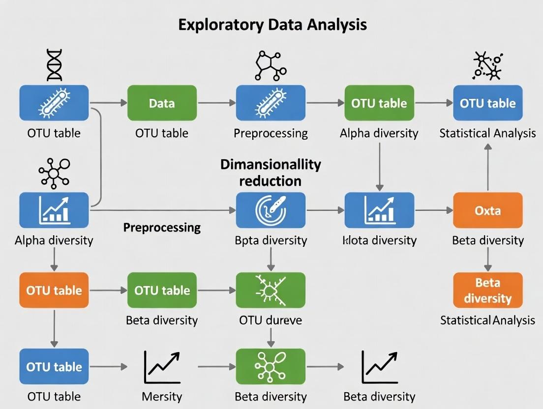
Exploratory Data Analysis (EDA) is the foundational step in transforming raw, high-dimensional microbiome sequencing data into actionable biological hypotheses. Framed within the broader thesis that rigorous EDA is indispensable for robust microbiome research, this technical guide details the methodologies, visualizations, and analytical protocols essential for researchers and drug development professionals navigating the complexity of microbial community data.
Microbiome data, typically generated via 16S rRNA amplicon or shotgun metagenomic sequencing, is characterized by extreme dimensionality (hundreds to thousands of taxa), compositionality (relative abundances sum to a constant), sparsity (many zero counts), and high technical variability. EDA provides the critical first lens through which researchers assess data quality, identify patterns, detect outliers, and inform downstream statistical modeling and hypothesis testing. Neglecting EDA risks deriving insights from artifacts or noise.
Foundational EDA Metrics and Quantitative Summaries
Key quantitative descriptors form the initial assessment of a microbiome dataset.
Table 1: Core Quantitative Metrics in Microbiome EDA
| Metric | Description | Typical Range/Interpretation | Tool/Formula |
|---|---|---|---|
| Sequencing Depth | Total reads per sample. | Inadequate depth (<10k reads for 16S) may omit rare taxa. | colSums(otu_table) |
| Alpha Diversity | Within-sample richness/evenness. | Shannon Index: 3.0-7.0; Observed OTUs: 100-1000s. | vegan::diversity() |
| Beta Diversity | Between-sample dissimilarity. | Bray-Curtis: 0 (identical) to 1 (max dissimilarity). | vegdist(matrix, method="bray") |
| Sample Read Distribution | Spread of total reads across samples. | High skew may indicate failed library prep. | Summary statistics (mean, median, CV) |
| Taxonomic Composition | Proportion of dominant phyla/classes. | Often: Firmicutes, Bacteroidetes dominate gut. | taxa_sums(physeq)/sum(taxa_sums) |
Table 2: Common Quality Control (QC) Filtering Thresholds
| Filtering Step | Typical Threshold | Rationale |
|---|---|---|
| Low-abundance OTU/ASV removal | Prevalence < 10% of samples & relative abundance < 0.01% | Reduces sparsity and noise from sequencing errors. |
| Low-depth sample removal | Reads < 30% of median sample depth | Excludes potential technical failures. |
| Contaminant removal | Frequency correlation with negative controls (p<0.1) | Uses decontam (R) to identify reagent/kit DNA. |
| Chimeric sequence removal | >99% similarity to parent sequences | Applied during DADA2/QIIME2 pipeline pre-EDA. |
Experimental Protocols for Key EDA-Centric Analyses
Protocol 3.1: Alpha and Beta Diversity Analysis with PERMANOVA
Objective: To quantify and statistically compare microbial diversity within and between pre-defined sample groups (e.g., disease vs. control).
Materials: Processed and filtered OTU/ASV table, sample metadata, R with vegan, phyloseq, ggplot2.
Procedure:
- Normalize: Rarefy data to even depth (if using non-phylogenetic metrics like Bray-Curtis) or use a variance-stabilizing transformation (e.g.,
DESeq2'svarianceStabilizingTransformation). - Calculate Alpha Diversity: Compute indices (Observed, Shannon, Simpson) for each sample. Visualize with boxplots grouped by condition.
- Statistical Test: Apply Wilcoxon rank-sum test (for two groups) or Kruskal-Wallis test (multiple groups) to alpha diversity indices.
- Calculate Beta Diversity: Generate a dissimilarity matrix (Bray-Curtis, Unweighted/Weighted UniFrac).
- Ordination: Perform Principal Coordinates Analysis (PCoA) on the matrix. Plot ordination using first two principal coordinates.
- PERMANOVA: Use the
adonis2function (vegan::adonis2(distance_matrix ~ Condition, data=metadata, permutations=999)) to test if group centroids differ significantly. - Homogeneity Check: Test dispersion homogeneity with
betadisper; a significant result confounds PERMANOVA.
Protocol 3.2: Differential Abundance Analysis via DESeq2
Objective: To identify taxa whose abundances are significantly associated with a phenotype, accounting for compositionality and over-dispersion.
Materials: Raw (non-rarefied) ASV/OTU count table, metadata, R with DESeq2, phyloseq.
Procedure:
- Phyloseq to DESeq2: Convert phyloseq object:
diagdds = phyloseq_to_deseq2(physeq, ~ Condition). - Model Fitting: Apply negative binomial model:
diagdds = DESeq(diagdds, test="Wald", fitType="parametric"). - Results Extraction: Retrieve results:
res = results(diagdds, contrast=c("Condition", "Treatment", "Control")). - Multiple Testing Correction: Apply Benjamini-Hochberg FDR correction. Filter results at
alpha = 0.05. - Visualization: Generate volcano plots (log2 fold-change vs. -log10(p-value)) or plot normalized counts of significant taxa.
Mandatory Visualizations: Workflows and Relationships
Diagram Title: Microbiome Analysis Pipeline with EDA Feedback
Diagram Title: Core EDA Modules for Microbiome Data
The Scientist's Toolkit: Research Reagent Solutions
Table 3: Essential Reagents and Tools for Microbiome EDA Workflows
| Item | Function in EDA/Research | Example Product/Kit |
|---|---|---|
| DNA Extraction Kit (with bead-beating) | Standardizes microbial lysis from complex matrices, a major source of pre-sequencing bias. Critical for comparing alpha diversity across studies. | Qiagen DNeasy PowerSoil Pro Kit, MoBio PowerLyzer |
| PCR Reagents & Barcoded Primers | Amplifies target region (e.g., V4 of 16S) with sample-specific barcodes for multiplexing. Efficiency impacts sequencing depth distribution. | KAPA HiFi HotStart ReadyMix, Illumina 16S Metagenomic Sequencing Library Prep |
| Negative Extraction Controls | Reagents-only controls used in wet-lab to identify kit/reagent contaminants for subsequent bioinformatic filtering (e.g., via decontam). |
Molecular grade water processed identically to samples |
| Mock Community Standards | Defined mixtures of known microbial genomes. Used to validate entire wet-lab to dry-lab pipeline, assess error rates, and calibrate EDA expectations. | ZymoBIOMICS Microbial Community Standards |
| Positive Sequencing Control (PhiX) | Spiked into Illumina runs for calibration, quality scoring, and identifying low-diversity samples (a potential sign of contamination). | Illumina PhiX Control v3 |
| Bioinformatics Pipelines | Software that processes raw FASTQ into feature tables, the substrate for EDA. Choice affects resolution (OTU vs. ASV). | QIIME 2, DADA2 (R), MOTHUR |
| Statistical Software & Packages | Primary environment for executing EDA protocols, visualization, and statistical testing. | R (phyloseq, vegan, DESeq2, ggplot2), Python (qiime2, scikit-bio, anndata) |
This guide outlines the foundational steps for preparing high-dimensional microbiome sequencing data for exploratory data analysis (EDA). Within the broader thesis of EDA for microbiome research, robust pre-processing is critical to transform raw sequence data into a reliable biological signal, enabling downstream statistical and ecological interpretation.
Quality Control (QC) and Trimming
The initial step involves assessing and improving the quality of raw sequencing reads (typically from 16S rRNA gene amplicon sequencing).
Key QC Metrics
Table 1: Common QC Metrics and Recommended Thresholds
| Metric | Description | Typical Threshold (16S Amplicons) | Tool Output Example |
|---|---|---|---|
| Per-base Sequence Quality | Phred score (Q) across all bases. | Q ≥ 30 (99.9% accuracy) | FastQC, MultiQC |
| Per-sequence Quality | Average quality per read. | Mean Q ≥ 25 | FastQC, MultiQC |
| Sequence Length | Distribution of read lengths post-trimming. | Expect uniformity based on amplicon length. | FastQC |
| Adapter Content | Presence of sequencing adapters. | ≤ 5% of reads | FastQC, cutadapt |
| Ambiguous Bases (N) | Proportion of uncalled bases. | 0% | Trimming/Filtering step |
| GC Content | Distribution of GC% per read. | Should match expected organism/target. | FastQC |
Experimental Protocol: QC and Trimming with DADA2 or QIIME 2
A. Tools Required: FastQC, MultiQC, Trimmomatic/cutadapt, DADA2 (R) or QIIME 2 (q2-demux, q2-quality-filter). B. Protocol Steps:
- Demultiplexing: Assign reads to samples based on barcodes (often done by sequencing facility).
- Initial QC: Run FastQC on all raw FASTQ files. Aggregate reports with MultiQC.
- Trimming:
- Remove Adapters: Use cutadapt or the
q2-cutadaptplugin to trim adapter sequences. - Trim Low-Quality Ends: Using DADA2's
filterAndTrim()function orq2-quality-filter. Typical parameters: trim left=10-20 bases (remove primers/initial low quality), truncate at first instance of Q<20-30.
- Remove Adapters: Use cutadapt or the
- Post-trimming QC: Run FastQC again on trimmed reads to confirm improvement.
Denoising and Sequence Variant Inference
Denoising corrects sequencing errors and infers the true biological sequences, moving beyond Operational Taxonomic Unit (OTU) clustering to Amplicon Sequence Variants (ASVs).
OTU vs. ASV Approaches
Table 2: Comparison of OTU Clustering and ASV Denoising
| Feature | OTU Clustering (97% similarity) | ASV Denoising (DADA2, Deblur, UNOISE3) |
|---|---|---|
| Definition | Clusters reads based on similarity threshold. | Infers exact biological sequences by error modeling. |
| Resolution | Lower (groups similar sequences). | Single-nucleotide resolution. |
| Reproducibility | Variable (depends on clustering method/database). | Highly reproducible. |
| Common Tools | VSEARCH, UCLUST, mothur. | DADA2, Deblur, UNOISE3. |
| Typical Output | OTU table (counts per OTU per sample). | ASV table (counts per exact sequence per sample). |
Experimental Protocol: Denoising with DADA2
A. Core Algorithm Steps: Error-rate learning → Dereplication → Sample inference → Denoising (error correction) → Chimera removal. B. Detailed Protocol (DADA2 in R):
- Learn Error Rates:
learnErrors(filtFs, multithread=TRUE)models the platform-specific error profile. - Dereplication:
derepFastq()combines identical reads, reducing computation. - Core Sample Inference:
dada(derep, err=error_rates, pool=FALSE)applies the error model to each sample, identifying true sequences. - Merge Paired-end Reads:
mergePairs()for paired-end data, ensuring high-quality overlaps. - Construct Sequence Table:
makeSequenceTable()creates the initial ASV-by-sample count matrix. - Remove Chimeras:
removeBimeraDenovo()identifies and removes PCR chimeras. - Output: A denoised ASV table (counts) and a FASTA file of ASV sequences.
Diagram Title: DADA2 Denoising Workflow for ASV Inference
ASV/OTU Table Creation and Basic Filtering
The final output of pre-processing is a feature-by-sample count matrix.
Table Structure and Post-Processing
Table 3: Structure of a Typical ASV/OTU Table
| Sample_1 | Sample_2 | Sample_3 | ... | Taxonomy | |
|---|---|---|---|---|---|
| ASV_001 | 1500 | 450 | 0 | ... | pFirmicutes; cBacilli; ... |
| ASV_002 | 0 | 1200 | 85 | ... | pBacteroidota; cBacteroidia; ... |
| ... | ... | ... | ... | ... | ... |
| Total Reads | 50,000 | 48,500 | 52,100 | ... |
Experimental Protocol: Table Refinement and Taxonomy Assignment
- Taxonomy Assignment: Assign taxonomy to ASV sequences using a reference database (e.g., SILVA, Greengenes, RDP). Tool:
assignTaxonomy()in DADA2 orq2-feature-classifierin QIIME 2. - Basic Filtering:
- Remove singletons/doubletons: Filter ASVs with total abundance ≤ 2 across all samples to reduce spurious noise.
- Prevalence filtering: Remove ASVs present in fewer than a threshold percentage of samples (e.g., 5%).
- Remove non-bacterial sequences: (for 16S data) Filter out sequences assigned to Archaea, Chloroplasts, Mitochondria.
- Normalization (for downstream EDA): Note: Normalization is part of EDA, not pre-processing, but is essential for comparison. Methods include rarefaction, Cumulative Sum Scaling (CSS), or relative abundance conversion.
Diagram Title: Post-Denoising Table Curation Steps
The Scientist's Toolkit: Essential Research Reagent Solutions
Table 4: Key Reagents and Materials for 16S rRNA Amplicon Sequencing Pre-Processing
| Item | Function in Pre-Processing Context | Example/Note |
|---|---|---|
| High-Fidelity DNA Polymerase | Critical for accurate PCR amplification with minimal bias during library prep. | KAPA HiFi HotStart, Q5 Hot Start. |
| Validated Primer Sets | Target-specific amplification of variable regions (V3-V4, V4). | 515F/806R, 341F/785R. Must be barcoded/indexed for multiplexing. |
| Standardized Mock Community DNA | Positive control for evaluating pipeline performance (denoising error, taxonomy). | ZymoBIOMICS Microbial Community Standard. |
| Negative Extraction Control Reagents | Identifies reagent/lab kit contamination for downstream filtering. | Sterile water or buffer taken through extraction. |
| Quantitation Kits (dsDNA-specific) | Accurate measurement of library concentration for pooling. | Qubit dsDNA HS Assay, Quant-iT PicoGreen. |
| Size Selection Beads | Cleanup of PCR products and final libraries to remove primer dimers. | AMPure XP, SPRIselect beads. |
| Sequencing Platform-Specific Kits | Generation of sequencing-ready libraries (e.g., adding adapters, indices). | Illumina Nextera XT, Swift Amplicon. |
| Bioinformatics Software Suites | De facto reagents for computational pre-processing. | QIIME 2 (plugins), DADA2 (R package), MOTHUR. |
Within the broader framework of Exploratory Data Analysis (EDA) for high-dimensional microbiome research, initial sanity checks are the critical first step to ensure data integrity and interpretability. These checks validate the raw sequencing output, identify gross anomalies, and set the stage for robust downstream analysis. This guide details the protocols for assessing library size distribution, sample heterogeneity, and batch effects.
Library Size Distribution Analysis
Purpose: To verify that sequencing depth (total reads per sample) is sufficient, comparable across samples, and free from failures.
Experimental Protocol: Quantifying Sequencing Depth
- Data Input: Start with the raw feature table (e.g., ASV/OTU table) or the output from
qiime2 demuxorDADA2. - Calculation: For each sample, sum the counts across all microbial features (taxa, genes). This yields the library size.
- Visualization: Generate a bar plot (samples on x-axis, library size on y-axis). Order samples by size.
- Statistical Summary: Calculate mean, median, standard deviation, and range. Flag samples where library size is below a pre-defined threshold (e.g., < 10,000 reads for 16S rRNA data) or is a statistical outlier (e.g., > 3 standard deviations from the mean).
Key Data Metrics & Interpretation
Table 1: Typical Library Size Metrics for 16S rRNA Amplicon Studies (Post-QC)
| Metric | Acceptable Range | Concerning Indication |
|---|---|---|
| Mean Library Size | Study-dependent; > 20,000 reads is often robust | Low mean may indicate poor sequencing run or over-dilution of pool. |
| Standard Deviation | Ideally < 50% of the mean | High variance suggests inconsistent sequencing performance or sample prep issues. |
| Minimum Sample Size | > 10,000 reads (conservative) | Samples with very low depth may need rarefaction or removal due to undersampling. |
| Outlier Samples | >3 SD from mean or visual inspection | May represent failed libraries or contamination; warrant investigation. |
Sample Heterogeneity Assessment
Purpose: To ensure the dataset captures expected biological variation and to identify potential sample swaps or extreme outliers.
Experimental Protocol: Beta Diversity Analysis
- Data Transformation: Rarefy the feature table to an even depth (if using metrics sensitive to depth like unweighted UniFrac) or use a compositional transform (e.g., CLR for Aitchison distance).
- Distance Matrix Calculation: Compute a beta diversity distance matrix (e.g., Bray-Curtis, Jaccard, Weighted/Unweighted UniFrac for taxonomy; Aitchison for composition).
- Ordination: Perform dimensionality reduction (e.g., Principal Coordinates Analysis - PCoA) on the distance matrix.
- Visualization: Plot samples in 2D/3D ordination space, colored by known metadata (e.g., body site, treatment group, donor).
- Statistical Test: Use PERMANOVA (Adonis) to test if pre-defined groups explain a significant proportion of variance. This validates expected heterogeneity.
Table 2: Common Beta Diversity Metrics for Sample Heterogeneity
| Metric | Best For | Interpretation of Spread |
|---|---|---|
| Bray-Curtis | General ecological dissimilarity (abundance-aware) | Tight clusters = low heterogeneity; dispersed points = high heterogeneity. |
| Unweighted UniFrac | Phylogenetic presence/absence differences | Captures deep phylogenetic divergence between communities. |
| Weighted UniFrac | Phylogenetic abundance-weighted differences | Emphasizes abundance changes in closely related taxa. |
| Aitchison Distance | Compositional data (requires CLR transform) | Robust to library size, focuses on relative log-ratios. |
Batch Effect Detection
Purpose: To identify and quantify non-biological technical variation introduced by sequencing run, extraction batch, or processing date.
Experimental Protocol: Multivariate Modeling & Visualization
- Metadata Annotation: Ensure metadata includes potential batch variables (e.g., sequencinglane, extractiondate, PCR_plate).
- Exploratory Ordination: As in Section 3, generate PCoA plots but color points by batch variable instead of biological group.
- Formal Statistical Testing: Use multivariate methods to partition variance:
- PERMANOVA: Test significance of batch variable.
- Distance-Based Redundancy Analysis (dbRDA): Constrain ordination by biological variable and visualize residuals for batch.
- Principal Variance Component Analysis (PVCA): Combine PCA and variance components analysis to quantify proportion of variance attributable to batch vs. biology.
- Batch-Corrected Visualization: Apply a batch correction method (e.g., ComBat-seq for counts, RUVseq, or limma's
removeBatchEffecton CLR-transformed data) and re-plot ordination.
Table 3: Batch Effect Detection Methods & Outputs
| Method | Input Data | Key Quantitative Output |
|---|---|---|
| PERMANOVA on Batch | Distance Matrix | R² and p-value for batch variable. R² > 0.05 often concerning. |
| PVCA | Normalized Feature Table | Variance proportion attributed to each batch and biological factor. |
| dbRDA | Distance Matrix + Model | Visual and statistical assessment of variance partition. |
| PCA on Controls | Features from Control Samples | Clustering of technical replicates from different batches indicates effect. |
The Scientist's Toolkit: Essential Research Reagents & Software
Table 4: Key Solutions for Microbiome Data Sanity Checks
| Item / Solution | Function in Sanity Checks | Example or Note |
|---|---|---|
| QIIME 2 / R phyloseq | Primary environment for data handling, normalization, and distance calculation. | qiime diversity core-metrics-phylogenetic or phyloseq::plot_richness(). |
| Negative Extraction Controls | Identifies reagent or environmental contamination affecting library sizes. | Used to filter contaminant ASVs from feature table. |
| Technical Replicates (Mock Community) | Distinguishes technical noise from biological variation; assesses sequencing accuracy. | ZymoBIOMICS or ATCC mock microbial standards. |
| R Packages: vegan, ape | Statistical testing (PERMANOVA, Mantel test) and distance matrix computation. | vegan::adonis2() for PERMANOVA. |
| Batch Correction Tools (ComBat-seq, RUVSeq) | Statistical removal of batch effects post-detection for downstream analysis. | sva::ComBat_seq() for count data. |
| Interactive Visualization (ggplot2, plotly) | Creation of publication-quality and exploratory plots for outlier detection. | ggplot2::geom_boxplot() for library size plots. |
| DNA/RNA Stabilization Buffer | Preserves sample integrity from collection to extraction, reducing batch variation. | RNAlater, OMNIgene•GUT, etc. |
| Standardized DNA Extraction Kit | Minimizes technical variation in library prep across batches. | DNeasy PowerSoil Pro Kit, MagAttract PowerSoil DNA Kit. |
Exploratory data analysis (EDA) for high-dimensional microbiome data presents unique statistical challenges. The data generated from 16S rRNA gene sequencing or shotgun metagenomics is inherently compositional, sparse, and zero-inflated. Ignoring these intrinsic properties can lead to biased inference, false discoveries, and invalid conclusions in downstream analyses, such as differential abundance testing or association studies in drug development. This technical guide provides an in-depth examination of these three core properties—sparsity, zero-inflation, and compositionality—framing them as the foundational first step in any robust microbiome EDA pipeline.
Sparsity
Sparsity refers to the condition where most entries in the data matrix are zero. In microbiome studies, this arises because any single sample contains only a subset of the vast microbial diversity present in an ecosystem.
Quantitative Characterization
Table 1: Typical Sparsity Metrics in Microbiome Datasets
| Dataset Type | Average Sparsity (% Zeros) | Range Across Studies | Typical Sample Depth (Reads) | Reference Year |
|---|---|---|---|---|
| 16S rRNA (Gut) | 85-95% | 75-99% | 10,000 - 100,000 | 2023 |
| Shotgun Metagenomic (Soil) | 90-98% | 88-99.5% | 10-50 million | 2024 |
| Viral Metagenome | 95-99.9% | 94-99.99% | 1-5 million | 2023 |
Experimental Protocol for Assessing Sparsity
- Data Input: Load Amplicon Sequence Variant (ASV) or Operational Taxonomic Unit (OTU) count table (samples x features).
- Filtering: Apply a minimum prevalence filter (e.g., retain features present in >10% of samples) to remove ultra-rare taxa. Note: This step is optional for pure sparsity assessment but common in practice.
- Calculation:
- Total entries = number of samples (N) × number of features (p)
- Zero count = Sum of all entries where count == 0
- Sparsity % = (Zero count / Total entries) × 100
- Visualization: Generate a heatmap of the log-transformed count matrix to visually inspect the pattern of non-zero entries.
Zero-Inflation
Zero-inflation goes beyond simple sparsity; it describes a situation where the observed frequency of zeros is greater than expected under a standard count distribution model (e.g., Poisson or Negative Binomial). Zeros can be either "biological" (the microbe is truly absent) or "technical" (the microbe is present but undetected due to limited sequencing depth).
Quantitative Characterization
Table 2: Sources and Prevalence of Zero Inflation
| Zero Type | Estimated Contribution | Primary Cause | Detection Clue |
|---|---|---|---|
| Technical (Dropout) | 20-60% of all zeros | Low biomass, library preparation bias, low sequencing depth | Correlated with low sample total read depth. |
| Biological (True Absence) | 40-80% of all zeros | Genuine absence of taxon in the niche, competitive exclusion | May be uncorrelated with sequencing depth; can be ecologically structured. |
Experimental Protocol for Zero-Inflation Modeling
- Model Fitting: For each taxon j, fit two models to its count vector across samples i:
- A standard count model (e.g., Negative Binomial):
Count_ij ~ NB(μ, dispersion). - A zero-inflated model (e.g., Zero-Inflated Negative Binomial - ZINB):
Count_ij ~ π * 0 + (1-π) * NB(μ, dispersion), where π is the probability of a structural zero.
- A standard count model (e.g., Negative Binomial):
- Goodness-of-Fit Test: Perform a likelihood-ratio test (LRT) between the nested models.
- Interpretation: A significant p-value from the LRT indicates the taxon's counts are zero-inflated, justifying the use of zero-inflated models for differential analysis.
Title: Decision Flow for Interpreting Zero Counts in Microbiome Data
Compositionality
Compositionality means that the data conveys only relative abundance information. Sequencing data is constrained sum (e.g., to total library size), so an increase in the relative abundance of one taxon necessitates an apparent decrease in others. This leads to spurious correlations if not accounted for.
Core Principle: The Aitchison Simplex
Microbiome count data resides in a simplex space where only relative information is valid. Analyses must use compositional methods, such as centered log-ratio (CLR) transformations, or employ models specifically designed for compositional data.
Experimental Protocol for Compositional Data Analysis
- Normalization (Optional but Common): Convert raw counts to proportions (total-sum scaling) or use a rarefaction procedure. Note: Modern approaches often use direct modeling with offsets instead.
- Transformation for Euclidean Space:
- CLR Transformation: For a sample vector x with D taxa, calculate
clr(x) = [ln(x1 / g(x)), ..., ln(xD / g(x))], whereg(x)is the geometric mean of x. - Handle zeros: Add a pseudo-count or use a more advanced method like the
compositionspackage'sclr()with multiplicative replacement.
- CLR Transformation: For a sample vector x with D taxa, calculate
- Downstream Analysis: Perform PCA on the CLR-transformed matrix (creating "Aitchison distance") or use a compositional regression model like ALDEx2 or a Dirichlet-Multinomial model.
Title: Workflow Contrast: Ignoring vs. Accounting for Compositionality
The Scientist's Toolkit: Research Reagent Solutions
Table 3: Essential Tools for Analyzing Sparse, Zero-Inflated, Compositional Data
| Item/Tool | Function/Benefit | Example Package/Software |
|---|---|---|
| Zero-Inflated Models (GLMM) | Models both technical zeros and count distribution simultaneously, providing unbiased effect estimates. | glmmTMB, pscl (R) |
| Compositional Transform | Projects constrained data into unconstrained Euclidean space for standard statistical methods. | compositions::clr(), microViz::transform_clr() (R) |
| Compositional Distance Metric | Measures dissimilarity between samples while respecting compositionality. | Aitchison Distance, Bray-Curtis (robust) |
| Feature-Specific Regularization | Handles high-dimensionality and sparsity by shrinking noise features' effects. | maaslin2 (LASSO), metagenomeSeq (CSS + zero-inflated Gaussian) |
| Bayesian Multiplicative Replacement | Handles zeros in compositional transforms without arbitrary pseudo-counts. | zCompositions::cmultRepl() (R) |
| Dirichlet-Multinomial Model | A probabilistic model for over-dispersed, compositional count data. | DirichletMultinomial (R/Bioconductor) |
| Co-occurrence Network Inference | Estimates microbial associations while correcting for compositionality and sparsity. | SPIEC-EASI, FlashWeave |
| Experimental Positive Controls | Spike-in synthetic microbial communities to distinguish technical from biological zeros. | ZymoBIOMICS Microbial Community Standards |
Navigating the Data: Essential Methods, Visualizations, and Hands-On Applications for Microbiome EDA
Within the framework of a thesis on Exploratory Data Analysis for High-Dimensional Microbiome Data Research, dimensionality reduction is a critical step. Microbiome datasets, generated via 16S rRNA gene sequencing or shotgun metagenomics, are characterized by thousands of operational taxonomic units (OTUs) or genes across a limited number of samples. This high-dimensional, sparse, and compositional nature poses significant challenges for visualization and interpretation. Core ordination techniques transform these complex datasets into lower-dimensional spaces, preserving ecological distances to reveal patterns, clusters, and outliers essential for hypothesis generation in research and drug development.
This whitepaper provides an in-depth technical guide to four principal ordination methods: Principal Coordinate Analysis (PCoA), Non-Metric Multidimensional Scaling (NMDS), and the modern manifold techniques t-Distributed Stochastic Neighbor Embedding (t-SNE) and Uniform Manifold Approximation and Projection (UMAP). We focus on their application, comparative strengths, and practical implementation in microbiome analysis.
Foundational Concepts & Distance Metrics
Ordination techniques require a measure of dissimilarity between samples. The choice of distance metric is paramount and should reflect the biological question.
Core Distance Metrics for Microbiome Data:
| Metric | Formula (for samples j and k) | Key Properties | Best Use Case |
|---|---|---|---|
| Bray-Curtis | $$BC{jk} = \frac{\sumi | x{ij} - x{ik} |}{\sumi (x{ij} + x_{ik})}$$ | Non-Euclidean, robust to outliers, handles zeros. Measures community composition dissimilarity. | General beta-diversity analysis comparing community structures. |
| Unweighted UniFrac | $$U_{jk} = \frac{\text{length of unshared branches}}{\text{total branch length of tree}}$$ | Incorporates phylogenetic tree. Sensitive to presence/absence of lineages. | Detecting deep phylogenetic community shifts. |
| Weighted UniFrac | $$W_{jk} = \frac{\text{sum (branch length × | abundance proportion difference |)}}{\text{total abundance-scaled branch length}}$$ | Incorporates phylogeny and relative abundance. | Detecting changes in abundant lineages. |
| Jaccard | $$J_{jk} = 1 - \frac{| A \cap B |}{| A \cup B |}$$ | Presence/Absence based. Ignores abundance. | Analyzing rare biosphere or strong turnover. |
| Aitchison | Euclidean distance on centered log-ratio (CLR) transformed data. | Proper metric for compositional data. Requires zero imputation (e.g., pseudocount). | Core method for analyzing compositional microbial abundance data. |
Core Ordination Techniques
Principal Coordinate Analysis (PCoA / Classical Multidimensional Scaling)
Methodology: PCoA aims to find a low-dimensional representation where the Euclidean distances between points approximate a given dissimilarity matrix. It performs an eigendecomposition of a double-centered distance matrix. The eigenvalues indicate the proportion of variance explained by each principal coordinate.
Protocol for Microbiome PCoA:
- Compute Dissimilarity: Calculate a full distance matrix (e.g., Bray-Curtis, UniFrac) from the OTU/ASV table.
- Transform Matrix: Perform double-centering:
G = -0.5 * (I - 11^T/n) * D^2 * (I - 11^T/n), whereDis the distance matrix. - Eigendecomposition: Solve
G = UΛU^T. Eigenvectors (U) are the coordinates. Eigenvalues (Λ) indicate variance. - Select Axes: Typically, plot the first 2-3 eigenvectors with the largest positive eigenvalues.
- Variance Interpretation: The percentage of variance explained by axis i is
(λ_i / Σ(λ_positive)) * 100.
Key Considerations: PCoA can handle non-Euclidean distances (like Bray-Curtis), but negative eigenvalues may arise, indicating the distance is not fully embeddable in Euclidean space. The variance explained is a crucial quantitative output.
Non-Metric Multidimensional Scaling (NMDS)
Methodology: NMDS is a rank-based method. It seeks a configuration where the ranking of distances between points in the low-dimensional space is as close as possible to the ranking of the original dissimilarities (monotonic relationship). It minimizes a stress function (e.g., Kruskal's stress) iteratively.
Protocol for Microbiome NMDS:
- Define Dimensions (k): Choose the number of ordination dimensions (k=2 or 3 for visualization).
- Initial Configuration: Generate a random starting configuration of points in k-dimensional space.
- Calculate Distances: Compute Euclidean distances between all points in the current configuration.
- Regress & Compare: Perform a monotonic regression (Shepard diagram) between the configuration distances and the original dissimilarities.
- Minimize Stress: Iteratively adjust point positions to minimize stress:
Stress = √[ Σ (d_orig - d_pred)^2 / Σ d_orig^2 ]. - Iterate: Use a numerical optimizer (e.g., gradient descent) over many iterations (e.g., 500) to find the best configuration.
- Assess Fit: Final stress value indicates goodness-of-fit: <0.05 = excellent, <0.1 = good, >0.2 = potentially misleading.
Key Considerations: NMDS is robust and makes no assumptions about the linear relationship between distance metrics. The final axes are arbitrary (rotatable) and must be interpreted relative to each other. Always run multiple random starts to avoid local minima.
t-SNE and UMAP
These are modern non-linear manifold learning techniques.
t-SNE (t-Distributed Stochastic Neighbor Embedding):
- Methodology: Models pairwise similarities in high-dimension as probabilities (using a Gaussian kernel) and in low-dimension as probabilities (using a Student's t-distribution). It minimizes the Kullback–Leibler divergence between these two distributions via gradient descent.
- Key Parameter:
perplexity, which balances local vs. global structure (typical range 5-50 for microbiome data). - Limitation: Cannot transform new data; global structure is not always preserved.
UMAP (Uniform Manifold Approximation and Projection):
- Methodology: Constructs a topological representation of the high-dimensional data (fuzzy simplicial complex) and optimizes a low-dimensional layout to be as topologically similar as possible. It uses cross-entropy as a cost function.
- Key Parameters:
n_neighbors(balances local/global structure) andmin_dist(controls clustering tightness). - Advantages: Faster, often better preserves global structure, and can transform new data.
Comparative Protocol for t-SNE/UMAP:
- Input: Start with a transformed feature table (e.g., CLR-transformed abundances, not a distance matrix).
- Parameter Selection: For UMAP:
n_neighbors=15-50,min_dist=0.1-0.5. For t-SNE:perplexity=30. - Dimensionality Pre-reduction: It is often advisable to first reduce dimensions via PCA (e.g., to 50 PCs) to denoise.
- Optimization: Run the algorithm (t-SNE or UMAP) on the PC scores or original features.
- Multiple Runs: Due to stochasticity, run multiple times with different random seeds to assess stability.
Quantitative Comparison of Techniques
| Technique | Input Type | Preserves | Key Output Metrics | Computational Load | Best for Microbiome... |
|---|---|---|---|---|---|
| PCoA | Distance Matrix | Global distances (optimally for Euclidean) | % Variance explained per axis (from eigenvalues). | Low | Hypothesis-driven analysis with interpretable variance. Standard for beta-diversity. |
| NMDS | Distance Matrix | Distance rank order (monotonic fit) | Stress value (goodness-of-fit). Iterations to convergence. | Medium | Exploratory analysis with any non-linear distance metric. |
| t-SNE | Feature Matrix (or Distance) | Local neighborhoods | KL Divergence (final cost). | High | Visualizing fine-grained clusters and sub-structure. |
| UMAP | Feature Matrix (or Distance) | Local & more global topology | Not typically a single fit metric. | Medium-High | Visualizing hierarchical clustering structure and patterns. |
The Scientist's Toolkit: Research Reagent Solutions
| Item/Category | Function in Microbiome Ordination Analysis |
|---|---|
| QIIME 2 (Core) | Plugin-based platform. qiime diversity pcoa, qiime diversity umap, and qiime emperor for visualization. Essential for standardized UniFrac/PCoA pipelines. |
| R (phyloseq & vegan) | phyloseq object manages OTU table, taxonomy, sample data, and tree. vegan::metaMDS() for NMDS, ape::pcoa() for PCoA. vegdist() computes distances (Bray-Curtis). |
| Python (scikit-bio, scikit-learn) | skbio.stats.ordination.pcoa and skbio.distance for PCoA. sklearn.manifold for t-SNE and UMAP. Enables integration into machine learning workflows. |
| FastTree / SEPP | Generates the phylogenetic trees required for calculating UniFrac distances. |
| GUniFrac R Package | Computes generalized UniFrac distances, offering a flexible parameter to balance abundance weighting. |
| Aitchison Imputation (e.g., zCompositions) | R package zCompositions provides methods (e.g., Bayesian-multiplicative replacement) to handle zeros for robust Aitchison distance calculation. |
| Emperor / ggOrdination | Visualization tools for creating interactive 3D plots (Emperor) or publication-quality static plots (ggplot2-based ggord, ggfortify). |
Experimental Workflow & Logical Pathway
The following diagram outlines the standard decision pathway for applying core ordination techniques in microbiome exploratory data analysis.
Diagram 1: Microbiome Ordination Technique Selection Workflow
Selecting the appropriate ordination technique is fundamental to effective exploratory data analysis in microbiome research. PCoA paired with a biologically relevant distance metric (e.g., Weighted UniFrac for abundance, Aitchison for composition) remains the gold standard for hypothesis-oriented beta-diversity assessment, providing clear variance metrics. NMDS offers robust, assumption-free exploration when distance metrics are non-linear. For uncovering complex, non-linear sub-structures within the data, t-SNE and UMAP are powerful, with UMAP generally preferred for its speed and better preservation of continuum relationships. Integrating these tools into a coherent analytical workflow, as guided by the research question and data properties, empowers researchers and drug developers to extract meaningful biological insights from the complexity of microbial ecosystems.
In the context of exploratory data analysis (EDA) for high-dimensional microbiome data, visualization is a critical bridge between raw sequencing output and biological insight. This technical guide details advanced plotting techniques essential for interpreting complex microbial community structures, their dynamics, and associations within a broader thesis on microbiome research and therapeutic discovery.
Foundational Visualization Techniques
Heatmaps
Heatmaps are indispensable for visualizing the relative abundance of microbial taxa (e.g., OTUs, ASVs) across multiple samples, revealing patterns of co-occurrence and sample clustering.
Experimental Protocol for Data Preparation:
- Data Input: Start with a filtered feature table (e.g., from QIIME 2, mothur) containing counts per taxon per sample.
- Normalization: Apply a compositional normalization like Centered Log-Ratio (CLR) transformation or convert to relative abundance (percentage). For differential abundance, use variance-stabilizing transformations (e.g., DESeq2).
- Filtering: Retain top N most abundant or most variable taxa to reduce dimensionality and improve clarity.
- Clustering: Perform hierarchical clustering on rows (taxa) and/or columns (samples) using distance metrics (Bray-Curtis, Euclidean) and linkage methods (Ward, average).
- Plotting: Map normalized abundance values to a color gradient. Include sidebars for sample metadata (e.g., disease state, treatment) and taxonomic classification.
Bar Plots
Bar plots, typically stacked, provide a straightforward view of taxonomic composition at a chosen level (e.g., Phylum, Genus) across sample groups.
Experimental Protocol:
- Aggregation: Aggregate sequence variants to the desired taxonomic level (Phylum, Class, etc.).
- Grouping: Average relative abundances within pre-defined sample groups (e.g., control vs. treated cohorts).
- Plotting: Generate stacked bars for each group. Use a consistent, color-blind friendly palette for taxa. Order taxa by overall abundance or a specific metric.
Violin Plots
Violin plots combine a box plot with a kernel density estimate to show the distribution, spread, and probability density of a specific metric (e.g., alpha diversity index, abundance of a key taxon) across sample groups.
Experimental Protocol for Alpha Diversity Visualization:
- Calculation: Compute alpha diversity indices (Shannon, Faith's PD, Observed Features) per sample using rarefied data.
- Metadata Integration: Merge diversity values with sample metadata grouping variable.
- Statistical Testing: Perform non-parametric tests (Kruskal-Wallis, pairwise Wilcoxon) between groups.
- Plotting: Plot the distribution of the diversity index per group, overlaying box plot elements (median, IQR) and individual data points. Annotate with statistical significance.
Network Graphs
Co-occurrence or correlation networks infer potential ecological interactions between microbial taxa from abundance patterns across samples.
Experimental Protocol for Network Inference:
- Correlation Calculation: Compute pairwise associations (SparCC, Spearman, Pearson) on CLR-transformed or proportionally normalized abundance data.
- Thresholding: Apply significance (p-value) and magnitude (correlation coefficient) thresholds to create an adjacency matrix.
- Network Construction: Define taxa as nodes and significant correlations as edges. Weight edges by correlation strength and sign (positive/negative).
- Topological Analysis: Calculate network properties (modularity, degree centrality, betweenness centrality).
- Visualization: Layout nodes using force-directed algorithms (Fruchterman-Reingold). Color nodes by taxonomy or module membership. Size nodes by degree centrality or abundance.
Data Presentation: Comparative Table of Visualization Techniques
| Visualization | Primary Use Case | Key Metrics Visualized | Data Preprocessing Needs | Common Tools/Packages |
|---|---|---|---|---|
| Heatmap | Pattern discovery, sample/taxon clustering | Relative abundance (transformed) | Normalization, filtering, clustering | R: pheatmap, ComplexHeatmap; Python: seaborn.clustermap |
| Bar Plot | Taxonomic composition summary | Mean relative abundance per group | Taxonomic aggregation, group averaging | R: ggplot2; Python: matplotlib, seaborn |
| Violin Plot | Distribution comparison across groups | Alpha diversity, specific taxon abundance | Index calculation, group assignment | R: ggplot2; Python: seaborn.violinplot |
| Network Graph | Interaction inference, system properties | Correlation coefficients, centrality measures | Correlation inference, thresholding | R: igraph, ggraph; Python: networkx, igraph |
Workflow Diagram
Microbiome Visualization Analysis Workflow
The Scientist's Toolkit: Key Reagents & Software
| Item | Function in Microbiome Visualization |
|---|---|
| QIIME 2 (Core 2024.5) | Primary pipeline for processing raw sequences into feature tables and performing initial diversity analyses. Provides q2-diversity and q2-taxa plugins for plotting. |
| R (v4.3+) with Packages | Statistical computing environment. phyloseq for data object management; ggplot2/ggpubr for static plots; vegan for ecological metrics; igraph for networks. |
| Python (v3.10+) with Libraries | Programming language for flexible analysis. biom-format for table handling; scikit-bio for diversity; matplotlib/seaborn for plotting; networkx for networks. |
| SparCC Algorithm | Tool for calculating robust correlations between compositional taxa, mitigating spurious correlations in microbiome data. |
| Graphviz Layout Engines | Used by igraph and networkx to generate clean, readable network graph layouts (e.g., neato, fdp). |
| ColorBrewer Palettes | Sets of colorblind-friendly, print-safe palettes (e.g., Set2, Set3, Paired) implemented in RColorBrewer and seaborn for categorical data (taxa). |
| Centered Log-Ratio (CLR) Transform | Essential normalization for compositional data prior to correlation analysis, available in the compositions R package or scikit-bio in Python. |
In the analysis of high-dimensional microbiome data, a fundamental challenge is the compositional nature of the data. Measurements, such as 16S rRNA gene amplicon sequencing reads or metagenomic counts, do not represent absolute abundances but rather relative proportions constrained to a constant sum (e.g., total library size). This compositional constraint induces spurious correlations and obscures true biological signals. Exploratory Data Analysis (EDA) for microbiome research must, therefore, begin with an appropriate transformation to address compositionality. This guide provides an in-depth technical overview of the Centered Log-Ratio (CLR), Additive Log-Ratio (ALR), and other key transformations, framed within the broader thesis that proper transformation is the critical first step for valid inference in microbial ecology and therapeutic development.
Core Transformations: Theory and Application
The following table summarizes the mathematical definitions, key properties, and primary use cases for the major compositional data transformations.
Table 1: Comparison of Core Compositional Data Transformations
| Transformation | Formula (for vector x with D parts) | Key Properties | Primary Use Case in Microbiome Research |
|---|---|---|---|
| Center Log-Ratio (CLR) | clr(x)_i = ln(x_i / g(x)) where g(x) is the geometric mean of all parts. |
Symmetric, isometric. Creates a Euclidean space. Parts sum to zero. | Default for many multivariate methods (PCA, PERMANOVA). Foundation for SparCC and ANCOM-BC. |
| Additive Log-Ratio (ALR) | alr(x)_i = ln(x_i / x_D) where x_D is an arbitrarily chosen reference part. |
Simple, asymmetric. Results in a (D-1)-dimensional real space. | Analyzing specific taxon ratios. Simpler interpretation vs. a chosen baseline (e.g., a common bacterium). |
| Isometric Log-Ratio (ILR) | ilr(x) = V^T * ln(x) where V is an orthonormal basis in the simplex. |
Isometric, orthonormal coordinates. Many possible balances. | Testing a priori phylogenetic or ecological balances (e.g., via PhILR). |
| Rarefaction | Random subsampling to an even sequencing depth. | Discards data, introduces variance. Not a transformation per se. | Largely deprecated for differential abundance. Sometimes used for alpha diversity estimation. |
| Total Sum Scaling (TSS) | x_i / sum(x) |
Simple normalization to proportions. Does not address compositionality. | Initial visualization. Input for downstream transformations (CLR, ALR). |
Experimental Protocol: A Standard Workflow for Compositional EDA
The following is a detailed methodology for conducting EDA on a typical 16S rRNA microbiome dataset (e.g., from QIIME 2 or mothur) with compositionality in mind.
Protocol 1: Core EDA Workflow with Compositional Transformations
Data Preprocessing & Import:
- Start with a Feature Table (OTU/ASV table) of raw counts and associated Metadata.
- Apply a prevalence filter (e.g., retain features present in >10% of samples).
- Pseudocount Addition: Add a small pseudocount (e.g., 1 or a fraction of the minimum positive count) to all zero values to enable log-transformation. Note: More sophisticated zero imputation (e.g., Bayesian or model-based) may be used in advanced pipelines.
Transformation & Normalization:
- Path A - For multivariate distance-based analysis: Apply the CLR transformation.
- Calculate the geometric mean
g(x)for each sample. - Compute
ln(x_i / g(x))for each taxon. - The resulting matrix is suitable for Aitchison distance calculation and ordination (PCA, PCoA).
- Calculate the geometric mean
- Path B - For specific hypothesis testing: Apply the ALR transformation using a stable, common taxon as the denominator.
- Compute
ln(x_i / x_reference)for all other taxai. - These log-ratios can be analyzed with standard statistical tests (e.g., t-tests, regression).
- Compute
- Path A - For multivariate distance-based analysis: Apply the CLR transformation.
Exploratory Visualization & Analysis:
- Perform Principal Component Analysis (PCA) on the CLR-transformed data.
- Generate biplots to visualize sample clustering and taxon loadings.
- Calculate and visualize Aitchison distances between sample groups.
- Use heatmaps of CLR-transformed abundances to identify taxon-wise patterns.
Downstream Statistical Inference:
- For differential abundance testing, employ compositionally aware tools (e.g., ANCOM-BC,
corncob, orDESeq2with careful interpretation) on the raw count data, as these models incorporate transformations internally.
- For differential abundance testing, employ compositionally aware tools (e.g., ANCOM-BC,
Visualizing the Analytical Workflow
Workflow for Compositional Analysis of Microbiome Data
Mathematical Relationship of ALR and CLR
The Scientist's Toolkit: Key Reagents & Computational Tools
Table 2: Essential Research Reagent Solutions for Compositional Microbiome Analysis
| Item / Tool | Function / Purpose | Key Considerations |
|---|---|---|
| 16S rRNA Gene Primer Sets (e.g., 515F/806R) | Amplify variable regions for sequencing. Defines taxonomic breadth. | Choice affects compositional summary. Use consistent set within study. |
| Mock Community Standards (e.g., ZymoBIOMICS) | Control for technical variation, batch effects, and calibration. | Essential for validating that observed log-ratio changes are biological. |
| QIIME 2 (qiime2.org) | End-to-end microbiome analysis platform. | Plugins like q2-composition offer ANCOM and other compositional methods. |
R Package: compositions |
Core suite for CoDA transformations (CLR, ALR, ILR). | Provides the fundamental clr(), alr(), ilr() functions. |
R Package: phyloseq / mia |
Data structure and tools for microbiome analysis. | Integrates with transformation packages for coherent EDA. |
R Package: ANCOMBC |
Differential abundance testing accounting for compositionality and sampling fraction. | State-of-the-art method for robust hypothesis testing. |
R Package: propr / ccrepe |
Measures pairwise (spurious) correlation and proportionality. | Identifies associations in compositional data (e.g., via ρp metric). |
| Graphviz (DOT Language) | Generates publication-quality diagrams of workflows and concepts. | Enables reproducible, code-based figure generation as used in this guide. |
Addressing compositionality via CLR, ALR, or ILR transformations is not an optional step but a mathematical necessity for valid exploratory data analysis in high-dimensional microbiome research. The choice of transformation should be guided by the biological question—whether it is an unsupervised exploration of community structure (CLR) or a focused analysis of specific taxon relationships (ALR/ILR). By integrating these transformations into a standardized EDA protocol, researchers and drug development professionals can uncover true biological signals, generate robust hypotheses, and build reliable models for therapeutic intervention and diagnostic development.
Exploratory data analysis (EDA) for high-dimensional microbiome data research necessitates robust statistical frameworks to describe microbial community structure. Within this thesis, diversity metrics serve as the cornerstone for initial hypothesis generation, enabling researchers to quantify and compare the compositional complexity within (alpha) and between (beta) samples. This guide provides a technical deep dive into the calculation, application, and interpretation of these foundational metrics.
Core Concepts & Quantitative Definitions
Alpha Diversity: Within-Sample Richness and Evenness
Alpha diversity measures the diversity of species within a single sample or habitat. Common metrics are summarized in Table 1.
Table 1: Common Alpha Diversity Metrics, Formulas, and Interpretation
| Metric | Formula | Sensitivity | Interpretation |
|---|---|---|---|
| Observed Features (Richness) | $S$ = Count of unique ASVs/OTUs | Insensitive to abundance | Pure count of species. Ignores evenness. |
| Shannon Index (H') | $H' = -\sum{i=1}^{S} pi \ln(p_i)$ | Moderate to abundant species | Combines richness and evenness. Increases with more species and more even abundances. |
| Simpson Index (λ) | $\lambda = \sum{i=1}^{S} pi^2$ | Dominant species | Probability two randomly selected individuals are the same species. Inverse (1-λ) is diversity. |
| Faith's Phylogenetic Diversity | $PD = \sum_{branch\ lengths}$ of phylogeny spanning all present species | Includes evolutionary distance | Sum of phylogenetic branch lengths represented in a community. |
| Pielou's Evenness (J') | $J' = \frac{H'}{\ln(S)}$ | Evenness only | Ratio of observed Shannon to maximum possible Shannon (if all species were equally abundant). |
Beta Diversity: Between-Sample Dissimilarity
Beta diversity quantifies the difference in species composition between samples. Key metrics and distances are in Table 2.
Table 2: Common Beta Diversity/Dissimilarity Metrics
| Metric | Formula (for samples j & k) | Quantitative vs. Qualitative | Phylogenetic? | ||||
|---|---|---|---|---|---|---|---|
| Jaccard Distance | $1 - \frac{ | A \cap B | }{ | A \cup B | }$ | Presence/Absence (Qualitative) | No |
| Bray-Curtis Dissimilarity | $1 - \frac{2 \sum \min(x{ij}, x{ik})}{\sum (x{ij} + x{ik})}$ | Abundance-weighted (Quantitative) | No | ||||
| Unweighted UniFrac | $\frac{\sum{i} bi | I(p{ij}>0) - I(p{ik}>0) | }{\sum{i} bi}$ | Presence/Absence | Yes | ||
| Weighted UniFrac | $\frac{\sum{i} bi | p{ij} - p{ik} | }{\sum{i} bi (p{ij} + p{ik})}$ | Abundance-weighted | Yes |
Experimental Protocols for Diversity Analysis
Standard Workflow for 16S rRNA Amplicon Data
Protocol: From Sequences to Diversity Matrices
- Sequence Processing & Denoising: Use DADA2, deblur, or QIIME 2 to correct errors, merge paired-end reads, remove chimeras, and generate an Amplicon Sequence Variant (ASV) table.
- Taxonomic Assignment: Assign taxonomy to ASVs using a reference database (e.g., SILVA, Greengenes).
- Phylogenetic Tree Construction: Generate a phylogenetic tree (e.g., with QIIME 2 via MAFFT/FastTree) for phylogenetic diversity metrics.
- Normalization (Rarefaction): Rarefy all samples to an even sequencing depth (e.g., the minimum read count) to correct for sampling heterogeneity. Note: Controversial; alternatives like DESeq2 or ANCOM exist for differential abundance.
- Metric Calculation: Using the normalized feature table (and tree), compute alpha diversity indices per sample and beta diversity distance matrices between all sample pairs.
- Statistical Testing & Visualization: Apply PERMANOVA (Adonis) on distance matrices for group comparisons. Visualize via PCoA (Principal Coordinates Analysis) plots.
Title: 16S Amplicon Diversity Analysis Workflow
Protocol for PERMANOVA Hypothesis Testing
Method: Permutational Multivariate Analysis of Variance (PERMANOVA) using the adonis2 function (R vegan package).
- Input: A beta diversity distance matrix (e.g., Bray-Curtis) and a sample metadata file with a grouping factor (e.g., Treatment vs. Control).
- Model Specification:
adonis2(distance_matrix ~ Group, data = metadata, permutations = 9999) - Procedure: The F-statistic is calculated based on the observed partitioning of variance. Group labels are randomly permuted (9,999 times) to generate a null distribution of F-statistics.
- Interpretation: The p-value is the proportion of permuted F-statistics greater than or equal to the observed F-statistic. A significant p-value (e.g., <0.05) indicates the group explains a non-random portion of the compositional variance.
The Scientist's Toolkit: Research Reagent Solutions
Table 3: Essential Materials for Microbiome Diversity Studies
| Item | Function & Application |
|---|---|
| QIIME 2 Core Distribution | Reproducible, extensible microbiome analysis pipeline for processing sequences and calculating diversity metrics. |
| DADA2 R Package | High-resolution sample inference from amplicon data, producing more accurate ASVs than OTU clustering. |
| SILVA or GTDB Database | Curated, aligned reference databases for accurate taxonomic classification of 16S/18S/ITS sequences. |
| PhyloSeq R Package | Data structure and specialized functions for integrating phylogenetic tree, OTU table, and sample data for analysis. |
| vegan R Package | Comprehensive suite of functions for ecological diversity analysis (PERMANOVA, PCoA, alpha/beta metrics). |
| Mock Community (ZymoBIOMICS) | Defined microbial community standard used to validate sequencing accuracy, bioinformatics pipeline, and diversity recovery. |
| MoBio/Qiagen PowerSoil Pro Kit | Industry-standard kit for high-yield, inhibitor-free microbial DNA extraction from complex samples (soil, stool). |
| Illumina MiSeq System & Reagents | Platform of choice for 16S rRNA gene amplicon sequencing (2x300 bp paired-end). |
Interpretation and Reporting Guidelines
Alpha Diversity: Report multiple complementary indices (e.g., Observed, Shannon). Use non-parametric tests (Kruskal-Wallis) for group comparisons. Visualize with boxplots. Beta Diversity: Always pair distance metric choice (Bray-Curtis vs. UniFrac) with the biological question. Report PERMANOVA R² and p-value. Visualize with PCoA plots, coloring points by group and optionally displaying confidence ellipses.
Title: Selecting a Beta Diversity Metric
In the context of a broader thesis on Exploratory Data Analysis (EDA) for high-dimensional microbiome research, differential abundance (DA) testing serves as a cornerstone for hypothesis generation. Following initial steps like alpha/beta diversity analysis and ordination, DA testing rigorously identifies which microbial taxa, genes, or pathways exhibit statistically significant differences in abundance between predefined sample groups (e.g., healthy vs. diseased, treated vs. control). The high-dimensional, sparse, and compositional nature of microbiome data (e.g., 16S rRNA amplicon sequencing or metagenomic shotgun data) presents unique statistical challenges, driving the development and refinement of specialized tools. This technical overview examines three predominant methodologies—DESeq2, edgeR, and ANCOM-BC—detailing their theoretical frameworks, protocols, and application in generating robust, testable biological hypotheses.
Theoretical Foundations
DESeq2: Employs a negative binomial generalized linear model (GLM) with shrinkage estimators for dispersion and fold change. It is particularly adept at handling over-dispersed count data with small sample sizes by borrowing information across genes/taxa. Originally developed for RNA-seq, its application to microbiome data requires careful consideration of normalization via median-of-ratios or alternative scaling factors.
edgeR: Also utilizes a negative binomial model, offering both a common, trended, and tagwise dispersion estimation. It features robust capabilities for a wide range of experimental designs through its GLM framework. The robust=TRUE option in estimateDisp helps mitigate the influence of outliers common in microbiome datasets.
ANCOM-BC: Addresses the compositional nature of microbiome data directly. It fits a linear model on the log-transformed observed abundances, accounting for the sampling fraction (sample-specific scaling factor) and correcting for bias through an empirical Bayes procedure. Its core principle is to avoid identifying differentially abundant features based solely on relative proportions, thereby controlling for false positives due to compositionality.
Quantitative Comparison of Tool Characteristics
Table 1: Core Algorithmic and Practical Characteristics of DA Tools
| Feature / Parameter | DESeq2 | edgeR | ANCOM-BC |
|---|---|---|---|
| Core Statistical Model | Negative Binomial GLM | Negative Binomial GLM | Linear Model with Bias Correction |
| Key Innovation | Adaptive dispersion shrinkage; LFC shrinkage | Robust dispersion estimation; Flexibility | Sampling fraction correction; Compositionality |
| Primary Normalization | Median-of-ratios (geometric mean) | Trimmed Mean of M-values (TMM) | Sample-specific sampling fraction estimation |
| Handling of Zeros/Sparsity | Internally handles zeros; can be sensitive | Handles zeros via pseudo-counts (prior.count) | Log-transform with pseudo-count; CLR options |
| Control for Compositionality | No explicit control (post-hoc analysis advised) | No explicit control (post-hoc analysis advised) | Explicitly models and corrects for it |
| Typical Output Metrics | log2FoldChange, p-value, adjusted p-value (padj) | logFC, p-value, FDR | logFC (W statistic), p-value, adjusted p-value |
| Best Suited For | Small sample sizes; high sensitivity | Complex designs; large sample sizes | Standard designs where compositionality is a primary concern |
Experimental Protocol for a Typical Microbiome DA Analysis
A generalized workflow for applying these tools to 16S rRNA amplicon sequencing data is outlined below.
Protocol: Differential Abundance Testing from ASV/OTU Table
Input Data Preparation:
- Start with a quality-filtered Amplicon Sequence Variant (ASV) or Operational Taxonomic Unit (OTU) count table (rows: features, columns: samples).
- Prepare a sample metadata table with grouping variables (e.g., DiseaseState, Treatment, Age).
Preprocessing & Filtering:
- Low-abundance Filtering: Remove features with very low counts (e.g., present in less than 10% of samples or with less than 10 total reads) to reduce noise. Thresholds are tool and dataset specific.
- Data Object Creation: Import the filtered count table and metadata into the tool-specific object:
- DESeq2:
DESeqDataSetFromMatrix(). - edgeR:
DGEList()followed bycalcNormFactors(). - ANCOM-BC:
ancombc2_data()or similar function from theANCOMBCpackage.
- DESeq2:
Model Specification & Testing:
- DESeq2: Specify the design formula (e.g.,
~ DiseaseState). RunDESeq()which estimates size factors, dispersions, fits models, and performs Wald or LRT tests. - edgeR: Define design matrix using
model.matrix(). Estimate dispersions withestimateDisp(), then fit GLM withglmFit()and test withglmLRT()orglmQLFTest(). - ANCOM-BC: Call the core function (e.g.,
ancombc2()) specifying the fixed effect formula (e.g.,DiseaseState). The function internally corrects for sampling fraction and performs testing.
- DESeq2: Specify the design formula (e.g.,
Result Extraction & Interpretation:
- Extract results tables with log-fold changes, p-values, and adjusted p-values (FDR, e.g., Benjamini-Hochberg).
- Apply significance thresholds (e.g.,
abs(logFC) > 1 & padj < 0.05). - Visualize results via Volcano plots, heatmaps, or bar plots of significant taxa.
Diagram 1: Generalized DA Testing Workflow for Microbiome Data
The Scientist's Toolkit: Essential Research Reagents & Materials
Table 2: Key Reagent Solutions for Microbiome DA Validation Experiments
| Reagent / Material | Function in Experimental Validation |
|---|---|
| DNA Extraction Kits (e.g., DNeasy PowerSoil Pro Kit) | Standardized, high-yield microbial genomic DNA isolation from complex samples (stool, soil, swabs) for sequencing library prep. |
| 16S rRNA Gene PCR Primers (e.g., 515F/806R for V4 region) | Amplify the target hypervariable region from extracted DNA for amplicon sequencing, defining the taxonomic resolution. |
| Quant-iT PicoGreen dsDNA Assay Kit | Fluorometric quantification of double-stranded DNA libraries post-amplification, ensuring accurate pooling for sequencing. |
| SPRIselect Beads | Size-selective magnetic beads for PCR clean-up, library size selection, and normalization prior to sequencing. |
| PhiX Control v3 | Spiked into sequencing runs for error rate monitoring and calibration, crucial for low-diversity microbiome libraries. |
| Mock Microbial Community DNA (e.g., ZymoBIOMICS D6300) | Positive control containing known, stable ratios of bacterial genomes to assess sequencing accuracy, bioinformatic pipeline performance, and potential technical bias in DA results. |
| QIIME 2, dada2, or USEARCH pipelines | Bioinformatic software suites for processing raw sequence data into the ASV/OTU count tables that serve as direct input for DESeq2, edgeR, or ANCOM-BC. |
Advanced Considerations and Hypothesis Generation
DA test results are the starting point for biological interpretation. Significant taxa should be evaluated in the context of:
- Effect Size: The magnitude of log-fold change indicates biological relevance beyond statistical significance.
- Taxonomic Identity: Linking ASVs to known genera or species (e.g., via SILVA or Greengenes databases) informs mechanistic hypotheses.
- Multiplicity Correction: The choice of FDR method impacts the stringency of the generated hypothesis list.
Diagram 2: From DA Results to Biological Hypothesis
DESeq2, edgeR, and ANCOM-BC represent distinct philosophical approaches to the same critical question in microbiome EDA: "What is differentially abundant?" DESeq2 and edgeR, as count-based models, offer high sensitivity and flexibility, while ANCOM-BC directly mitigates the fundamental issue of compositionality. The choice of tool should be guided by experimental design, data characteristics, and the specific biological questions at hand. Within an EDA framework, the judicious application of these tools, coupled with rigorous experimental design and appropriate controls, transforms high-dimensional microbial census data into a focused set of testable biological hypotheses, driving subsequent confirmatory research in microbiology and drug development.
Solving Real-World Problems: Troubleshooting Common Pitfalls and Optimizing Your Microbiome EDA Workflow
In high-dimensional microbiome data research, exploratory data analysis (EDA) is the critical first step for generating hypotheses and understanding microbial community dynamics. However, the biological signal is often obscured by technical artifacts (batch effects) and confounding biological variables (confounders). This whitepaper provides a technical guide to identifying, diagnosing, and correcting these issues to ensure robust downstream analysis and interpretation.
Identification and Diagnosis
Principal Component Analysis (PCA) and Visualization
Batch effects are identified by associating principal components (PCs) of variation with technical batches rather than biological conditions.
Experimental Protocol (PCA for Batch Diagnosis):
- Perform rarefaction or use a variance-stabilizing transformation (e.g., DESeq2's
varianceStabilizingTransformationfor count data) on the Amplicon Sequence Variant (ASV) or Operational Taxonomic Unit (OTU) table. - Compute PCA on the transformed data (using Euclidean distance) or on a suitable distance matrix (e.g., Aitchison distance for compositional data via a CLR transformation).
- Color-code the PCA plot (PC1 vs. PC2, PC1 vs. PC3) by batch identifier (e.g., sequencing run, extraction plate) and by primary biological condition (e.g., disease state).
- Statistically assess the association between PCs and metadata variables using PERMANOVA (adonis2 function in R's
veganpackage).
- Perform rarefaction or use a variance-stabilizing transformation (e.g., DESeq2's
Quantitative Data Summary: Table 1: Example PERMANOVA Results Demonstrating a Batch Effect.
Factor R² Value p-value Interpretation Sequencing Run 0.32 0.001 Strong technical variation. Disease State 0.15 0.001 Meaningful biological signal. Patient Age 0.04 0.12 Not a major driver of variance.
Diagram 1: Workflow for Identifying Batch Effects.
Identifying Confounding Variables
Confounders are covariates that correlate with both the independent variable of interest and the dependent outcome.
Experimental Protocol (Correlation Analysis):
- Calculate alpha-diversity indices (e.g., Shannon, Faith's PD) and beta-diversity distances (e.g., Weighted UniFrac) from the microbiome data.
- Test for associations between these diversity metrics and all available metadata (e.g., age, BMI, diet, antibiotic history, sample storage time) using linear models or non-parametric tests (e.g., Spearman correlation).
- For categorical variables of interest (e.g., case vs. control), use statistical tests (t-test, Wilcoxon) to check for significant differences in potential confounder distributions between groups.
Quantitative Data Summary: Table 2: Analysis of Potential Confounders in a Case-Control Study.
Variable Correlation with Shannon (p-value) Difference Case vs. Control (p-value) Suspected Confounder? Antibiotic Use (last 3mo) <0.001 0.002 Yes Sample Storage Time (days) 0.012 0.03 Yes Patient Age 0.15 0.45 No BMI 0.08 0.32 No
Correction Strategies
Batch Effect Correction Methods
Protocol for ComBat (Empirical Bayes):
- Input a transformed and normalized feature table (e.g., CLR-transformed counts).
- Specify the batch variable (e.g., sequencing run) and optional biological covariates to preserve (e.g., disease status) using the
sva::ComBatfunction in R. - The algorithm fits a model to the data, estimates batch effect parameters, and removes them via an empirical Bayes framework.
- Validate by re-running PCA and PERMANOVA to confirm reduction in batch association.
Protocol for MMUPHin (Meta-analysis Methods):
- Use the
MMUPHin::adjust_batchfunction with the normalized feature table, batch ID, and biological covariates. - This method simultaneously corrects for discrete batches and continuous confounders.
- It is particularly designed for microbiome data and can integrate multiple studies.
- Use the
Quantitative Data Summary: Table 3: Comparison of Batch Correction Methods on Simulated Microbiome Data.
Correction Method Median Reduction in Batch R² (%) Preservation of Biological Signal R² (%) Software Package ComBat 85 95 sva(R)MMUPHin 82 97 MMUPHin(R/Python)limma (removeBatchEffect) 78 90 limma(R)No Correction 0 100 -
Accounting for Confounders in Study Design & Analysis
- In Analysis: Linear Modeling with Covariates
- Use multivariate models that include both the variable of interest and confounders. For differential abundance analysis with tools like
DESeq2orMaAsLin2, add confounders (e.g.,~ antibiotic_use + storage_time + disease_state) to the design formula. - For beta-diversity, use
adonis2with a sequential model to partition variance (adonis2(distance_matrix ~ antibiotic_use + disease_state, data=meta)).
- Use multivariate models that include both the variable of interest and confounders. For differential abundance analysis with tools like
Diagram 2: Logical Relationship of a Confounding Variable.
The Scientist's Toolkit: Research Reagent Solutions
Table 4: Essential Materials and Tools for Microbiome Batch Effect Management.
| Item | Function / Rationale |
|---|---|
| Mock Microbial Community Standards | Controlled mixtures of known microbial genomes. Included in each batch to track and correct for technical variability in sequencing efficiency and bias. |
| Extraction Kit Negative Controls | Reagent-only controls to identify contaminating bacterial DNA introduced during DNA extraction, a major batch-specific confounder. |
| PCR Negative Controls | Water controls for PCR to detect reagent or environmental contamination during amplification. |
| Indexed Sequencing Primers (Dual-Indexing) | Unique dual barcodes for each sample to minimize index hopping (crosstalk) between samples and batches on Illumina platforms. |
| Standardized Storage Buffers | Consistent use of DNA/RNA stabilization buffers (e.g., Zymo DNA/RNA Shield) to minimize batch effects arising from sample degradation. |
| Bioinformatic Pipelines (QIIME 2, DADA2) | Standardized, reproducible pipelines for sequence processing to avoid introducing "pipeline batch effects." Use fixed version numbers. |
| Reference Databases (SILVA, GTDB) | Consistent, version-controlled taxonomic databases for classification to ensure comparability across analysis batches. |
In high-dimensional microbiome research, zero-inflated count data presents a fundamental analytical challenge. This sparsity arises from a complex mixture of biological absence and technical limitations (e.g., sequencing depth, DNA extraction efficiency). Within the broader thesis on Exploratory Data Analysis (EDA) for such data, addressing zeros is not a pre-processing step but a core determinant of downstream ecological inference, biomarker discovery, and therapeutic target identification. This whitepaper provides a technical guide to contemporary strategies for understanding, mitigating, and modeling sparsity.
Deconstructing Sparsity: Biological vs. Technical Zeros
Sparsity in microbiome datasets is multi-factorial. A critical first step in EDA is to conceptually and technically dissect its origins.
Table 1: Sources of Zeros in Amplicon Sequence Variant (ASV) / Operational Taxonomic Unit (OTU) Tables
| Source Category | Specific Cause | EDA Diagnostic Cue |
|---|---|---|
| Biological | True absence in the sampled environment | Correlated with host phenotype or environmental gradient. |
| Technical | Insufficient sequencing depth | Strong positive correlation between sample read depth and observed richness. |
| Technical | DNA extraction bias | Failure to detect known, spike-in control organisms. |
| Technical | PCR amplification bias | Primer mismatch; variance in GC content affecting amplification. |
| Undersampling | Rarefaction (in the statistical sense) | High proportion of singletons/doubletons; species accumulation curves not plateauing. |
Diagram Title: Causal Network for Zero-Inflated Microbiome Data
Core Strategies and Experimental Protocols
Rarefaction (Subsampling)
Protocol: Library Size Normalization by Rarefaction
- Input: Raw ASV/OTU count table (samples x features).
- Determine Threshold: Calculate the minimum sequencing depth across all samples intended for analysis. Alternatively, define a threshold that retains a pre-specified percentage of samples (e.g., 90%).
- Subsampling: For each sample independently, randomly draw without replacement a number of sequences equal to the chosen threshold from the multinomial distribution defined by the sample's original count proportions.
- Iteration (Optional): Repeat step 3 multiple times (e.g., 100-1000x) to generate a consensus, averaged count table or to propagate uncertainty.
- Output: A rarefied count table with uniform total reads per sample.
Limitations: Discards valid data, increases variance, and is sensitive to the chosen threshold.
Model-Based Alternatives
A. Variance-Stabilizing Transformations (VST)
- Protocol (DESeq2's VST):
- Estimate size factors (s_j) for each sample j:
s_j = median_{i} (k_{ij} / (Π_{v=1}^{m} k_{iv})^{1/m}). - Fit a generalized linear model (GLM) for each feature i:
K_{ij} ~ NB(μ_{ij}, α_i)whereμ_{ij} = s_j q_{ij}. - Estimate a dispersion-mean trend
α_i(μ)across all features. - Transform counts to a continuous scale:
k'_{ij} = ∫^{k_{ij}} 1 / sqrt(μ + α(μ) μ^2) dμ, approximating homoscedastic variance.
- Estimate size factors (s_j) for each sample j:
- Advantage: Uses all data, accounts for mean-variance relationship.
B. Bayesian or Probabilistic Imputation
- Protocol (Dirichlet-Multinomial or Zero-Inflated Gaussian):
- Model Specification: Assume counts arise from a hierarchical process:
p ~ Dirichlet(α)(community proportion), thenK ~ Multinomial(p, N). - Parameter Estimation: Use Markov Chain Monte Carlo (MCMC) or variational inference to estimate posterior distributions of the hyperparameters
α. - Imputation: For a true zero (deemed technical), the posterior estimate of
pprovides a non-zero probability. Replace zeros with expected values or draw from the posterior predictive distribution.
- Model Specification: Assume counts arise from a hierarchical process:
- Advantage: Formally separates technical zeros from biological states.
Diagram Title: Decision Workflow for Addressing Microbiome Zeros
Comparative Analysis of Methods
Table 2: Quantitative Comparison of Sparsity-Handling Methods (Simulated Dataset Example)
| Method | Avg. Features Retained | Beta-diversity Distortion* (PC1 %Var Change) | False Positive Rate in Diff. Abundance | Computational Cost (Relative CPU Time) |
|---|---|---|---|---|
| No Treatment | 100% | +12.5% | 0.35 | 1.0 |
| Rarefaction | 65% | +2.1% | 0.08 | 1.2 |
| VST (DESeq2) | 100% | +1.8% | 0.06 | 3.5 |
| CSS Normalization | 100% | +3.5% | 0.10 | 1.5 |
| ANCOM-BC | 100% | N/A | 0.05 | 8.0 |
| Bayesian Imputation | 100% | +0.5% | 0.07 | 25.0 |
*Measured as absolute change in the percentage of variance explained by the first principal coordinate after treatment versus a gold-standard null dataset.
The Scientist's Toolkit: Research Reagent Solutions
Table 3: Essential Reagents and Tools for Sparsity-Focused Microbiome Research
| Item | Function | Example Product/Citation |
|---|---|---|
| Mock Community Standards | Distinguishes technical zeros (dropouts) from biological zeros. | ZymoBIOMICS Microbial Community Standard (D6300). |
| Internal Spike-in DNA | Controls for and corrects variation in extraction & PCR efficiency. | Spike-in of known quantity of an organism absent in host (e.g., Pseudomonas fluorescens in gut studies). |
| Inhibitor Removal Buffers | Reduces PCR inhibition, a source of false zeros. | OneStep PCR Inhibitor Removal Kit (Zymo Research). |
| High-Fidelity Polymerase | Reduces amplification bias, improving detection of low-abundance taxa. | Q5 High-Fidelity DNA Polymerase (NEB). |
| Duplex-Specific Nuclease | Reduces host (e.g., human) DNA, increasing microbial sequencing depth. | NEBNext Microbiome DNA Enrichment Kit. |
| Blocking Primers/Oligos | Reduces amplification of abundant non-target DNA (e.g., host chloroplasts). | PNA (Peptide Nucleic Acid) clamps for human mitochondrial DNA. |
| Bioinformatics Pipelines | Implements rigorous quality filtering and read curation to minimize artificial zeros. | DADA2, deblur, or QIIME 2 with strict chimera removal. |
In the context of a thesis on Exploratory Data Analysis (EDA) for high-dimensional microbiome data research, optimizing computational workflows is paramount. Researchers and drug development professionals routinely face datasets comprising millions of sequencing reads, thousands of operational taxonomic units (OTUs) or amplicon sequence variants (ASVs), and hundreds of clinical metadata variables. Efficient handling is essential for scalable, reproducible, and timely insights.
Core Strategies for Efficiency
Data Ingestion & Storage
High-throughput 16S rRNA or shotgun metagenomic sequencing can yield raw data files (FASTQ) exceeding tens of gigabytes per sample. Efficient workflows begin at ingestion.
Key R/Python Solutions:
- R:
data.table::fread()for rapid reading of tabular data;Biostrings(Bioconductor) for streaming nucleotide data. - Python:
pandas.read_csv()withchunksizeordtypeparameters;Daskfor parallel reading; specialized bioinformatics libraries likeBiopython.
In-Memory vs. Out-of-Memory Computation
When data exceeds available RAM, out-of-memory strategies become necessary.
| Strategy | R Implementation | Python Implementation | Best For Microbiome Data When... |
|---|---|---|---|
| Chunking | readr::read_csv_chunked(), RSQLite |
pandas.read_csv(chunksize=) |
Iterative preprocessing, filtering, or aggregation. |
| Compression | fst, qs formats for fast serialization. |
feather, parquet formats via pyarrow. |
Storing intermediate processed tables (e.g., OTU table). |
| Database | dbplyr front-end to SQLite, PostgreSQL. |
sqlite3, SQLAlchemy with pandas. |
Querying and joining large sample metadata with taxonomic assignments. |
| Sparse Matrices | Matrix package (sparse matrix support). |
scipy.sparse modules. |
Storing OTU/ASV abundance tables (typically >90% zeros). |
Parallel and High-Performance Computing
Leveraging multiple CPU cores significantly speeds up permutation tests, bootstrap resampling, and distance matrix calculations common in microbiome analysis.
Experimental Protocol for Parallelizing Distance Matrix Calculation:
- Objective: Calculate Bray-Curtis dissimilarity for a 5000-sample x 1000-ASV table.
- Tool: R's
parallelpackage withmclapply(Unix) orparLapply(Windows). - Protocol:
a. Load normalized ASV table (as a matrix).
b. Split the sample indices into
nchunks, wherenis the number of available cores (e.g.,detectCores() - 1). c. Define a function that computes Bray-Curtis for a subset of sample pairs. d. Usemclapplyto apply this function to each chunk in parallel. e. Combine the results into a single distance matrix object. - Benchmark: A 5000x1000 matrix computation time reduced from ~45 minutes (serial) to ~7 minutes on an 8-core machine.
Workflow Visualization for Microbiome EDA
Title: High-Dimensional Microbiome Data Analysis Workflow
The Scientist's Toolkit: Research Reagent Solutions
| Item | Function in Microbiome Computational Workflow |
|---|---|
| QIIME 2 (Python) | A plugin-based, reproducible platform that wraps numerous tools for end-to-end microbiome analysis from raw sequences to statistical results. |
| DADA2 (R Package) | A key reagent for modeling and correcting Illumina-sequenced amplicon errors, directly inferring sample ASVs with high resolution. |
| phyloseq (R Package) | The central S4 object class and toolbox for handling and analyzing microbiome data, integrating OTU table, taxonomy, metadata, and phylogeny. |
| MetaPhlAn / HUMAnN (Python) | Tools for profiling microbial community composition and metabolic potential from shotgun metagenomic sequencing data. |
| ANCOM-BC (R Package) | A state-of-the-art statistical method for differential abundance testing, addressing compositionality and false-discovery rates. |
| SpeedySeq (R Package) | An optimized re-implementation of common phyloseq operations using data.table and tidyverse, dramatically improving speed. |
| LEfSe (Python/Conda) | Identifies biomarkers (features) that are statistically different between biological conditions, emphasizing biological consistency. |
| MaAsLin 2 (R Package) | A multivariate statistical framework to find associations between clinical metadata and microbial multivariate meta-omics features. |
Optimized EDA Protocol for Large Microbiome Datasets
Protocol: Efficient Alpha and Beta Diversity Analysis on 10,000 Samples
- Data Input: Load a sparse ASV count matrix and sample metadata into an R
phyloseqobject, usingspeedyseqfor faster manipulation. - Preprocessing: Apply a prevalence filter (retain ASVs in >1% of samples) using
phyloseq::filter_taxa()to reduce matrix size. - Normalization: Perform rarefaction (if even sampling depth is critical) using parallelized subsampling or use a variance-stabilizing transformation like
DESeq2(out-of-memory capable). - Alpha Diversity: Calculate indices (Shannon, Faith's PD) in parallel by splitting the
phyloseqobject by sample groups and usingparallel::mclapply. - Beta Diversity: Compute Bray-Curtis and Unifrac distances using the
veganandGUniFracpackages. For largen, calculate distances on a random subset of 2000 samples for initial PCoA, or useSmartPCAfrom EIGENSOFT. - Visualization: Use
ggplot2for static plots. For interactive exploration of large point clouds (>5000 samples), generateplotlyorscattermoregraphics.
Performance Benchmarks:
| Operation (on 10k x 5k ASV table) | Base R/Python Time | Optimized Workflow Time | Key Optimization |
|---|---|---|---|
| Rarefaction to 10k reads/sample | ~120 min | ~18 min | Parallelized chunk processing with future.apply. |
| Bray-Curtis Distance Matrix | >180 min (Memory Error) | ~25 min | Using vegan's optimized C code & sparse matrix input. |
| PCoA Ordination (on BC matrix) | ~15 min | ~3 min | Using RSpectra for fast partial eigenvalue decomposition. |
| PERMANOVA (1000 permutations) | ~90 min | ~12 min | Parallelized permutation testing across 16 cores. |
Within the framework of exploratory data analysis (EDA) for high-dimensional microbiome research, a primary challenge is the discrimination of genuine biological signals from technical artifacts. This guide details systematic approaches for identifying, quantifying, and mitigating technical confounders, thereby ensuring robust biological inference in drug development and translational research.
High-throughput sequencing of microbial communities generates complex datasets where biological variation is often entangled with technical noise. Over-interpretation of artifacts as biology can derail research validity and drug discovery pipelines. This whitepaper, situated within a thesis on EDA for microbiome data, provides a technical roadmap for this critical distinction.
Technical artifacts arise throughout the experimental workflow, from sample collection to bioinformatic processing.
Table 1: Primary Sources of Technical Artifacts in Microbiome Studies
| Experimental Stage | Common Artifacts | Potential Misinterpretation |
|---|---|---|
| Sample Collection & Storage | Nucleic acid degradation, cross-contamination, preservative bias. | Spurious correlations with clinical metadata; false rare taxa. |
| DNA Extraction | Differential lysis efficiency, reagent contamination (kitome), humic acid carryover. | Biased community composition; false positive for contaminant taxa. |
| PCR Amplification | Primer bias, chimera formation, GC-content bias, cycle number effects. | Distorted alpha/beta diversity; artificial sequences. |
| Sequencing | Batch effects, index hopping, low-quality reads, phasing/pre-phasing. | Batch-correlated "signals"; inflated error rates. |
| Bioinformatics | Inadequate quality filtering, database errors, parameter choices in clustering. | Altered taxonomic profiles; erroneous OTU/ASV definitions. |
Experimental Protocols for Artifact Detection & Control
Protocol for Negative and Positive Controls
- Purpose: To identify contamination and assess reagent purity.
- Methodology:
- Include extraction negatives (no sample, reagents only) in every batch.
- Include PCR negatives (no template DNA) for every primer set.
- Utilize positive controls (mock microbial communities with known composition, e.g., ZymoBIOMICS suite).
- Sequence all controls alongside experimental samples on the same flow cell.
- Bioinformatically track sequences from controls. Taxa present in negatives are likely contaminants; quantify their abundance.
- Compare observed vs. expected composition in mock communities to quantify technical bias and limit of detection.
Protocol for Batch Effect Assessment
- Purpose: To determine if variation is driven by technical batches rather than biology.
- Methodology:
- Randomly replicate a subset of samples across different extraction kits, personnel, sequencing runs, and library preparation dates.
- Perform Principal Coordinates Analysis (PCoA) on a robust beta-diversity metric (e.g., UniFrac).
- Statistically test for association between primary PCoA axes and batch variables using PERMANOVA.
- If batch effect is significant (>10% of variance), apply batch-correction methods (e.g., ComBat-seq, RUVseq) before downstream biological analysis.
Protocol for Investigating PCR Artifacts
- Purpose: To assess and mitigate amplification bias and chimera formation.
- Methodology:
- Cycle Number Titration: Amplify a subset of samples at different PCR cycles (e.g., 25, 30, 35). A strong increase in diversity with higher cycles suggests chimera-driven inflation.
- Replicate PCRs: Perform technical PCR replicates. High dissimilarity between replicates indicates stochastic amplification bias.
- Use of UMI: Incorporate Unique Molecular Identifiers (UMIs) during reverse transcription or initial amplification to enable accurate deduplication and error correction.
Data Analysis & Visualization Strategies
EDA must incorporate artifact screening.
Table 2: Key EDA Metrics for Artifact Identification
| EDA Metric/Tool | Calculation/Action | Interpretation of Concerning Result |
|---|---|---|
| Sample Read Depth | Total sequences per sample after quality control. | Large variation can drive diversity metrics; correlates with batch. |
| Contaminant Prevalence | Frequency of negative control taxa in true samples. | Taxa with prevalence/abundance correlated with negative controls are suspect. |
| Rarefaction Curves | Observed richness vs. sequencing depth. | Failure to plateau suggests undersampling or high artifact load. |
| PCoA with Batch Overlay | Ordination colored by technical variables. | Clear clustering by batch, run date, or extraction kit. |
| Taxa-Batch Correlation | Spearman correlation of taxon abundance with batch. | Strong, significant correlations for many taxa with technical variables. |
Visualization of the Artifact Investigation Workflow
Title: Microbiome Data Artifact Investigation Workflow
Title: Technical Artifact Sources Across the Microbiome Pipeline
The Scientist's Toolkit: Research Reagent Solutions
Table 3: Essential Reagents & Materials for Artifact Control
| Item | Function & Role in Artifact Control |
|---|---|
| Mock Microbial Community Standards (e.g., ZymoBIOMICS Gut Microbiome Standard) | Serves as a positive control with known composition to quantify technical bias, accuracy, and limit of detection across the entire workflow. |
| DNA Extraction Kit Negative | The reagents from the extraction kit processed without a sample. Essential for identifying contaminating DNA ("kitome") introduced during extraction. |
| PCR Grade Water | Used as the negative template control (NTC) during amplification. Detects contamination in master mix reagents, primers, or the laboratory environment. |
| Unique Molecular Identifiers (UMIs) | Short random nucleotide tags added to each molecule pre-amplification. Enables precise deduplication to correct for PCR amplification bias and sequencing errors. |
| Phasing/Pre-phasing Control Dye (for Illumina platforms) | Allows the sequencer to calibrate and correct for signal miscalibrations within each cycle, reducing substitution errors (a sequencing artifact). |
| Balanced Nucleotide Index (Barcode) Kits | Indexes with balanced base composition minimize base-calling errors and reduce the risk of index hopping (sample cross-talk), a major batch artifact. |
| Human DNA Blockers/Blocking Primers | Reduces host DNA reads in host-associated microbiome studies, increasing the proportion of microbial reads and reducing noise. |
| Process Monitoring Software (e.g., MiniKraken, FastQC) | Real-time or post-run analysis tools to quickly identify outlier samples, contamination, or technical failures based on sequence composition and quality. |
A rigorous, control-based EDA framework is non-negotiable for high-dimensional microbiome research. By systematically implementing the experimental protocols, analytical checks, and visualization strategies outlined herein, researchers can robustly distinguish technical artifacts from biological signals. This discipline is foundational for generating reliable data that can inform mechanistic studies and fuel successful drug development pipelines.
Within high-dimensional microbiome research, reproducibility is the cornerstone of scientific validity. Exploratory data analysis (EDA) of such datasets—characterized by thousands of operational taxonomic units (OTUs), amplicon sequence variants (ASVs), and functional pathways—introduces unique challenges. This guide details rigorous practices for managing code, computational environments, and metadata to ensure that EDA findings in microbiome studies are transparent, reproducible, and actionable for drug development and translational science.
Code Management for Reproducible Analysis
Version Control with Git: All analytical code, from raw sequence processing to statistical EDA, must be managed in a version-controlled repository (e.g., GitHub, GitLab). Commit messages should reference specific experimental batches or dataset versions.
Literate Programming: Implement analyses using literate programming tools (e.g., Jupyter Notebooks, R Markdown). These documents intertwine narrative, code, and outputs, explicitly documenting the EDA thought process.
Key Practices:
- Modularize Code: Separate data preprocessing, visualization, and statistical testing into reusable functions/scripts.
- Code Review: Enforce peer review of analysis code before publication.
- Persistent Identifiers: Use Digital Object Identifiers (DOIs) for released code via platforms like Zenodo.
Computational Environment Capture
The complexity of microbiome analysis pipelines (QIIME 2, mothur, DADA2) and their dependencies necessitates strict environment control.
Containerization: Package the complete operating system environment.
- Docker/Singularity: Create containers that encapsulate the OS, all software, libraries, and the exact versions used. This guarantees identical execution across systems.
Package Management: For lighter-weight capture, use language-specific environment managers.
- Conda: Use
environment.ymlfiles to specify all Python/R packages and versions. - renv (R) / pip + virtualenv (Python): Track precise package versions.
Table 1: Environment Management Tools Comparison
| Tool | Scope | Best For | Key Command for Export |
|---|---|---|---|
| Docker | Full OS, kernel, apps | Complex, multi-tool pipelines (e.g., QIIME2 + custom R scripts) | docker commit / Dockerfile |
| Conda | Packages, interpreter | Python/R-centric workflows within a lab | conda env export > environment.yml |
| renv | R packages only | R-focused statistical EDA and visualization | renv::snapshot() |
Metadata Management: The MIxS Standard
For microbiome EDA, sample metadata is as critical as the sequence data itself. Adherence to the Minimum Information about any (x) Sequence (MIxS) standards is a best practice.
Essential Components:
- Sample-associated Metadata: Environmental package (e.g., MIMARKS for marker genes), including collection location, date, host health status, pH, temperature.
- Sequencing-assay Metadata: Platform (Illumina HiSeq/NovaSeq), read length, sequencing depth (average 50k-100k reads/sample for robust beta-diversity analysis).
- Processing Metadata: Parameters for trimming, denoising (e.g., DADA2 error model), chimera removal, and taxonomy database (e.g., SILVA v138, Greengenes v13.8) with version.
Table 2: Critical Metadata Fields for High-Dimensional Microbiome EDA
| Field Category | Example Fields | Importance for EDA Reproducibility |
|---|---|---|
| Wet Lab | DNA extraction kit (e.g., MoBio PowerSoil), PCR primers (515F/806R), amplification cycles | Accounts for technical bias in observed community structure. |
| Sequencing | Instrument model, run ID, average read depth per sample | Essential for diagnosing batch effects and normalizing. |
| Bioinformatics | ASV/OTU clustering threshold (97%), taxonomy classifier confidence threshold (0.7), rarefaction depth (e.g., 10,000 reads) | Directly determines the input matrix for all downstream analyses. |
Experimental Protocols: A Reproducible 16S rRNA EDA Workflow
Protocol Title: Reproducible Exploratory Data Analysis of 16S rRNA Microbiome Data from Raw Sequences to Beta-Diversity.
Objective: To generate and explore a sample-by-ASV feature table from multiplexed raw sequencing reads, incorporating provenance tracking at each step.
Materials & Reagent Solutions:
- Raw Data: Demultiplexed paired-end FASTQ files (
.fastq.gz). - Bioinformatics Pipeline: QIIME 2 (version 2023.9) via a Docker container.
- Reference Database: SILVA 138 SSU Ref NR99 taxonomy classifier (pre-fitted for QIIME 2).
- Metadata File: Sample information TSV file formatted per MIxS standards.
Detailed Methodology:
- Environment Activation: Launch the versioned QIIME 2 Docker container:
docker run -v $(pwd):/data qiime2/core:2023.9. - Data Import: Import FASTQs into a QIIME 2 artifact (
demux.qza) while recording manifest file. - Sequence Processing: Denoise with DADA2, specifying truncation lengths based on quality plots:
qiime dada2 denoise-paired --i-demultiplexed-seqs demux.qza --p-trunc-len-f 220 --p-trunc-len-r 200 --o-table table.qza --o-representative-sequences rep-seqs.qza --o-denoising-stats stats.qza. - Taxonomy Assignment: Classify ASVs:
qiime feature-classifier classify-sklearn --i-classifier silva-138-99-nb-classifier.qza --i-reads rep-seqs.qza --o-classification taxonomy.qza. - Exploratory Analysis:
- Alpha-Rarefaction: Create a depth-dependent richness curve to determine a rational rarefaction depth.
- Core Metrics: Run
qiime diversity core-metrics-phylogeneticat the chosen depth (e.g., 10,000 sequences/sample) to generate a suite of alpha (Shannon, Faith PD) and beta (Bray-Curtis, Jaccard, Unweighted/Weighted UniFrac) diversity results. - Visualization: Generate Emperor PCoA plots for beta-diversity ordinations.
Provenance Tracking: QIIME 2 automatically records a complete, executable provenance graph for every output artifact, which is critical for audit and reproduction.
The Scientist's Toolkit: Research Reagent Solutions
Table 3: Essential Research Reagents & Materials for Reproducible Microbiome EDA
| Item | Function in Microbiome Research | Example Product/Brand |
|---|---|---|
| Standardized DNA Extraction Kit | Minimizes bias in lysis efficiency across sample types (stool, soil, swab). Critical for cross-study comparison. | MoBio PowerSoil Pro Kit, DNeasy Blood & Tissue Kit |
| Mock Community Control | Defined mix of known microbial genomes. Used to validate entire wet-lab and bioinformatic pipeline, estimating error rates. | ZymoBIOMICS Microbial Community Standard |
| PCR Primer Set | Amplifies target hypervariable region (e.g., V4 of 16S). Choice directly influences observed community composition. | 515F (Parada) / 806R (Apprill) for 16S V4 |
| Positive Control DNA | Ensures PCR reagents and conditions are functional. | Escherichia coli Genomic DNA |
| Negative Extraction Control | Water processed through extraction. Identifies contamination from reagents or kit. | Nuclease-Free Water |
| Indexed Adapter Kit | Allows multiplexing of samples in a single sequencing run. Unique dual indices (UDIs) are essential to mitigate index hopping in Illumina platforms. | Illumina Nextera XT Index Kit v2 |
Visualizations
Diagram 1: Reproducible Microbiome EDA Workflow with Provenance
Diagram 2: Components of a Reproducible Research Bundle
Ensuring Robustness: Validation Frameworks and Comparative Analysis of Microbiome EDA Findings
In high-dimensional microbiome research, exploratory data analysis (EDA) is pivotal for generating hypotheses from complex, unstructured datasets. However, the risk of identifying spurious patterns is high. Internal validation techniques, such as cross-validation and permutation tests, provide a critical framework for assessing the reliability of exploratory findings without requiring an independent validation cohort. This guide details their application within microbiome studies, focusing on robustness and reproducibility.
Core Concepts in Internal Validation
Cross-Validation (CV): A resampling technique used to evaluate model stability and predictive performance by partitioning data into training and testing sets multiple times. Permutation Tests: Non-parametric procedures that assess the statistical significance of an observed metric (e.g., classification accuracy, cluster separation) by comparing it to a null distribution generated by randomly shuffling labels.
Methodological Protocols
k-Fold Cross-Validation for Feature Selection
This protocol evaluates the stability of microbial taxa identified as important predictors.
- Input: Normalized OTU/ASV table (samples x taxa), associated metadata (e.g., disease state).
- Procedure: a. Randomly partition the dataset into k (typically 5 or 10) equally sized folds. b. For fold i: - Hold out fold i as the test set. - Use the remaining k-1 folds as the training set. - Perform feature selection (e.g., via LASSO regression or Random Forest importance) on the training set only. - Record the top N selected features (e.g., top 20 taxa). c. Repeat for all k folds.
- Output: A list of selected features for each fold. Stability is measured by the frequency of a feature's appearance across all folds.
Permutation Test for Differential Abundance
This protocol validates the significance of differential abundance findings from tools like DESeq2 or LEfSe.
- Input: Microbial count table, group labels (e.g., Healthy vs. IBD).
- Procedure: a. Compute the observed test statistic (e.g., log2 fold-change, LefSe LDA score) for each taxon. b. For permutation p (e.g., 1 to 1000): - Randomly shuffle the group labels across all samples. - Recompute the test statistic for each taxon using the permuted labels. - Record the maximum absolute statistic across all taxa for this permutation. c. Construct a null distribution from the p maximum statistics. d. For each taxon, calculate the p-value as the proportion of permuted statistics exceeding the observed statistic.
- Output: False discovery rate (FDR)-corrected p-values for each taxon, controlling for multiple testing.
Data Presentation
Table 1: Comparative Analysis of Internal Validation Methods
| Method | Primary Goal | Key Output | Advantages | Limitations | Typical Use in Microbiome EDA |
|---|---|---|---|---|---|
| k-Fold CV | Assess model/feature stability | Frequency of feature selection across folds | Simple, estimates predictive performance | Computationally intensive for large k | Validating taxa linked to a phenotype |
| Permutation Test | Assess statistical significance | Empirical p-value against a null distribution | Non-parametric, makes fewer assumptions | Very computationally intensive | Confirming differential abundance |
| Leave-One-Out CV | Maximum use of data for training | Performance metric for each sample | Low bias, deterministic | High variance, high compute cost | Small sample size studies (n<50) |
| Monte Carlo CV | Flexible validation | Distribution of performance metrics | More efficient than k-Fold for large n | Overlap between training sets | General model validation |
Table 2: Example Output from a 10-Fold CV on a Random Forest Classifier (IBD vs. Healthy)
| Taxon (ASV ID) | Selection Frequency (10 folds) | Mean Decrease in Gini (Avg. across folds) | Associated Genus |
|---|---|---|---|
| ASV_001 | 10/10 | 15.7 ± 2.3 | Faecalibacterium |
| ASV_045 | 9/10 | 12.1 ± 3.1 | Bacteroides |
| ASV_128 | 8/10 | 9.5 ± 4.0 | Prevotella |
| ASV_089 | 7/10 | 8.2 ± 2.8 | Ruminococcus |
| ASV_332 | 2/10 | 3.1 ± 1.5 | Akkermansia |
Visualization of Workflows
Title: k-Fold Cross-Validation Workflow for Microbiome EDA
Title: Permutation Test for Differential Abundance Analysis
The Scientist's Toolkit: Research Reagent Solutions
Table 3: Essential Tools for Internally Validated Microbiome EDA
| Item | Function in Validation | Example/Note |
|---|---|---|
| High-Performance Computing (HPC) Cluster | Enables parallel processing of computationally intensive permutation tests and repeated CV. | Essential for 10,000+ permutations on metagenomic datasets. |
| R/Python Statistical Environment | Provides libraries for implementing validation schemes and statistical testing. | R: caret, mlr3, permute. Python: scikit-learn, statsmodels. |
| Normalization Software | Prepares raw sequence count data for stable analysis across resampled datasets. | Tools like DESeq2 (median of ratios), edgeR (TMM), or QIIME 2. |
| Feature Selection Wrapper Scripts | Automates the process of applying selection algorithms within each CV fold. | Custom scripts in bash or snakemake to call tools like LEfSe or MaAsLin2. |
| FDR Control Library | Corrects for multiple hypothesis testing following permutation analysis. | R: p.adjust (methods="BH"). Python: statsmodels.stats.multitest. |
| Reproducibility Framework | Tracks random seeds, code versions, and environment states for exact replication. | Jupyter notebooks, R Markdown, or workflow managers (Nextflow, Snakemake). |
| Visualization Package | Generates plots of null distributions, CV stability, and result summaries. | R: ggplot2, ComplexHeatmap. Python: matplotlib, seaborn. |
In the broader thesis on Exploratory Data Analysis for High-Dimensional Microbiome Data Research, external validation is a critical, non-negotiable step. It moves findings beyond cohort-specific artifacts, grounding them in the broader ecological and clinical reality. Public data repositories provide the substrate for this validation, offering vast amounts of contextualized, raw, and processed data for comparison, meta-analysis, and hypothesis generation. This guide details the technical workflow for leveraging these resources.
The field hosts several major repositories, each with distinct data types, scopes, and access mechanisms. The table below provides a current comparison.
Table 1: Core Public Microbiome Data Repositories
| Repository | Primary Focus | Data Type Held | Key Quantitative Metric (as of 2024) | Direct Access Method |
|---|---|---|---|---|
| Qiita (https://qiita.ucsd.edu) | Multi-omics, emphasis on 16S rRNA & shotgun | Raw sequences, processed BIOM tables, metadata | > 500,000 samples; > 800 public studies | Web UI, Qiita API (REST) |
| MG-RAST (https://www.mg-rast.org) | Metagenomics & Metatranscriptomics | Raw reads, functional annotations (SEED, KO), taxonomy | ~450,000 metagenomes; > 250 Tbp | Web UI, MG-RAST API, direct FTP |
| European Nucleotide Archive (ENA) | Archival for raw nucleotide sequences | Raw FASTQ, submitted metadata | ~1.5M microbiome-related samples | Web UI, ENA API, Aspera/FTP |
| SRA (NCBI) | Archival for raw nucleotide sequences | Raw FASTQ, SRA metadata | > 10 Petabases of NGS data | SRA Toolkit, AWS/GCP mirrors |
| curatedMetagenomicData | Curated, uniformly processed human shotgun data | Species/Pathway abundance, metadata | > 17,000 samples from > 100 studies | R/Bioconductor package |
Core Experimental Protocol for Repository-Based Validation
This protocol outlines a standard workflow for using public data to validate a novel finding (e.g., a microbial taxon associated with a disease).
Protocol: Cross-Study Validation via Public Repositories
Define Validation Target: Precisely define the feature(s) for validation (e.g., Faecalibacterium prausnitzii abundance decrease in Inflammatory Bowel Disease (IBD)).
Repository Query and Selection:
- Use repository search interfaces with controlled vocabulary (e.g., "Crohn's disease," "gut microbiome," "16S").
- Inclusion Criteria: Document and apply criteria for study selection: compatible sequencing region (V4 for 16S), similar host phenotype (e.g., treatment-naïve IBD), acceptable sample type (stool), and minimum sample size (e.g., n>50 per group).
- Download Metadata and Data: For selected studies, download both the standardized metadata (e.g., using the EMP or MIxS templates) and the processed abundance tables (e.g., BIOM files) or raw FASTQs.
Data Harmonization (Critical Step):
- Metadata Curation: Map all study-specific metadata terms to a common schema (e.g., "CD" and "Crohn's" to "Crohn_disease").
- Taxonomic/Functional Harmonization: If using processed data, collapse taxonomy to a common level (e.g., Genus). For functional data (from MG-RAST), map annotations to a single ontology like KEGG Orthology (KO).
Re-analysis and Statistical Comparison:
- Process raw FASTQs through a standardized pipeline (e.g., DADA2 for 16S, KneadData/HUMAnN3 for shotgun) if necessary.
- Reproduce the primary analysis from your original study (e.g., differential abundance analysis using DESeq2 or ANCOM-BC) on the external dataset(s).
- Compare effect size (e.g., Log2 Fold Change) and directionality. Formal meta-analysis can be performed using random-effects models.
Contextual Interpretation: Interpret validation results in light of technical (sequencing depth) and biological (cohort demographics, geography) confounders recorded in the metadata.
Diagram: Workflow for External Validation Using Repositories
The Scientist's Toolkit: Research Reagent Solutions
Table 2: Essential Tools for Repository-Based Analysis
| Item/Category | Function & Relevance to Repository Work |
|---|---|
| QIIME 2 (2024.2) | Plugin-based platform for importing, processing, and analyzing microbiome data from repositories (especially Qiita). Essential for reproducibility. |
| R/Bioconductor Packages (phyloseq, curatedMetagenomicData, DESeq2) | phyloseq integrates OTU tables, taxonomy, and metadata for analysis. curatedMetagenomicData provides instant access to harmonized human shotgun data. DESeq2 is standard for robust differential abundance testing. |
| MG-RAST API Client (Python/R) | Enables programmatic query, submission, and retrieval of data and annotations from MG-RAST, automating large-scale validation workflows. |
| SRA Toolkit (v3.0.5) | Command-line tools (prefetch, fasterq-dump) to download raw sequencing data from the SRA/ENA for custom re-processing. |
| KneadData & HUMAnN 3.6 | Standardized pipeline for quality control and functional profiling of shotgun metagenomes downloaded from repositories. Enables cross-study functional comparison. |
| Jupyter / RStudio | Interactive computational notebooks to document every step of the validation analysis, ensuring transparency and reproducibility. |
Signaling Pathway: Data Flow in an Integrated Validation Pipeline
The diagram below illustrates the logical flow and integration points between local findings, public repositories, and analytical engines.
Diagram: Integrated Validation Pipeline Architecture
This guide is framed within a broader thesis on Exploratory Data Analysis (EDA) for high-dimensional microbiome data research. The analysis of microbial communities via techniques like 16S rRNA gene sequencing produces complex, compositional, and sparse datasets. Effective EDA in this field hinges on the informed choice of data transformation and distance metric, which directly influences downstream conclusions about beta-diversity, cohort stratification, and biomarker discovery. This whitepaper provides a technical benchmarking methodology to empirically compare these choices, ensuring analytical robustness in research aimed at therapeutic development and mechanistic insight.
Foundational Concepts
Transformations adjust raw sequence count data to address compositionality (where relative abundances sum to a constant) and heteroscedasticity. Common transformations include:
- Total Sum Scaling (TSS) / Relative Abundance: Converts counts to proportions.
- Centered Log-Ratio (CLR): Applies a log-transform after dividing each component by the geometric mean of the composition. Handles compositionality but creates a singular covariance matrix.
- PhILR (Phylogenetic Isometric Log-Ratio): Uses a phylogenetic tree to transform data into an orthogonal space, preserving inter-point distances.
Distance/Dissimilarity Metrics quantify the (dis)similarity between two samples. Choice is critical for ordination (PCoA, NMDS) and PERMANOVA.
- Bray-Curtis: Operates on relative abundance data; sensitive to composition and abundance.
- Jaccard: Presence/absence-based, focusing on shared taxa.
- Unweighted/Weighted UniFrac: Incorporate phylogenetic information; weighted includes abundance.
Experimental Protocol for Benchmarking
A standardized protocol is proposed to compare transformation-metric pairings.
Step 1: Data Simulation & Curation
- Use a grounded simulator like
SPARSimorSparseDOSSAto generate synthetic microbiome datasets with known cluster structures, differential abundance, and realistic sparsity. - Alternatively, curate a publicly available case-control dataset with a confirmed biological signal (e.g., from QIITA or the American Gut Project).
Step 2: Data Processing Pipeline
- Raw Data Input: ASV/OTU table, taxonomy, metadata.
- Preprocessing: Apply a consistent low-prevalence filter (e.g., features present in <10% of samples) and a minimum count filter.
- Transformation Module: Apply each transformation (TSS, CLR, PhILR, etc.) to the filtered count table.
- Distance Calculation Module: Compute a selected distance matrix from each transformed (or untransformed) table. Include metrics appropriate for the transformation (e.g., Euclidean for CLR, Bray-Curtis for TSS).
- Downstream Analysis: For each distance matrix, perform:
- Ordination: Principal Coordinates Analysis (PCoA).
- Cluster Validation: Calculate silhouette scores against known sample groupings.
- Statistical Power: Perform PERMANOVA (Adonis) to estimate variance explained (
R²) and p-value for the primary factor of interest.
Step 3: Evaluation Metrics
- Signal Strength:
R²from PERMANOVA. - Cluster Separation: Average silhouette width.
- Ordination Quality: Stress value (for NMDS) or percentage of variance explained by top PCoA axes.
- Computational Efficiency: Execution time for the full pipeline.
Diagram Title: Microbiome Benchmarking Workflow
Benchmarking Results: Comparative Tables
Table 1: Comparison of Transformation-Metric Pairings on a Simulated Dataset
| Transformation | Distance Metric | PERMANOVA R² (Signal) | Silhouette Width (Separation) | Top 2 PCoA % Variance |
|---|---|---|---|---|
| CLR | Euclidean | 0.42 | 0.51 | 68.5% |
| TSS | Bray-Curtis | 0.38 | 0.47 | 59.2% |
| PhILR | Euclidean | 0.35 | 0.43 | 72.1% |
| TSS | Jaccard | 0.29 | 0.31 | 52.7% |
| Untransformed | Weighted UniFrac | 0.40 | 0.49 | 61.8% |
Note: Data simulated with two distinct microbial clusters. CLR+Euclidean effectively captured the simulated gradient.
Table 2: Performance on a Public IBD Dataset (Case vs Control)
| Transformation | Distance Metric | PERMANOVA R² | p-value | Runtime (s)* |
|---|---|---|---|---|
| CLR | Euclidean | 0.18 | 0.001 | 1.2 |
| TSS | Bray-Curtis | 0.15 | 0.001 | 0.8 |
| TSS | Weighted UniFrac | 0.20 | 0.001 | 15.7 |
| TSS | Unweighted UniFrac | 0.09 | 0.001 | 16.1 |
| PhILR | Euclidean | 0.16 | 0.001 | 3.4 |
*Runtime for distance calculation on 500 samples.*
Diagram Title: Distance Metric Decision Logic
The Scientist's Toolkit: Research Reagent Solutions
| Item | Function in Microbiome Benchmarking |
|---|---|
| QIIME 2 | A plugin-based, reproducible platform for microbiome analysis from raw reads to distance matrices. Essential for standardized processing. |
| phyloseq (R) | An R/Bioconductor object and package for handling and analyzing high-throughput microbiome census data. Core for transformation and visualization. |
| scikit-bio (Python) | A Python library providing data structures, algorithms, and educational resources for bioinformatics. Used for calculating distance matrices. |
| SparCC / SPIEC-EASI | Tools designed for inferring microbial associations from compositional data. Used to generate realistic correlated synthetic data for benchmarking. |
| GUniFrac R Package | Provides Generalized UniFrac distances, allowing for tuning the emphasis on abundant vs. rare lineages. |
| vegan R Package | Contains essential functions for ecological diversity analysis (PERMANOVA, ordination, Mantel test). |
| Synthetic Data Generators (SPARSim) | Generates realistic, spike-in capable synthetic microbiome datasets for controlled method validation. |
Within the broader thesis on Exploratory Data Analysis (EDA) for high-dimensional microbiome data, a critical challenge lies in moving from correlation to causation. Initial 16S rRNA or shotgun metagenomic sequencing identifies microbial taxa and genes associated with a phenotype. However, these findings require validation through integration with host-response and functional data. This guide details methodologies for validating putative microbiome signals by integrating with metabolomic and host transcriptomic datasets, a cornerstone for robust biological inference in therapeutic development.
Foundational Concepts & Rationale
Microbiome-derived signals can influence the host through two primary, interconnected mechanisms: the production of microbial metabolites and the modulation of host gene expression. Isolating these pathways is key to validation.
- Microbiome-Metabolome Axis: Microbial communities directly produce or modify metabolites (e.g., short-chain fatty acids, bile acids, tryptophan derivatives). Detecting these metabolites in host biofluids or tissues corroborates the functional output of microbial signals.
- Microbiome-Transcriptome Axis: Microbial components (e.g., Lipopolysaccharides, flagellin) or their metabolites can bind to host receptors (e.g., Toll-like receptors, GPCRs), triggering specific transcriptional programs. Correlating microbial abundance with host gene expression patterns validates the host's physiological response.
Core Experimental Workflows
The following workflows outline sequential and parallel approaches for multi-omics validation.
Sequential Discovery-to-Validation Workflow
This pipeline begins with high-dimensional microbiome EDA and progresses to targeted validation.
Diagram Title: From Microbiome EDA to Multi-Omics Validation
Parallel Multi-Omics Cohort Study Design
For prospective studies, parallel data generation enables network-based integration.
Diagram Title: Parallel Multi-Omics Cohort Study Design
Detailed Methodological Protocols
Protocol: Validating SCFA-Producing Microbes with Targeted Metabolomics
Aim: Confirm that a microbe (Faecalibacterium prausnitzii) identified via differential abundance analysis is associated with increased fecal butyrate levels.
Sample Preparation:
- Homogenize 50 mg of frozen stool in 500 µL of cold PBS.
- Add internal standards (e.g., d4-acetate, d9-butyrate).
- Centrifuge at 15,000× g for 15 min at 4°C. Collect supernatant.
- Derivatize with N,O-Bis(trimethylsilyl)trifluoroacetamide (BSTFA) at 70°C for 30 min.
Instrumentation (GC-MS):
- Column: DB-5MS capillary column (30 m × 0.25 mm × 0.25 µm).
- Temperature Gradient: 50°C (2 min) → 10°C/min → 250°C (5 min).
- Detection: Selected Ion Monitoring (SIM) for butyrate (m/z 89, 145).
Data Integration:
- Quantify butyrate concentration (µg/g stool) using standard curves.
- Perform Spearman correlation between F. prausnitzii relative abundance (from 16S data) and butyrate concentration. A significant positive correlation (ρ > 0.6, p < 0.01) validates the functional link.
Protocol: Correlating Microbial Abundance with Host Mucosal Transcriptome
Aim: Validate that Akkermansia muciniphila abundance correlates with upregulation of intestinal tight junction genes.
Paired Sample Processing:
- From the same biopsy: split sample for (a) microbial DNA extraction and (b) host RNA extraction.
- Microbiome: Use bead-beating lysis, then qPCR with A. muciniphila-specific primers (e.g., Amuc1651F/R) or 16S sequencing.
- Host Transcriptome: Extract total RNA with TRIzol, DNase treat. Quality check (RIN > 7). Perform stranded mRNA-seq (Illumina).
Bioinformatic & Statistical Integration:
- Map RNA-seq reads to human genome (GRCh38) with STAR. Quantify gene expression (TPM).
- Identify differentially expressed genes (DEGs) in samples with high vs. low Akkermansia (DESeq2, FDR < 0.05).
- Perform Gene Set Enrichment Analysis (GSEA) on DEGs using the "GOTIGHTJUNCTION" gene set. A normalized enrichment score (NES) > 1.5 and FDR < 0.1 validates the host pathway link.
The Scientist's Toolkit: Research Reagent Solutions
| Reagent / Kit / Material | Primary Function in Validation Workflow |
|---|---|
| QIAamp PowerFecal Pro DNA Kit | Simultaneous inhibitor removal and microbial lysis from complex stool for robust metagenomic sequencing. |
| MiSeq Reagent Kit v3 (600-cycle) | Standardized reagent cartridges for 16S rRNA gene amplicon sequencing (e.g., V3-V4 region). |
| Nextera XT DNA Library Prep Kit | Rapid, low-input preparation of shotgun metagenomic sequencing libraries. |
| RNeasy PowerMicrobiome Kit | Co-isolation of high-quality microbial and host RNA from a single sample for paired analysis. |
| TruSeq Stranded mRNA LT Kit | Library preparation for host transcriptomics, preserving strand information. |
| Mass Spectrometry Grade Acetonitrile | Essential mobile phase for LC-MS metabolomics, ensuring minimal ion suppression. |
| Deuterated Internal Standards (d4-acetate, d9-butyrate) | Enables precise absolute quantification of SCFAs in complex biological matrices via GC-MS. |
| PBS (pH 7.4), RNAlater, -80°C Storage | Critical for preserving sample integrity and halting microbial activity post-collection. |
RStudio with phyloseq, DESeq2, mixOmics |
Core open-source software packages for EDA, differential analysis, and multi-omics integration. |
Data Integration & Statistical Approaches
| Method | Data Types Integrated | Purpose | Key Output Metric | Software/Tool |
|---|---|---|---|---|
| Spearman/Pearson Correlation | Relative abundance + Metabolite conc. | Test linear/non-linear pairwise relationships between a microbe and a metabolite/host gene. | Correlation coefficient (ρ/r), p-value | R: cor.test, Hmisc |
| Sparse Canonical Correlation Analysis (sCCA) | Two high-dimensional datasets (e.g., Microbiome + Transcriptome) | Identify correlated latent components driving both data types, with feature selection. | Canonical loadings, correlation of latent components | R: mixOmics |
| Multi-Block PLS (MB-PLS) | >2 datasets (e.g., Microbiome, Metabolome, Transcriptome) | Model relationship between multiple 'X' blocks and a phenotypic 'Y' variable. | Variable Importance in Projection (VIP) scores | R: mixOmics, ropls |
| Multi-Omic Integration via Clustering | Any multi-omic data | Identify patient subgroups driven by concordant patterns across omics layers. | Cluster consistency (silhouette width), survival differences | R: iClusterPlus, MOFA2 |
| Maximal Information Coefficient (MIC) | Any paired measurements | Detect complex, non-linear associations missed by linear correlation. | MIC score (0 to 1) | R: minerva |
Case Study: IBD Multi-Omics Validation
Initial EDA Finding: Reduced F. prausnitzii in Crohn's Disease (CD) patients. Validation Approach:
- Metabolomic Validation: Targeted GC-MS showed corresponding reduction in butyrate (p<0.001) in CD stool.
- Host Transcriptomic Validation: Mucosal RNA-seq from paired biopsies revealed a negative correlation between F. prausnitzii abundance and expression of pro-inflammatory cytokines (IL12A, IL23A; ρ = -0.71, p=0.003) and a positive correlation with anti-inflammatory PPAR-γ signaling (GSEA NES=1.8, FDR=0.04).
Diagram Title: F. prausnitzii Anti-inflammatory Validation Pathway
Validation of high-dimensional microbiome discoveries is a non-negotiable step for translational research. Strategic integration with metabolomic and host transcriptomic data moves beyond association, providing testable mechanistic hypotheses essential for drug target identification and biomarker development. This multi-omics validation framework, grounded in rigorous EDA principles, establishes a credible path from microbial correlation to host-relevant causation.
Within high-dimensional microbiome research, Exploratory Data Analysis (EDA) primarily identifies statistical associations between microbial features and host phenotypes. The critical translational challenge lies in strategically framing these associative findings to formulate and prioritize testable causal hypotheses. This guide outlines a rigorous framework to transition from microbiome EDA outputs to the design of downstream mechanistic experiments and clinical interventions.
A Conceptual Framework for Transition
Table 1: Translational Framework from Association to Causation
| EDA Association Phase | Causal Hypothesis Generation | Downstream Study Design | Primary Goal |
|---|---|---|---|
| Taxon (e.g., Faecalibacterium) abundance correlated with disease remission. | The taxon produces metabolites that modulate host immune pathways. | In vitro immune cell co-culture with bacterial supernatant. | Establish biological plausibility. |
| Microbial Shannon diversity inversely associated with inflammation marker (CRP). | Increased diversity stabilizes gut barrier, reducing systemic inflammation. | Gnotobiotic mouse model colonized with high- vs. low-diversity consortia. | Demonstrate causality in a living system. |
| Specific microbial gene pathway (e.g., butyrate synthesis) is depleted in patients. | Restoration of this metabolic function ameliorates disease pathology. | Intervention with probiotic or prebiotic targeting the pathway. | Test therapeutic potential. |
Diagram Title: Path from Microbiome EDA to Intervention Studies
Experimental Protocols for Mechanistic Validation
Protocol: Gnotobiotic Mouse Model for Causality Testing
Objective: To determine if a microbial community or specific bacterium identified in EDA can cause a phenotypic change.
- Animal Housing: Maintain germ-free C57BL/6J mice in flexible film isolators.
- Consortium Derivation: Prepare bacterial suspensions from donor stool (human or animal). For a specific taxon, use an isolated bacterial strain cultured anaerobically.
- Colonization: Orally gavage 200µl of bacterial suspension or sterile vehicle control into 6-8 week-old germ-free mice (n=10/group).
- Phenotypic Monitoring: Monitor host phenotype (e.g., weight, glucose tolerance) weekly. Collect fecal samples for 16S rRNA sequencing to verify engraftment.
- Terminal Analysis: At sacrifice (e.g., 8 weeks), collect serum for inflammation markers (ELISA for TNF-α, IL-6), intestinal tissue for histology (H&E staining), and mucosal scrapings for host transcriptomics (RNA-seq).
Protocol: Metabolite-Mediated Host Cell Response Assay
Objective: To test if microbial metabolites mediate the host cell response suggested by EDA.
- Bacterial Metabolite Preparation: Culture the bacterium of interest in appropriate medium anaerobically. Harvest supernatant via 0.22µm filtration. Use spent medium as control.
- Host Cell Culture: Maintain human intestinal epithelial cell line (e.g., Caco-2) or primary immune cells (e.g., peripheral blood mononuclear cells - PBMCs).
- Stimulation: Treat cells with bacterial supernatant (e.g., 10% v/v) or purified metabolite (e.g., butyrate, 1mM) for 24 hours.
- Downstream Analysis:
- qPCR: Extract RNA, synthesize cDNA, perform qPCR for target genes (e.g., IL10, DEFB1). Calculate fold-change using 2^−ΔΔCt method.
- Western Blot: Lyse cells, separate proteins via SDS-PAGE, transfer to membrane, and probe for proteins of interest (e.g., phosphorylated NF-κB, ZO-1).
The Scientist's Toolkit: Research Reagent Solutions
Table 2: Essential Reagents for Mechanistic Microbiome Studies
| Item | Function & Application | Example Product/Catalog |
|---|---|---|
| Anaerobic Chamber | Provides oxygen-free atmosphere for culturing obligate anaerobic gut bacteria. | Coy Laboratory Products Vinyl Anaerobic Chamber |
| Gnotobiotic Isolators | Sterile housing for germ-free and defined-flora animal models. | Class Biologically Clean Ltd. Flexible Film Isolators |
| 16S rRNA Sequencing Kit | Amplifies and prepares the hypervariable regions of the 16S gene for microbial community profiling. | Illumina 16S Metagenomic Sequencing Library Prep |
| Cytokine ELISA Kit | Quantifies specific host inflammatory proteins (e.g., IL-6, TNF-α) in serum or cell culture supernatant. | R&D Systems DuoSet ELISA Kits |
| Mucolytic Agent (e.g., DTT) | Breaks down mucus to improve bacterial cell recovery from stool or intestinal tissue samples. | Dithiothreitol (DTT), Sigma D0632 |
| Cell Culture-Adapted Bacterial Strains | Well-characterized strains suitable for in vitro host-microbe interaction studies. | ATCC BAA-535 (Akkermansia muciniphila) |
| Butyrate (Sodium Salt) | A key microbial metabolite used to directly test host cell responses (anti-inflammatory, barrier function). | Sodium butyrate, Sigma 303410 |
| Transwell Permeable Supports | Used to culture epithelial cells as polarized monolayers for gut barrier integrity assays (TEER measurement). | Corning Costar Transwells, 0.4µm pore |
Data Integration & Causal Inference Criteria
Table 3: Applying Epidemiological Criteria to Microbiome EDA Findings
| Criterion | Application to Microbiome Data | Supporting Experimental Approach |
|---|---|---|
| Strength | Large effect size (e.g., High Odds Ratio). | Dose-response in animal models. |
| Consistency | Replicated across multiple independent cohorts. | Meta-analysis of public datasets. |
| Specificity | Association is unique to a specific taxon/pathway. | Selective depletion/addition experiments. |
| Temporality | The microbial change precedes the outcome. | Longitudinal sampling and time-series analysis. |
| Biological Gradient | Dose-response relationship. | Varying colonization levels in gnotobiotic models. |
| Plausibility | Coherent with known biological mechanisms. | Literature mining and pathway analysis (e.g., KEGG). |
| Coherence | Does not conflict with known facts of disease. | Integrate with host genomics and clinical data. |
| Experiment | Altering the exposure changes the outcome. | Definitive test: Fecal Microbiota Transplantation (FMT) or targeted intervention trials. |
Diagram Title: Example Microbial Metabolite Signaling Pathway
Designing Intervention Studies Based on EDA
The culmination of this pipeline is the design of targeted interventions. An EDA finding of depleted Bifidobacterium in Irritable Bowel Syndrome (IBS) patients, coupled with supportive mechanistic data, directly leads to the rationale for a probiotic intervention trial.
Table 4: Key Elements of a Microbiome-Targeted Intervention Trial Protocol
| Element | Consideration | Example Specification |
|---|---|---|
| Primary Endpoint | Must be clinically relevant and hypothesis-driven. | Change in IBS-Symptom Severity Score (IBS-SSS) at week 8. |
| Secondary Endpoints | Include mechanistic insights. | Fecal butyrate level (GC-MS), plasma IL-10 (ELISA), microbial abundance (shotgun metagenomics). |
| Arm Design | Include appropriate control. | Arm 1: Bifidobacterium longum BB536 (5x10^9 CFU/day). Arm 2: Placebo (maltodextrin). |
| Blinding | Double-blind to reduce bias. | Investigators and participants blinded to allocation. |
| Stratification | Account for major confounders. | Stratify randomization by baseline symptom severity (mild vs. moderate). |
| Sample Collection | Standardized for multi-omics. | Stool, blood, and clinical metadata at baseline, week 4, week 8, and follow-up. |
| Adherence Monitoring | Pill count and/or fecal quantitative PCR for the probiotic strain. | Strain-specific qPCR to verify engraftment. |
Conclusion
Exploratory Data Analysis is not merely a preliminary step but the cornerstone of rigorous microbiome science, transforming complex, high-dimensional data into actionable biological understanding. By mastering foundational principles, applying robust methodological toolkits, proactively troubleshooting analytical pitfalls, and employing stringent validation frameworks, researchers can ensure their findings are both technically sound and biologically meaningful. The future of microbiome research in biomedicine hinges on this rigorous analytical foundation, paving the way for discovering novel biomarkers, therapeutic targets, and personalized interventions. Moving forward, the integration of EDA with machine learning and causal inference frameworks will be crucial for unlocking the full translational potential of the human microbiome.